

Scientific images may be resized, cropped, or adjusted for brightness or contrast for presentation purposes. These updates are limited to presentation and do not affect the underlying scientific interpretation of the data. Where available, additional source data may be provided upon request.
Key Product Details
Species Reactivity
Validated:
Cited:
Applications
Validated:
Cited:
Label
Antibody Source
Product Specifications
Immunogen
Ala31-Thr879
Accession # O14514
Specificity
Clonality
Host
Isotype
Endotoxin Level
Scientific Data Images for Human BAI1 Antibody
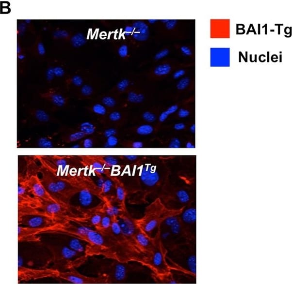
Detection of Mouse BAI1 by Immunocytochemistry/Immunofluorescence
Bai1Tg reduces accumulation of apoptotic corpses in Mertk−/− mice post-torsion. (A) Sertoli cell expression of Bai1, BAI1 signaling pathway genes, and Mertk were analyzed by quantitative RT-PCR. Sertoli cells were isolated from Mertk+/+ (n = 4) and Mertk−/− (n = 2) mice and were cultured for 3 days to expand them prior to RNA isolation. Error bars are standard error of mean (SEM). (B) Representative images of isolated Sertoli cells from Mertk−/− and Mertk−/−Bai1Tg mice were stained for BAI1 to confirm surface expression of the Bai1Tg. (C) Mice (8–12 weeks-old) underwent testicular torsion surgery to induce ischemic injury. Testicular cross sections from sham and torsion testes were stained for cleaved caspase 3 (CC3) (black arrowheads). Images are of representative tubule cross sections from matched sham and torsion testes. (D) The number of CC3 positive cells per tubule cross section was determined by analyzing the entire testicular cross section. Each mouse is represented by individual data points within the bars. Mertk+/+ (n = 5) Mertk−/− (n = 8) Mertk−/−Bai1Tg (n = 9). Error bars represent SEM. Statistical analysis was performed with a Wilcoxon rank-sum test. *p < 0.05, ***p < 0.001. Image collected and cropped by CiteAb from the following publication (https://www.nature.com/articles/s41598-017-15191-1), licensed under a CC-BY license. Not internally tested by R&D Systems.Applications for Human BAI1 Antibody
Neutralization
Formulation, Preparation, and Storage
Purification
Reconstitution
Reconstitute at 0.2 mg/mL in sterile PBS. For liquid material, refer to CoA for concentration.
Formulation
Shipping
Stability & Storage
- 12 months from date of receipt, -20 to -70 °C as supplied.
- 1 month, 2 to 8 °C under sterile conditions after reconstitution.
- 6 months, -20 to -70 °C under sterile conditions after reconstitution.
Calculators
Background: BAI1
References
- Nishimori, H. et al. (1997) Oncogene 15:2145.
- Koh, J.T. et al. (2001) Brain Res. Mol. Brain Res. 87:223.
- Mori, K. et al. (2002) Neurosci. Res. 43:69.
- Kaur, B. et al. (2003) Am. J. Pathol. 162:19.
- Park, D. et al. (2007) Nature 450:430.
- Duda, D.G. et al. (2002) Br. J. Cancer 86:490.
- Lee, J.H. et al. (2001) Int. J. Oncol. 18:355.
- Fukushima, Y. et al. (1998) Int. J. Oncol. 13:967.
- Kaur, B. et al. (2005) Oncogene 24:3632.
- Koh, J.T. et al. (2004) Exp. Cell Res. 294:172.
- Kaur, B. et al. (2009) Cancer Res. 69:1212.
- Kang, X. et al. (2006) Cancer Gene Ther. 13:385.
- Hatanaka, H. et al. (2000) Int. J. Mol. Med. 5:181.
Long Name
Alternate Names
Gene Symbol
UniProt
Additional BAI1 Products
Product Documents for Human BAI1 Antibody
Certificate of Analysis
To download a Certificate of Analysis, please enter a lot or batch number in the search box below.
Note: Certificate of Analysis not available for kit components.
Product Specific Notices for Human BAI1 Antibody
For research use only
Related Research Areas
Citations for Human BAI1 Antibody
 Powered by Bioz
Customer Reviews for Human BAI1 Antibody
There are currently no reviews for this product. Be the first to review Human BAI1 Antibody and earn rewards!
Have you used Human BAI1 Antibody?
Submit a review and receive an Amazon gift card!
$25/€18/£15/$25CAN/¥2500 for a review with an image
$10/€7/£6/$10CAN/¥1110 for a review without an image
Submit a review