

Scientific images may be resized, cropped, or adjusted for brightness or contrast for presentation purposes. These updates are limited to presentation and do not affect the underlying scientific interpretation of the data. Where available, additional source data may be provided upon request.
Key Product Details
Species Reactivity
Applications
Label
Antibody Source
Product Specifications
Immunogen
Phe34-Ser230
Accession # P98173
Specificity
Clonality
Host
Isotype
Scientific Data Images for Human FAM3A Antibody
Detection of FAM3A by Immunocytochemistry/ Immunofluorescence
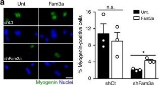
Fam3a rescues impaired commitment of Stat3 knockout (KO) muscle stem cells (MuSCs) in vitro and in vivo. a Immunofluorescence analysis and quantification of myogenin expression in MuSCs infected with shCt or shFam3a coding lentiviruses and cultured in growth conditions for 72 h in the presence or absence of recombinant Fam3a (1000 ng/ml) (n = 3 independent experiments). Scale bar 25 μm. b Immunofluorescence analysis and quantification of myogenin expression in control and Stat3 KO MuSCs cultured in growth conditions for 72 h in the presence or absence of recombinant Fam3a (1000 ng/ml) (n = 5 independent experiments). Scale bar 25 μm. c–e Measurement of the oxygen consumption rate (OCR) and the extracellular acidification rate (ECAR) of Stat3 KO MuSCs cultured in growth conditions for 3 days in the presence or absence of recombinant Fam3a (1000 ng/ml) (n = 5 independent experiments). The Cell Mito Stress Test was performed using the Seahorse XFp Analyzer. f Scheme of the experimental design for the intramuscular Fam3a treatment in vivo. g, h Immunofluorescence analysis and quantification of Pax7- and myogenin-positive cells in transversal sections of the tibialis anterior muscles of Pax7-CreERWT;Stat3f/f and Pax7-CreER;Stat3f/f male mice 6 days after barium chloride injury. Vehicle or 1 μg of Fam3a recombinant protein was injected intramuscularly 1 day after inducing injury (n = 3–4 animals). Scale bar 25 μm. Data are represented as mean ± SEM (Student’s t test or two-way analysis of variance; *p < 0.05, **p < 0.01, ****p < 0.0001) Image collected and cropped by CiteAb from the following open publication (https://pubmed.ncbi.nlm.nih.gov/30996264), licensed under a CC-BY license. Not internally tested by R&D Systems.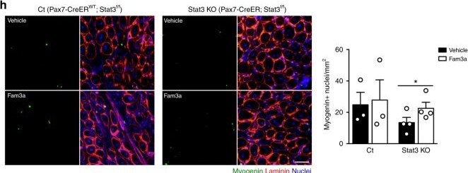
Detection of FAM3A by Immunohistochemistry
Fam3a rescues impaired commitment of Stat3 knockout (KO) muscle stem cells (MuSCs) in vitro and in vivo. a Immunofluorescence analysis and quantification of myogenin expression in MuSCs infected with shCt or shFam3a coding lentiviruses and cultured in growth conditions for 72 h in the presence or absence of recombinant Fam3a (1000 ng/ml) (n = 3 independent experiments). Scale bar 25 μm. b Immunofluorescence analysis and quantification of myogenin expression in control and Stat3 KO MuSCs cultured in growth conditions for 72 h in the presence or absence of recombinant Fam3a (1000 ng/ml) (n = 5 independent experiments). Scale bar 25 μm. c–e Measurement of the oxygen consumption rate (OCR) and the extracellular acidification rate (ECAR) of Stat3 KO MuSCs cultured in growth conditions for 3 days in the presence or absence of recombinant Fam3a (1000 ng/ml) (n = 5 independent experiments). The Cell Mito Stress Test was performed using the Seahorse XFp Analyzer. f Scheme of the experimental design for the intramuscular Fam3a treatment in vivo. g, h Immunofluorescence analysis and quantification of Pax7- and myogenin-positive cells in transversal sections of the tibialis anterior muscles of Pax7-CreERWT;Stat3f/f and Pax7-CreER;Stat3f/f male mice 6 days after barium chloride injury. Vehicle or 1 μg of Fam3a recombinant protein was injected intramuscularly 1 day after inducing injury (n = 3–4 animals). Scale bar 25 μm. Data are represented as mean ± SEM (Student’s t test or two-way analysis of variance; *p < 0.05, **p < 0.01, ****p < 0.0001) Image collected and cropped by CiteAb from the following open publication (https://pubmed.ncbi.nlm.nih.gov/30996264), licensed under a CC-BY license. Not internally tested by R&D Systems.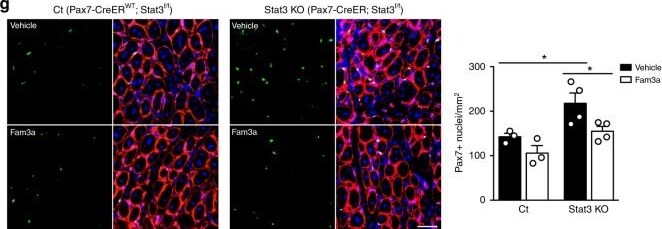
Detection of FAM3A by Immunohistochemistry
Fam3a rescues impaired commitment of Stat3 knockout (KO) muscle stem cells (MuSCs) in vitro and in vivo. a Immunofluorescence analysis and quantification of myogenin expression in MuSCs infected with shCt or shFam3a coding lentiviruses and cultured in growth conditions for 72 h in the presence or absence of recombinant Fam3a (1000 ng/ml) (n = 3 independent experiments). Scale bar 25 μm. b Immunofluorescence analysis and quantification of myogenin expression in control and Stat3 KO MuSCs cultured in growth conditions for 72 h in the presence or absence of recombinant Fam3a (1000 ng/ml) (n = 5 independent experiments). Scale bar 25 μm. c–e Measurement of the oxygen consumption rate (OCR) and the extracellular acidification rate (ECAR) of Stat3 KO MuSCs cultured in growth conditions for 3 days in the presence or absence of recombinant Fam3a (1000 ng/ml) (n = 5 independent experiments). The Cell Mito Stress Test was performed using the Seahorse XFp Analyzer. f Scheme of the experimental design for the intramuscular Fam3a treatment in vivo. g, h Immunofluorescence analysis and quantification of Pax7- and myogenin-positive cells in transversal sections of the tibialis anterior muscles of Pax7-CreERWT;Stat3f/f and Pax7-CreER;Stat3f/f male mice 6 days after barium chloride injury. Vehicle or 1 μg of Fam3a recombinant protein was injected intramuscularly 1 day after inducing injury (n = 3–4 animals). Scale bar 25 μm. Data are represented as mean ± SEM (Student’s t test or two-way analysis of variance; *p < 0.05, **p < 0.01, ****p < 0.0001) Image collected and cropped by CiteAb from the following open publication (https://pubmed.ncbi.nlm.nih.gov/30996264), licensed under a CC-BY license. Not internally tested by R&D Systems.Applications for Human FAM3A Antibody
Western Blot
Sample: Recombinant Human FAM3A
Formulation, Preparation, and Storage
Purification
Reconstitution
Reconstitute at 0.2 mg/mL in sterile PBS. For liquid material, refer to CoA for concentration.
Formulation
Shipping
Stability & Storage
- 12 months from date of receipt, -20 to -70 °C as supplied.
- 1 month, 2 to 8 °C under sterile conditions after reconstitution.
- 6 months, -20 to -70 °C under sterile conditions after reconstitution.
Calculators
Background: FAM3A
Long Name
Alternate Names
Entrez Gene IDs
Gene Symbol
UniProt
Additional FAM3A Products
Product Documents for Human FAM3A Antibody
Certificate of Analysis
To download a Certificate of Analysis, please enter a lot or batch number in the search box below.
Note: Certificate of Analysis not available for kit components.
Product Specific Notices for Human FAM3A Antibody
For research use only
Related Research Areas
Customer Reviews for Human FAM3A Antibody
There are currently no reviews for this product. Be the first to review Human FAM3A Antibody and earn rewards!
Have you used Human FAM3A Antibody?
Submit a review and receive an Amazon gift card!
$25/€18/£15/$25CAN/¥2500 for a review with an image
$10/€7/£6/$10CAN/¥1110 for a review without an image
Submit a review
Protocols
Find general support by application which include: protocols, troubleshooting, illustrated assays, videos and webinars.
- Cellular Response to Hypoxia Protocols
- R&D Systems Quality Control Western Blot Protocol
- Troubleshooting Guide: Western Blot Figures
- Western Blot Conditions
- Western Blot Protocol
- Western Blot Protocol for Cell Lysates
- Western Blot Troubleshooting
- Western Blot Troubleshooting Guide
- View all Protocols, Troubleshooting, Illustrated assays and Webinars