
Lysophosphatidic acid receptor 1, LPAR1, also known as EDG-2, is a G protein-coupled receptor that binds the lipid signaling molecule lysophosphatidic acid (LPA). EDG molecules are G protein-coupled receptors that bind plasma lysophospholipids. The EDG family consists of two subfamilies; the S1P (sphingosine-1-phosphate) subfamily consisting of EDG-1, 3, 5, 6, and 8, and the LPA subfamily that contains EDG-2, 4 and 7. The S1P family regulates essential cellular processes such as proliferation, migration, cytoskeletal organization, and differentiation.
Human LPAR1/LPA1/EDG-2 Antibody
R&D Systems | Catalog # AF9963

Scientific images may be resized, cropped, or adjusted for brightness or contrast for presentation purposes. These updates are limited to presentation and do not affect the underlying scientific interpretation of the data. Where available, additional source data may be provided upon request.
Key Product Details
Species Reactivity
Validated:
Human
Cited:
Human, Mouse
Applications
Validated:
Western Blot
Cited:
Immunohistochemistry, Western Blot
Label
Unconjugated
Antibody Source
Polyclonal Goat IgG
Loading...
Product Specifications
Immunogen
E. coli-derived recombinant human LPAR-1 CD33 protein
containing the sequences for the four extracellular domains, amino acids (aa) 1-50, aa112-125,
aa190-205 and aa281-294
Accession # Q92633
Accession # Q92633
Specificity
Detects human LPAR-1/LPA
Clonality
Polyclonal
Host
Goat
Isotype
IgG
Scientific Data Images for Human LPAR1/LPA1/EDG-2 Antibody
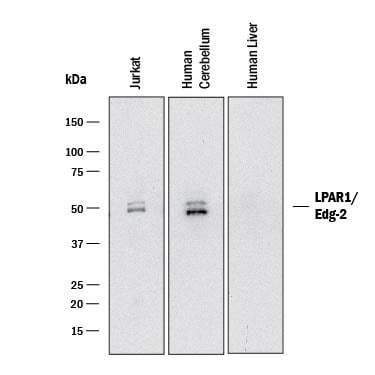
Detection of Human LPAR1/LPA1/EDG-2 by Western Blot.
Western blot shows lysates of Jurkat human acute T cell leukemia cell line, human brain (cerebellum) tissue, and human liver tissue (negative control). PVDF membrane was probed with 2 µg/mL of Goat Anti-Human LPAR1/LPA1/EDG-2 Antigen Affinity-purified Polyclonal Antibody (Catalog # AF9963) followed by HRP-conjugated Anti-Goat IgG Secondary Antibody (Catalog # HAF017). Specific bands were detected for LPAR1/LPA1/EDG-2 at approximately 48-51 kDa (as indicated). This experiment was conducted under reducing conditions and using Immunoblot Buffer Group 1.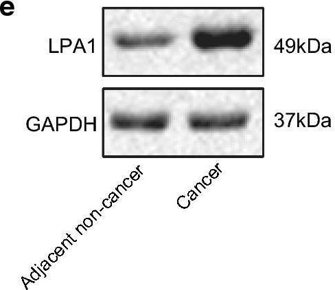
Detection of LPAR1/LPA1/EDG-2 by Western Blot
miR-367 downregulates LPA1 expression. a Bioinformatics prediction of binding sites for miR-367 and LPA1. b Luminescence activity of LPA1 by dual luciferase reporter gene assay. c RIP assay detection of the binding of miR-367 and LPA1 to Ago2. d Detection of relative expression of LPA1 in cancer tissues and adjacent normal tissues by RT-qPCR. e Analysis of protein level of LPA1 in cancer and adjacent normal tissues by western blot analysis. f Statistical expression of protein level of LPA1. g Correlation analysis between miR-367 and LPA1. h RT-qPCR results of the relative expression level of LPA1 in each group of cells. i Western blot analysis of LPA1 protein levels in each group of cells. j Statistical protein expression level of LPA1. Measurement data were expressed as mean ± standard deviation. Data in compliance with normal distribution and homogeneity between two groups were compared using t test. Comparisons among multiple groups were conducted by one-way ANOVA followed by Tukey’s post hoc test. *p < 0.05 vs. normal tissues or cells; #p < 0.05 vs. Inhibitor NC The experiment was repeated three times independently Image collected and cropped by CiteAb from the following open publication (https://pubmed.ncbi.nlm.nih.gov/33024414), licensed under a CC-BY license. Not internally tested by R&D Systems.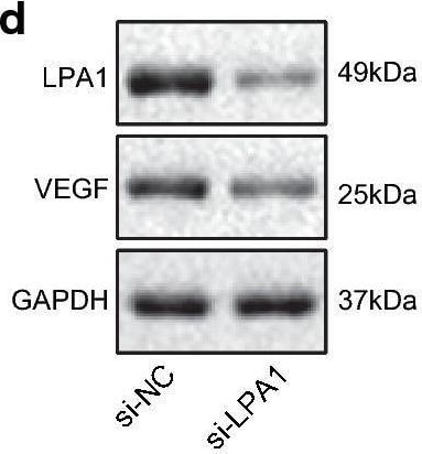
Detection of LPAR1/LPA1/EDG-2 by Western Blot
Low expression of LPA1 inhibits tumorigenic ability of ovarian cancer in nude mice. a Volume of mice tumor. b Weight of mice tumor. c RT-qPCR detection results of mRNA levels of related factors in tumor tissues. d Western blot analysis of the protein levels of related factors in tumor tissues. e Statistical graph of protein levels of related factors. f Immunohistochemistry for detecting CD34 expression (200×). Measurement data were expressed as mean ± standard deviation. Data in compliance with normal distribution and homogeneity between two groups were compared using t test. Comparisons among multiple groups were conducted by one-way ANOVA followed by Tukey’s post hoc test. Statistics of tumor volume at different time points was analyzed using repeated measures ANOVA. *p < 0.05 vs. normal tissues or cells. The experiment was repeated three times independently Image collected and cropped by CiteAb from the following open publication (https://pubmed.ncbi.nlm.nih.gov/33024414), licensed under a CC-BY license. Not internally tested by R&D Systems.Applications for Human LPAR1/LPA1/EDG-2 Antibody
Application
Recommended Usage
Western Blot
2 µg/mL
Sample: Jurkat human acute T cell leukemia cell line and Human brain (cerebellum) tissue
Sample: Jurkat human acute T cell leukemia cell line and Human brain (cerebellum) tissue
Formulation, Preparation, and Storage
Purification
Antigen Affinity-purified
Reconstitution
Reconstitute at 0.5 mg/mL in sterile PBS. For liquid material, refer to CoA for concentration.
Loading...
Formulation
Lyophilized from a 0.2 μm filtered solution in PBS with Trehalose. *Small pack size (SP) is supplied either lyophilized or as a 0.2 µm filtered solution in PBS.
Shipping
Lyophilized product is shipped at ambient temperature. Liquid small pack size (-SP) is shipped with polar packs. Upon receipt, store immediately at the temperature recommended below.
Stability & Storage
Use a manual defrost freezer and avoid repeated freeze-thaw cycles.
- 12 months from date of receipt, -20 to -70 °C as supplied.
- 1 month, 2 to 8 °C under sterile conditions after reconstitution.
- 6 months, -20 to -70 °C under sterile conditions after reconstitution.
Calculators
Background: LPAR1/LPA1/EDG-2
Long Name
Lysophosphatidic Acid Receptor 1
Alternate Names
EDG2, GPCR26, GPR26, LPA1, rec.1.3, vzg-1
Gene Symbol
LPAR1
UniProt
Additional LPAR1/LPA1/EDG-2 Products
Product Documents for Human LPAR1/LPA1/EDG-2 Antibody
Certificate of Analysis
To download a Certificate of Analysis, please enter a lot or batch number in the search box below.
Note: Certificate of Analysis not available for kit components.
Product Specific Notices for Human LPAR1/LPA1/EDG-2 Antibody
For research use only
Related Research Areas
Citations for Human LPAR1/LPA1/EDG-2 Antibody
 Powered by Bioz
Customer Reviews for Human LPAR1/LPA1/EDG-2 Antibody
There are currently no reviews for this product. Be the first to review Human LPAR1/LPA1/EDG-2 Antibody and earn rewards!
Have you used Human LPAR1/LPA1/EDG-2 Antibody?
Submit a review and receive an Amazon gift card!
$25/€18/£15/$25CAN/¥2500 for a review with an image
$10/€7/£6/$10CAN/¥1110 for a review without an image
Submit a review
Protocols
Find general support by application which include: protocols, troubleshooting, illustrated assays, videos and webinars.
- Cellular Response to Hypoxia Protocols
- R&D Systems Quality Control Western Blot Protocol
- Troubleshooting Guide: Western Blot Figures
- Western Blot Conditions
- Western Blot Protocol
- Western Blot Protocol for Cell Lysates
- Western Blot Troubleshooting
- Western Blot Troubleshooting Guide
- View all Protocols, Troubleshooting, Illustrated assays and Webinars
Loading...