
Annexin A13 (ANXA13), also known as Annexin XIII and Intestine-Specific Annexin, is a member of the Annexin protein family. Two isoforms have been identified resulting from alternate splicing. Isoform A consists of 316 amino acids and has a predicted molecular weight of 36 kDa whereas isoform B is composed of 357 amino acids and has a predicted molecular weight of 40 kDa. Annexins are a family of Calcium-dependent phospholipid-binding proteins that are preferentially located on the cytosolic face of the plasma membrane. The Annexin’s have a molecular weight of approximately 35 to 40 kDa and consist of a unique amino terminal domain followed by a homologous C-terminal core domain containing the calcium-dependent phospholipid-binding sites. The C terminal domain is comprised of four 60‑70 amino acid repeats, known as annexin repeats or an endonexin fold (Annexin A6 contains 8 annexin repeats). The four annexin repeats form a highly alpha -helical, tightly packed disc known as the annexin domain, which binds to phospholipids in the membrane in a calcium-dependent manner. Members of the annexin family play a role in cytoskeletal interactions, phospholipase inhibition, regulation of cellular growth, and intracellular signal transduction pathways. The specific function of Annexin A13 has not been determined but it is associated with the plasma membrane of undifferentiated, proliferating endothelial cells and differentiated villus enterocytes.

Scientific images may be resized, cropped, or adjusted for brightness or contrast for presentation purposes. These updates are limited to presentation and do not affect the underlying scientific interpretation of the data. Where available, additional source data may be provided upon request.
Key Product Details
Species Reactivity
Validated:
Cited:
Applications
Validated:
Cited:
Label
Antibody Source
Product Specifications
Immunogen
Met1-His316
Accession # P27216
Specificity
Clonality
Host
Isotype
Scientific Data Images for Annexin A13 Antibody
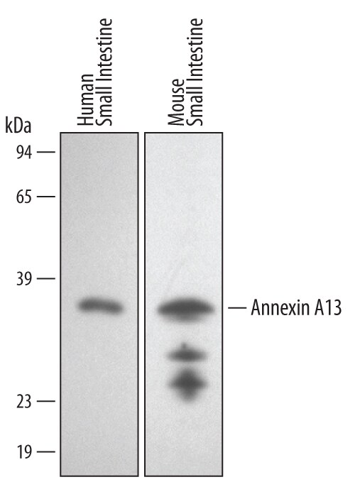
Detection of Human/Mouse Annexin A13 by Western Blot.
Western blot shows lysates of human and mouse small intestine tissue. PVDF membrane was probed with 1 µg/mL of Human/Mouse Annexin A13 Antigen Affinity-purified Polyclonal Antibody (Catalog # AF4149) followed by HRP-conjugated Anti-Goat IgG Secondary Antibody (Catalog # HAF017). A specific band was detected for Annexin A13 at approximately 36 kDa (as indicated). This experiment was conducted under reducing conditions and using Immunoblot Buffer Group 1.Applications for Annexin A13 Antibody
Western Blot
Sample: Human and mouse small intestine tissue
Formulation, Preparation, and Storage
Purification
Reconstitution
Reconstitute at 0.2 mg/mL in sterile PBS. For liquid material, refer to CoA for concentration.
Formulation
Shipping
Stability & Storage
- 12 months from date of receipt, -20 to -70 °C as supplied.
- 1 month, 2 to 8 °C under sterile conditions after reconstitution.
- 6 months, -20 to -70 °C under sterile conditions after reconstitution.
Calculators
Background: Annexin A13
Alternate Names
Gene Symbol
UniProt
Additional Annexin A13 Products
Product Documents for Annexin A13 Antibody
Certificate of Analysis
To download a Certificate of Analysis, please enter a lot or batch number in the search box below.
Note: Certificate of Analysis not available for kit components.
Product Specific Notices for Annexin A13 Antibody
For research use only
Related Research Areas
Citations for Annexin A13 Antibody
 Powered by Bioz
Customer Reviews for Annexin A13 Antibody
There are currently no reviews for this product. Be the first to review Annexin A13 Antibody and earn rewards!
Have you used Annexin A13 Antibody?
Submit a review and receive an Amazon gift card!
$25/€18/£15/$25CAN/¥2500 for a review with an image
$10/€7/£6/$10CAN/¥1110 for a review without an image
Submit a review
Protocols
Find general support by application which include: protocols, troubleshooting, illustrated assays, videos and webinars.
- Cellular Response to Hypoxia Protocols
- R&D Systems Quality Control Western Blot Protocol
- Troubleshooting Guide: Western Blot Figures
- Western Blot Conditions
- Western Blot Protocol
- Western Blot Protocol for Cell Lysates
- Western Blot Troubleshooting
- Western Blot Troubleshooting Guide
- View all Protocols, Troubleshooting, Illustrated assays and Webinars