
LDHA (lactate dehydrogenase A chain; also LDH-M and PIG-19) is a 34-36 kDa member of the LDH family of enzymes. It is a part of a cytoplasmic complex that is found principally in hepatocytes and skeletal muscle. The LDHA complex catalyzes the conversion of pyruvate to lactate, thereby generating NAD, the final step in anerobic glycolysis. This NAD is essential for the subsequent generation of ATP. Human LDHA is 332 amino acids (aa) in length. It contains an N-terminal coenzyme binding region, a central catalytic site, and at least nine utilized Lys acetylation and two Tyr phosphorylation sites. It also undergoes ISGylation where a 17 kDa product of the ISG15 gene is covalently attached to LDHA in a ubiquitin-like manner. LDHA forms tetramers composed of two dimers of varying composition. The tetramer may be homotetrameric (four monomers), or contain from one to three substitute LDHB monomers typically found in heart muscle. There are multiple splice variants. One possesses a five aa substitution for aa 237-332, a second contains a 45 aa substitution for aa 230-332, a third shows a deletion of aa 82-139, while a fourth utilizes an alternative start site 29 aa upstream of the standard site. Over aa 2-92, human LDHA shares 93% aa sequence identity with mouse LDHA.
Lactate Dehydrogenase A/LDHA Antibody
R&D Systems | Catalog # AF7304

Scientific images may be resized, cropped, or adjusted for brightness or contrast for presentation purposes. These updates are limited to presentation and do not affect the underlying scientific interpretation of the data. Where available, additional source data may be provided upon request.
Key Product Details
Species Reactivity
Validated:
Cited:
Applications
Validated:
Cited:
Label
Antibody Source
Product Specifications
Immunogen
Ala2-Val92
Accession # P00338
Specificity
Clonality
Host
Isotype
Scientific Data Images for Lactate Dehydrogenase A/LDHA Antibody
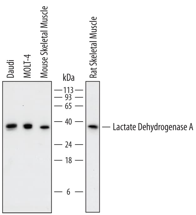
Detection of Human, Mouse, and Rat Lactate Dehydrogenase A/LDHA by Western Blot.
Western blot shows lysates of Daudi human Burkitt's lymphoma cell line, MOLT-4 human acute lymphoblastic leukemia cell line, mouse skeletal muscle tissue, and rat skeletal muscle tissue. PVDF membrane was probed with 0.5 µg/mL of Sheep Anti-Human/Mouse/Rat Lactate Dehydrogenase A/LDHA Antigen Affinity-purified Polyclonal Antibody (Catalog # AF7304) followed by HRP-conjugated Anti-Sheep IgG Secondary Antibody (Catalog # HAF016). A specific band was detected for Lactate Dehydrogenase A/LDHA at approximately 36 kDa (as indicated). This experiment was conducted under reducing conditions and using Immunoblot Buffer Group 1.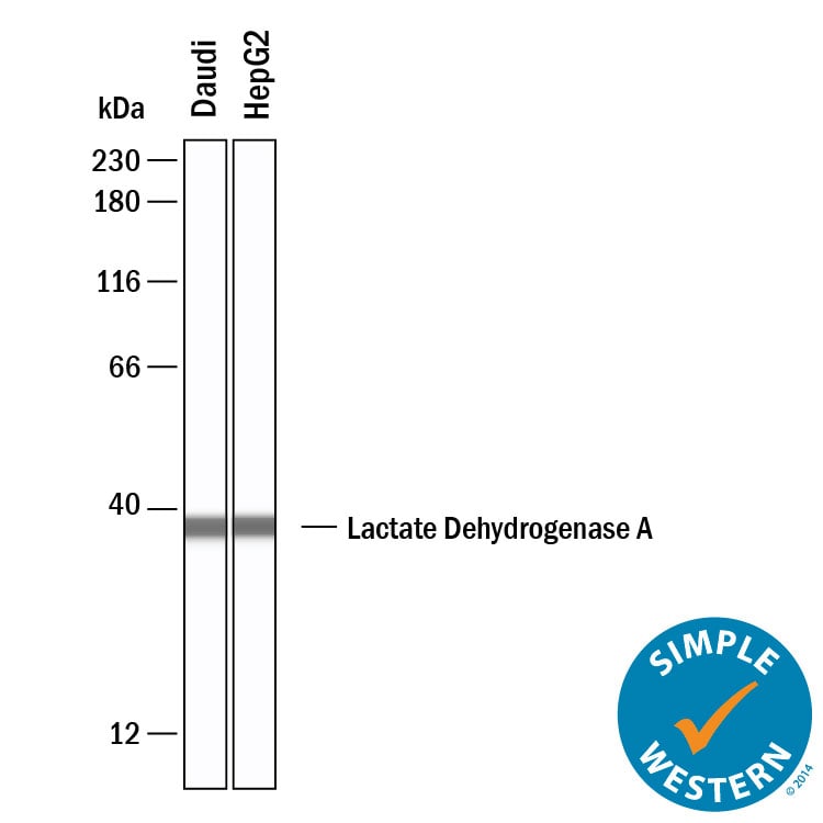
Detection of Human Lactate Dehydrogenase A/LDHA by Simple WesternTM.
Simple Western shows lysates of Daudi human Burkitt's lymphoma cell line and HepG2 human hepatocellular carcinoma cell line, loaded at 0.5 mg/ml. A specific band was detected for Lactate Dehydrogenase A/LDHA at approximately 38 kDa (as indicated) using 50 µg/mL of Sheep Anti-Human/Mouse/Rat Lactate Dehydrogenase A/LDHA Antigen Affinity-purified Polyclonal Antibody (Catalog # AF7304). This experiment was conducted under reducing conditions and using the 12-230 kDa separation system.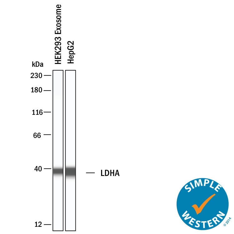
Detection of Human Lactate Dehydrogenase A/LDHA by Simple WesternTM.
Simple Western shows lysates of Exosome Standards (HEK293 cell line) (NBP3-11684) and HepG2 human hepatocellular carcinoma cell line, loaded at 0.5 mg/ml. A specific band was detected for Lactate Dehydrogenase A/LDHA at approximately 38 kDa (as indicated) using 50 µg/mL of Sheep Anti-Human/Mouse/Rat Lactate Dehydrogenase A/LDHA Antigen Affinity-purified Polyclonal Antibody (Catalog # AF7304). This experiment was conducted under reducing conditions and using the 12-230 kDa separation system.Applications for Lactate Dehydrogenase A/LDHA Antibody
Simple Western
Sample: Daudi human Burkitt's lymphoma cell line and HepG2 human hepatocellular carcinoma cell line
Western Blot
Sample: Daudi human Burkitt's lymphoma cell line, MOLT‑4 human acute lymphoblastic leukemia cell line, mouse skeletal muscle tissue, and rat skeletal muscle tissue
Formulation, Preparation, and Storage
Purification
Reconstitution
Sterile PBS to a final concentration of 0.2 mg/mL. For liquid material, refer to CoA for concentration.
Formulation
Shipping
Stability & Storage
- 12 months from date of receipt, -20 to -70 °C as supplied.
- 1 month, 2 to 8 °C under sterile conditions after reconstitution.
- 6 months, -20 to -70 °C under sterile conditions after reconstitution.
Calculators
Background: Lactate Dehydrogenase A/LDHA
Alternate Names
Gene Symbol
UniProt
Additional Lactate Dehydrogenase A/LDHA Products
Product Documents for Lactate Dehydrogenase A/LDHA Antibody
Certificate of Analysis
To download a Certificate of Analysis, please enter a lot or batch number in the search box below.
Note: Certificate of Analysis not available for kit components.
Product Specific Notices for Lactate Dehydrogenase A/LDHA Antibody
For research use only
Related Research Areas
Citations for Lactate Dehydrogenase A/LDHA Antibody
 Powered by Bioz
Customer Reviews for Lactate Dehydrogenase A/LDHA Antibody
There are currently no reviews for this product. Be the first to review Lactate Dehydrogenase A/LDHA Antibody and earn rewards!
Have you used Lactate Dehydrogenase A/LDHA Antibody?
Submit a review and receive an Amazon gift card!
$25/€18/£15/$25CAN/¥2500 for a review with an image
$10/€7/£6/$10CAN/¥1110 for a review without an image
Submit a review
Protocols
Find general support by application which include: protocols, troubleshooting, illustrated assays, videos and webinars.
- Cellular Response to Hypoxia Protocols
- R&D Systems Quality Control Western Blot Protocol
- Troubleshooting Guide: Western Blot Figures
- Western Blot Conditions
- Western Blot Protocol
- Western Blot Protocol for Cell Lysates
- Western Blot Troubleshooting
- Western Blot Troubleshooting Guide
- View all Protocols, Troubleshooting, Illustrated assays and Webinars