
NADPH Oxidase 4/NOX4, also known as Renal NADPH Oxidase/RENOX and Kidney Oxidase 1/KOX1, is a 66 to 75 kDa member of the NOX family of NADPH oxidases. NOX family members are a major source of reactive oxygen species (ROS), including hydrogen peroxide and superoxide, and are critical mediators of redox signaling. NOX4 is strongly expressed in the kidney, especially in the proximal convoluted tubule cells of the renal cortex, and may function as an oxygen sensor that regulates the synthesis of erythropoietin. Expression is also high in endothelial cells, and because aortas from deficient mice exhibit increased inflammation, hypertrophy and endothelial dysfunction, NOX4 may function to protect the vasculature during ischemic or inflammatory stress. Human NOX4 is 578 amino acids (aa) in length, with several additional smaller isoforms identified. Over aa 450-578, human NOX4 has 92% sequence identity with mouse NOX4, and 91% identity with rat NOX4.

Scientific images may be resized, cropped, or adjusted for brightness or contrast for presentation purposes. These updates are limited to presentation and do not affect the underlying scientific interpretation of the data. Where available, additional source data may be provided upon request.
Key Product Details
Species Reactivity
Validated:
Cited:
Applications
Validated:
Cited:
Label
Antibody Source
Product Specifications
Immunogen
Lys450-Ser578
Accession # Q9NPH5
Specificity
Clonality
Host
Isotype
Scientific Data Images for Human Nox4 Antibody
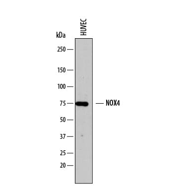
Detection of Human Nox4 by Western Blot.
Western blot shows lysates of HUVEC human umbilical vein endothelial cells. PVDF membrane was probed with 2 µg/mL of Sheep Anti-Human Nox4 Antigen Affinity-purified Polyclonal Antibody (Catalog # AF8158) followed by HRP-conjugated Anti-Sheep IgG Secondary Antibody (Catalog # HAF016). A specific band was detected for Nox4 at approximately 75 kDa (as indicated). This experiment was conducted under reducing conditions and using Immunoblot Buffer Group 1.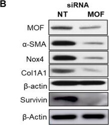
Detection of Nox4 by Western Blot
Silencing Mof decreases H4K16Ac levels, and down-regulates multiple pro-fibrotic genes that are associated with H4K16Ac in IPF lung fibroblasts. Representative figures of IPF lung fibroblasts with siRNA non-targeting (NT) control or Mof. (A) Histone H4K16Ac and H4K20Me3 levels were examined by western blots with histone extracts. H4 is loading control. Mof expression in these cells is shown in B. (B) The whole cell lysate were collected for western blots (Survivin is on a separate blot, the Mof knockdown was shown in online Figure S3B); and (C) RNA were collected for real-time RT-PCR to examine pro-fibrotic genes alpha -SMA, Nox4, Col1A1 and survivin expression at protein (B) or mRNA (C) levels. (D) Schematic of the relative locations of the PCR primers sets used to examine the association of H4K16Ac and the promoter regions with the genes listed in E-H by ChIP assays, primer sequences are listed in Table S2. (E-H) Primary IPF lung fibroblasts with siRNA NT or Mof (treated as in A), were subject to ChIP assays. DNA was immunoprecipitated with H4K16Ac antibody. The relative levels of PCR products were represented by bars of the association of H4K16Ac with promoter region of alpha -SMA (E), Nox4 (F), Col1A1 (G) or Survivin (H). Quantitative PCR data were analyzed by 2 delta delta Ct method, normalized to input DNA, expressed as fold changes relative to primer set A or B. Negative control represents IgG pull-down. The values are expressed as mean ± SD from average of three independent experiments of one representative cell line. *P < 0.05, siRNA Mof vs siRNA NT control. Image collected and cropped by CiteAb from the following open publication (https://pubmed.ncbi.nlm.nih.gov/34976199), licensed under a CC-BY license. Not internally tested by R&D Systems.Applications for Human Nox4 Antibody
Western Blot
Sample: HUVEC human umbilical vein endothelial cells
Reviewed Applications
Read 1 review rated 3 using AF8158 in the following applications:
Formulation, Preparation, and Storage
Purification
Reconstitution
Reconstitute at 0.2 mg/mL in sterile PBS. For liquid material, refer to CoA for concentration.
Formulation
Shipping
Stability & Storage
- 12 months from date of receipt, -20 to -70 °C as supplied.
- 1 month, 2 to 8 °C under sterile conditions after reconstitution.
- 6 months, -20 to -70 °C under sterile conditions after reconstitution.
Calculators
Background: Nox4
Long Name
Alternate Names
Gene Symbol
UniProt
Additional Nox4 Products
Product Documents for Human Nox4 Antibody
Certificate of Analysis
To download a Certificate of Analysis, please enter a lot or batch number in the search box below.
Note: Certificate of Analysis not available for kit components.
Product Specific Notices for Human Nox4 Antibody
For research use only
Related Research Areas
Citations for Human Nox4 Antibody
 Powered by Bioz
Customer Reviews for Human Nox4 Antibody (1)
Have you used Human Nox4 Antibody?
Submit a review and receive an Amazon gift card!
$25/€18/£15/$25CAN/¥2500 for a review with an image
$10/€7/£6/$10CAN/¥1110 for a review without an image
Submit a review
Customer Images
-
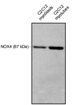
Application: Western BlotSample Tested: Differentiated C2C12 myoblast cells and C2C12 mouse myoblast cell lineSpecies: MouseVerified Customer | Posted 11/24/2017Representative Western blot image showing detection of NOX4 in C2C2C12 mouse myoblasts and myotubes. For myoblasts and myotubes, 6 ug of total protein were loaded.Anti-NOX4 - 1:500 Anti-sheep HRP - 1:1000

There are no reviews that match your criteria.
Protocols
Find general support by application which include: protocols, troubleshooting, illustrated assays, videos and webinars.
- Cellular Response to Hypoxia Protocols
- R&D Systems Quality Control Western Blot Protocol
- Troubleshooting Guide: Western Blot Figures
- Western Blot Conditions
- Western Blot Protocol
- Western Blot Protocol for Cell Lysates
- Western Blot Troubleshooting
- Western Blot Troubleshooting Guide
- View all Protocols, Troubleshooting, Illustrated assays and Webinars

