
TNF receptor 1 (TNF RI; also called TNF R-p55/p60 and TNFRSF1A) is a type I transmembrane protein member of the TNF receptor superfamily member, designated TNFRSF1A (1, 2). Both TNF RI and TNF RII (TNFRSF1B) are widely expressed and contain four TNF- alpha trimer-binding cysteine-rich domains (CRD) in their extracellular domains (ECD). However, TNF RI is thought to mediate most of the cellular effects of TNF-alpha (3). It is essential for proper development of lymph node germinal centers and Peyer’s patches, and for combating intracellular pathogens such as Listeria (1 - 3). TNF RI is also a receptor for TNF-beta /TNFSF1B (lymphotoxin-alpha ) (4). TNF RI is present on the cell surface as a trimer of 55 kDa subunits (4, 5). TNF-alpha induces sequestering of TNF RI in lipid rafts, where it activates NF kappa B and is cleaved by ADAM-17/TACE (9, 10). Release of the 28 - 34 kDa TNF RI ECD also occurs constitutively and in response to products of pathogens such as LPS, CpG DNA or S. aureus protein A (1, 6 - 8). Full-length TNF RI may also be released in exosome-like vesicles (11). Release helps to resolve inflammatory reactions, since it down-regulates cell surface TNF RI and provides soluble TNF RI to bind TNF-alpha (6, 12, 13). Exclusion from lipid rafts causes endocytosis of TNF RI complexes and induces apoptosis (1). Human TNF RI is a 455 amino acid (aa) protein that contains a 21 aa signal sequence, a 190 aa ECD with a PLAD domain(5) that mediates constitutive trimer formation, followed by the four CRD, a 23 aa transmembrane domain, and a 221 aa cytoplasmic sequence that contains a neutral sphingomyelinase activation domain and a death domain (15). The ECD of human TNF RI shows 80%, 80%, 73%, 69% and 70% aa identity with dog, cat, pig, rat and mouse TNF RI, respectively; it shows 23% aa identity with the ECD of TNF RII.
Human TNF RI/TNFRSF1A Antibody
R&D Systems | Catalog # AB-225-PB

Scientific images may be resized, cropped, or adjusted for brightness or contrast for presentation purposes. These updates are limited to presentation and do not affect the underlying scientific interpretation of the data. Where available, additional source data may be provided upon request.
Key Product Details
Species Reactivity
Validated:
Cited:
Applications
Validated:
Cited:
Label
Antibody Source
Product Specifications
Immunogen
Specificity
Clonality
Host
Isotype
Endotoxin Level
Scientific Data Images for Human TNF RI/TNFRSF1A Antibody
Detection of TNF RI/TNFRSF1A by Western Blot
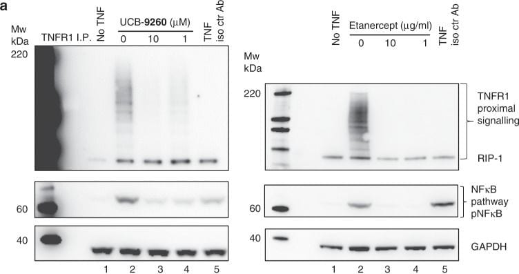
Inhibition of TNF signalling in cells.a Western blots measuring RIP1 ubiquitination following TNFR1 immunoprecipitation from Jurkat cells treated with TNF ± UCB-9260 or etanercept. pNFkB expression was assessed, along with GAPDH for loading control. An isotype control antibody was included as an immunoprecipitation control (lane 5). b TNF (10 pM) or anti-TNFR1 agonist antibody (0.3 mg/mL)-driven NFkB activity was measured following UCB-9260 treatment in Hek-293 cells using a reporter gene assay system. Representative concentration-response curves are shown. Percentage inhibitions for compound dilutions were calculated between a DMSO control and maximum inhibition (by excess biologic anti-TNF, or NFkB inhibitor–TPCA-1). Untransformed data shown in Supplementary Fig. 10c. c UCB-9260 inhibition of TNF (human or mouse) dependent cytotoxicity was measured in mouse L929 cells. Representative concentration-response curves are shown. Percentage inhibition was calculated between unstimulated wells (maximum signal), and wells with TNF, DMSO and actinomycin D (minimum signal). Untransformed data shown in Supplementary Fig. 10d. Source data are provided as a Source Data file. Image collected and cropped by CiteAb from the following open publication (https://pubmed.ncbi.nlm.nih.gov/31857588), licensed under a CC-BY license. Not internally tested by R&D Systems.Applications for Human TNF RI/TNFRSF1A Antibody
Agonist Activity
The ED50 for this effect is typically 10-15 μg/mL for L-929 cells or 5-10 μg/mL for A549 cells.
Western Blot
Sample: Recombinant Human TNF RI/TNFRSF1A (Catalog # 636-R1)
Formulation, Preparation, and Storage
Purification
Reconstitution
Reconstitute at 1 mg/mL in sterile PBS.
Formulation
Shipping
Stability & Storage
- 12 months from date of receipt, -20 to -70 °C as supplied.
- 1 month, 2 to 8 °C under sterile conditions after reconstitution.
- 6 months, -20 to -70 °C under sterile conditions after reconstitution.
Calculators
Background: TNF RI/TNFRSF1A
References
- Pfeffer, K. (2003) Cytokine Growth Factor Rev. 14:185.
- Hehlgans, T. and K. Pfeffer (2005) Immunology 115:1.
- Peschon, J.J. et al. (1998) J. Immunol. 160:943.
- Banner, D.W et al. (1993) Cell 73: 431.
- Chan, F.K. et al. (2000) Science 288:2351.
- Xanthoulea, S. et al. (2004) J. Exp. Med. 200:367.
- Jin, L. et al. (2000) J. Immunol. 165:5153.
- Gomez, M.I. et al. (2006) J. Biol. Chem. 281:20190.
- Legler, D.F. et al. (2003) Immunity 18:655.
- Tellier, E. et al. (2006) Exp. Cell Res. 312:3969.
- Islam, A. et al. (2006) J. Biol. Chem. 281:6860.
- Garton, K.J. et al. (2006) J. Leukoc. Biol. 79:1105.
- McDermott, M.F. et al. (1999) Cell 97:133.
- Schneider-Brachert, W. et al. (2004) Immunity 21:415.
- Schall, T.J. et al. (1990) Cell 61:361.
Long Name
Alternate Names
Gene Symbol
Additional TNF RI/TNFRSF1A Products
Product Documents for Human TNF RI/TNFRSF1A Antibody
Certificate of Analysis
To download a Certificate of Analysis, please enter a lot or batch number in the search box below.
Note: Certificate of Analysis not available for kit components.
Product Specific Notices for Human TNF RI/TNFRSF1A Antibody
For research use only
Citations for Human TNF RI/TNFRSF1A Antibody
 Powered by Bioz
Customer Reviews for Human TNF RI/TNFRSF1A Antibody
There are currently no reviews for this product. Be the first to review Human TNF RI/TNFRSF1A Antibody and earn rewards!
Have you used Human TNF RI/TNFRSF1A Antibody?
Submit a review and receive an Amazon gift card!
$25/€18/£15/$25CAN/¥2500 for a review with an image
$10/€7/£6/$10CAN/¥1110 for a review without an image
Submit a review
Protocols
Find general support by application which include: protocols, troubleshooting, illustrated assays, videos and webinars.
- Cellular Response to Hypoxia Protocols
- R&D Systems Quality Control Western Blot Protocol
- Troubleshooting Guide: Western Blot Figures
- Western Blot Conditions
- Western Blot Protocol
- Western Blot Protocol for Cell Lysates
- Western Blot Troubleshooting
- Western Blot Troubleshooting Guide
- View all Protocols, Troubleshooting, Illustrated assays and Webinars
Associated Pathways


