
Ubiquilin-1 (also known as DA41 and PLIC-1/Protein Linking IAP with Cytoskeleton 1) is a 62-70 kDa intracellular member of the UBL-UBA family of proteins. Consistent with its name, it ubiquitously expressed, and is particularly prominent in skeletal muscle and neurons. Multiple functions have been attributed to ubiquilin-1. For instance, it is now known to be a key player in APP processing. On the cleavage side, ubiquilin-1 binds to PS/presenilin, a component of the gamma -secretase complex that cleaves APP. Ubiquilin-1 appears to target both ubiquitinated single chain PS to the proteosome, and aggregated PS to the aggresome. With respect to APP, ubiquilin-1 serves as a molecular chaperone, binding to and inhibiting APP aggregation. On cells expressing CD47, Ubiquilin-1 acts as an intermediary between integrins, the cytoskeleton and CD47, promoting cell spreading. And in neurons, ubiquilin-1 stabilizes both membrane and GABA subunit stability, enhancing inhibitory synaptic transmission. Human ubiquilin-1 is 589 amino acids (aa) in length. It contains one ubiquitin-like domain (aa 37-111), an S5a binding site (aa 72-105) and a UBA binding region (aa 548-585). There are multiple splice variants, ranging from 19-59 kDa in size. One shows a deletion of aa 418-445, a second contains a three aa substitution for aa 151-589, a third possesses a 29 aa substitution for aa 112-589, and a fourth shows a deletion of aa 61-237 coupled to a second deletion of aa 361‑589. Over aa 546-585, human and mouse ubiquilin-1 are identical in amino acid sequence.

Scientific images may be resized, cropped, or adjusted for brightness or contrast for presentation purposes. These updates are limited to presentation and do not affect the underlying scientific interpretation of the data. Where available, additional source data may be provided upon request.
Key Product Details
Species Reactivity
Applications
Label
Antibody Source
Product Specifications
Immunogen
Arg546-Gly585
Accession # Q9UMX0
Specificity
Clonality
Host
Isotype
Scientific Data Images for Human Ubiquilin 1 Antibody
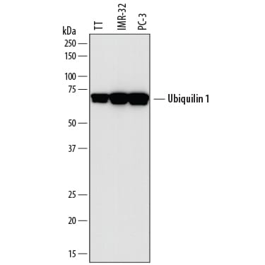
Detection of Human Ubiquilin 1 by Western Blot.
Western blot shows lysates of TT human medullary thyroid cancer cell line, IMR-32 human neuroblastoma cell line, and PC-3 human prostate cancer cell line. PVDF membrane was probed with 2 µg/mL of Sheep Anti-Human Ubiquilin 1 Antigen Affinity-purified Polyclonal Antibody (Catalog # AF7969) followed by HRP-conjugated Anti-Sheep IgG Secondary Antibody (Catalog # HAF016). A specific band was detected for Ubiquilin 1 at approximately 65-70 kDa (as indicated). This experiment was conducted under reducing conditions and using Immunoblot Buffer Group 1.Applications for Human Ubiquilin 1 Antibody
Western Blot
Sample: TT human medullary thyroid cancer cell line, IMR‑32 human neuroblastoma cell line, and PC‑3 human prostate cancer cell line
Formulation, Preparation, and Storage
Purification
Reconstitution
Sterile PBS to a final concentration of 0.2 mg/mL. For liquid material, refer to CoA for concentration.
Formulation
Shipping
Stability & Storage
- 12 months from date of receipt, -20 to -70 °C as supplied.
- 1 month, 2 to 8 °C under sterile conditions after reconstitution.
- 6 months, -20 to -70 °C under sterile conditions after reconstitution.
Calculators
Background: Ubiquilin 1
Alternate Names
Gene Symbol
UniProt
Additional Ubiquilin 1 Products
Product Documents for Human Ubiquilin 1 Antibody
Certificate of Analysis
To download a Certificate of Analysis, please enter a lot or batch number in the search box below.
Note: Certificate of Analysis not available for kit components.
Product Specific Notices for Human Ubiquilin 1 Antibody
For research use only
Customer Reviews for Human Ubiquilin 1 Antibody
There are currently no reviews for this product. Be the first to review Human Ubiquilin 1 Antibody and earn rewards!
Have you used Human Ubiquilin 1 Antibody?
Submit a review and receive an Amazon gift card!
$25/€18/£15/$25CAN/¥2500 for a review with an image
$10/€7/£6/$10CAN/¥1110 for a review without an image
Submit a review
Protocols
Find general support by application which include: protocols, troubleshooting, illustrated assays, videos and webinars.
- Cellular Response to Hypoxia Protocols
- R&D Systems Quality Control Western Blot Protocol
- Troubleshooting Guide: Western Blot Figures
- Western Blot Conditions
- Western Blot Protocol
- Western Blot Protocol for Cell Lysates
- Western Blot Troubleshooting
- Western Blot Troubleshooting Guide
- View all Protocols, Troubleshooting, Illustrated assays and Webinars