MAT1/2A Antibody - BSA Free
Novus Biologicals | Catalog # NB110-94162
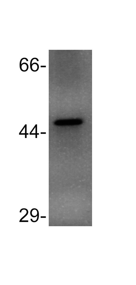
![Western Blot: MAT1/2A AntibodyBSA Free [NB110-94162]](https://resources.rndsystems.com/images/products/MAT1-2A-Antibody-Western-Blot-NB110-94162-img0003.jpg "Western Blot: MAT1/2A AntibodyBSA Free [NB110-94162]")
Key Product Details
Species Reactivity
Validated:
Human, Rat, Bovine, Mouse (Negative), Primate, Zebrafish
Cited:
Mouse
Applications
Validated:
Western Blot, Simple Western
Cited:
Western Blot
Label
Unconjugated
Antibody Source
Polyclonal Rabbit IgG
Format
BSA Free
Loading...
Product Specifications
Immunogen
Synthetic peptide made to an internal portion of the human MAT2A protein (within residues 100-200). [Swiss-Prot# P31153]
Reactivity Notes
Orangutan. Does not react with mouse.
Clonality
Polyclonal
Host
Rabbit
Isotype
IgG
Theoretical MW
43 kDa.
Disclaimer note: The observed molecular weight of the protein may vary from the listed predicted molecular weight due to post translational modifications, post translation cleavages, relative charges, and other experimental factors.
Disclaimer note: The observed molecular weight of the protein may vary from the listed predicted molecular weight due to post translational modifications, post translation cleavages, relative charges, and other experimental factors.
Scientific Data Images for MAT1/2A Antibody - BSA Free
Western Blot: MAT1/2A AntibodyBSA Free [NB110-94162]
Western Blot: MAT1/2A Antibody [NB110-94162] - Detection of MAT2A in HepG2 whole cell lysates using NB110-94162.![Simple Western: MAT1/2A AntibodyBSA Free [NB110-94162]](https://resources.rndsystems.com/images/products/MAT1-2A-Antibody-Simple-Western-NB110-94162-img0004.jpg "Simple Western: MAT1/2A AntibodyBSA Free [NB110-94162]")
Simple Western: MAT1/2A AntibodyBSA Free [NB110-94162]
Simple Western: MAT1/2A Antibody [NB110-94162] - Simple Western lane view shows a specific band for MAT1/2 A in 0.5 mg/ml of HepG2 lysate. This experiment was performed under reducing conditions using the 12-230 kDa separation system.
Western Blot: MAT1/2A Antibody - BSA Free [NB110-94162] -
C1 metabolic pathway and characterization of R6/2 mice. A Folate, Met and BH4 cycles in plants and animals and their associated metabolism. Red lines stand for mammal specific, green lines stand for plant specific while black lines stand for both. All enzymes with protein levels examined by immunoblotting are marked in red. B mHtt protein aggregates in cortex and striatum regions were detected with anti-Htt antibody (mEM48) in 4-week-old male R6/2 and NCAR mice. C, D Quantification analysis of immunoblotting results of GTPCH, DHFR, QDPR, MS, MAT1/2A, AHCY, MTHFR, TPH2, TH, nNOS and ChAT (n = 7). The band intensity of each protein from western blotting D was normalized with gamma -tubulin on the same blot. The ratio was further calculated against NCAR whose relative expression level was set as 1. All data plotted are the average (n = 7) +/- SD. Only one representative western blotting of gamma -tubulin is shown. Original blots of above proteins before cropping are presented in Fig. S11. E Contents of BH4 and BH2, and their ratio in brain tissues and plasma (n = 4, average +/- SD). *p < 0.05; **p < 0.01. ***p < 0.001. Abbreviations used for enzymes: AHCYS-adenosylhomocysteine hydrolase, ChAT choline acetyltransferase, DHFR dihydrofolate reductase, GTPCH GTP cyclohydrolase I, MAT1/2A methionine adenosyltransferase, MS methionine synthase, MTHFR methylene-tetrahydrofolate reductase, nNOS neuronal nitric oxide synthase, QDPR quinoid dihydropteridine reductase, TH tyrosine hydroxylase (Tyr), TPH2 tryptophan hydroxylase Image collected and cropped by CiteAb from the following open publication (https://pubmed.ncbi.nlm.nih.gov/36251090), licensed under a CC-BY license. Not internally tested by Novus Biologicals.Applications for MAT1/2A Antibody - BSA Free
Application
Recommended Usage
Simple Western
1:10
Western Blot
2 ug/ml
Application Notes
This MAT1/2A antibody is useful for Western blot, where a band is seen at approx. 43 kDa.
In Simple Western only 10 - 15 uL of the recommended dilution is used per data point.
See Simple Western Antibody Database for Simple Western validation: Tested in HepG2 lysate 0.5 mg/mL, separated by Size, antibody dilution of 1:10, apparent MW was 74 kDa. Separated by Size-Wes, Sally Sue/Peggy Sue.
The observed molecular weight of the protein may vary from the listed predicted molecular weight due to post translational modifications, post translation cleavages, relative charges, and other experimental factors.
Reviewed Applications
Read 1 review rated 5 using NB110-94162 in the following applications:
Formulation, Preparation, and Storage
Purification
Immunogen affinity purified
Formulation
PBS and 30% Glycerol
Format
BSA Free
Preservative
0.1% Sodium Azide
Concentration
1 mg/ml
Shipping
The product is shipped with polar packs. Upon receipt, store it immediately at the temperature recommended below.
Stability & Storage
Store at 4C short term. Aliquot and store at -20C long term. Avoid freeze-thaw cycles.
Background: MAT1/2A
Alternate Names
AdoMet synthase 2, AMS2, EC 2.5.1.6, MAT 2, MATA2SAMS2adoMet synthetase 2, MATII, MAT-II, Methionine adenosyltransferase 2, Methionine adenosyltransferase II, methionine adenosyltransferase II, alpha, S-adenosylmethionine synthase isoform type-2, S-adenosylmethionine synthetase isoform type-2
Gene Symbol
MAT2A
Additional MAT1/2A Products
Product Documents for MAT1/2A Antibody - BSA Free
Certificate of Analysis
To download a Certificate of Analysis, please enter a lot or batch number in the search box below.
Product Specific Notices for MAT1/2A Antibody - BSA Free
This product is for research use only and is not approved for use in humans or in clinical diagnosis. Primary Antibodies are guaranteed for 1 year from date of receipt.
Citations for MAT1/2A Antibody - BSA Free
 Powered by Bioz
Customer Reviews for MAT1/2A Antibody - BSA Free (1)
5 out of 5
1 Customer Rating
Have you used MAT1/2A Antibody - BSA Free?
Submit a review and receive an Amazon gift card!
$25/€18/£15/$25CAN/¥2500 Yen for a review with an image
$10/€7/£6/$10CAN/¥1110 Yen for a review without an image
Submit a review
Customer Images
Showing
1
-
1 的
1 review
Showing All
Filter By:
-
Application: Western BlotSample Tested: Whole cell lysate prepared from RT-4 cellsSpecies: HumanVerified Customer | Posted 12/14/2014Western blot for MAT1/2A in RT-4 cells

There are no reviews that match your criteria.
Protocols
View specific protocols for MAT1/2A Antibody - BSA Free (NB110-94162):
MAT1/2A Antibody:
Western Blot Protocol
1. Perform SDS-PAGE (4-12% MOPS) on samples to be analyzed, loading 40 ug of total protein per lane.
2. Transfer proteins to Nitrocellulose according to the instructions provided by the manufacturer of the transfer
apparatus.
3. Rinse membrane with dH2O and then stain the blot using Ponceau S for 1-2 minutes to access the transfer of proteins onto the nitrocellulose membrane. Rinse the blot in water to remove excess stain and mark the lane locations and locations of molecular weight markers using a pencil.
4. Rinse the blot in TBS for approximately 5 minutes.
5. Block the membrane using 5% NFDM + 1% BSA in TBS + Tween, 1 hour at RT.
6. Rinse the membrane in dH2O and then wash the membrane in wash buffer [TBS + 0.1% Tween] 3 times for 10 minutes each.
7. Dilute the rabbit anti-MAT1/2A primary antibody (NB 110-94162) in blocking buffer and incubate 1 hour at room temperature.
8. Rinse the membrane in dH2O and then wash the membrane in wash buffer [TBS + 0.1% Tween] 3 times for 10 minutes each.
9. Apply the diluted rabbit-IgG HRP-conjugated secondary antibody in blocking buffer (as per manufacturers
instructions) and incubate 1 hour at room temperature.
10. Wash the blot in wash buffer [TBS + 0.1% Tween] 3 times for 10 minutes each (this step can be repeated as required to reduce background).
11. Apply the detection reagent of choice in accordance with the manufacturers instructions (Pierce ECL).
Note: Tween-20 can be added to the blocking or antibody dilution buffer at a final concentration of 0.05-0.2%, provided it does not interfere with antibody-antigen binding.
Find general support by application which include: protocols, troubleshooting, illustrated assays, videos and webinars.
- Cellular Response to Hypoxia Protocols
- R&D Systems Quality Control Western Blot Protocol
- Troubleshooting Guide: Western Blot Figures
- Western Blot Conditions
- Western Blot Protocol
- Western Blot Protocol for Cell Lysates
- Western Blot Troubleshooting
- Western Blot Troubleshooting Guide
- View all Protocols, Troubleshooting, Illustrated assays and Webinars
Loading...