Axl (Ufo, Ark), Dtk (Sky, Tyro3, Rse, Brt), and Mer (human and mouse homologues of chicken c-Eyk) constitute a subfamily of the receptor tyrosine kinases (1, 2). The extracellular domains of these proteins contain two Ig-like motifs and two fibronectin type III motifs. This characteristic topology is also found in neural cell adhesion molecules and in receptor tyrosine phosphatases. The mouse Axl cDNA encodes an 888 amino acid (aa) precursor that includes an 18 aa signal sequence, a 427 aa extracellular domain, a 21 aa transmembrane segment, and a 422 aa cytoplasmic domain. The extracellular domains of mouse and human Axl share 81% aa sequence identity. These receptors bind the vitamin K-dependent protein growth arrest specific gene 6 (Gas6) which is structurally related to the anticoagulation factor protein S. Binding of Gas6 induces receptor autophosphorylation and downstream signaling pathways that can lead to cell proliferation, migration, or the prevention of apoptosis (3). This family of tyrosine kinase receptors is involved in hematopoiesis, embryonic development, tumorigenesis, and regulation of testicular functions.

Key Product Details
Species Reactivity
Validated:
Mouse
Cited:
Human, Mouse, Transgenic Mouse, Xenograft
Applications
Validated:
Immunohistochemistry, Western Blot
Cited:
Immunohistochemistry, Immunohistochemistry-Frozen, Western Blot, Neutralization, Flow Cytometry, Immunocytochemistry, Immunoprecipitation, Cell Culture, In vivo assay, Functional Assay
Label
Unconjugated
Antibody Source
Polyclonal Goat IgG
Loading...
Product Specifications
Immunogen
Mouse myeloma cell line NS0-derived recombinant mouse Axl
His20-Pro443
Accession # Q80YQ3
His20-Pro443
Accession # Q80YQ3
Specificity
Detects mouse Axl in direct ELISAs and Western blots. In direct ELISAs and Western blots, approximately 10% cross‑reactivity with recombinant human Axl is observed.
Clonality
Polyclonal
Host
Goat
Isotype
IgG
Scientific Data Images for Mouse Axl Antibody
Axl in Mouse Kidney.
Axl was detected in immersion fixed paraffin-embedded sections of mouse kidney using Goat Anti-Mouse Axl Antigen Affinity-purified Polyclonal Antibody (Catalog # AF854) at 3 µg/mL for 1 hour at room temperature followed by incubation with the Anti-Goat IgG VisUCyte™ HRP Polymer Antibody (Catalog # VC004). Before incubation with the primary antibody, tissue was subjected to heat-induced epitope retrieval using Antigen Retrieval Reagent-Basic (Catalog # CTS013). Tissue was stained using DAB (brown) and counterstained with hematoxylin (blue). Specific staining was localized to cell membranes in convoluted tubules. View our protocol for IHC Staining with VisUCyte HRP Polymer Detection Reagents.
Detection of Mouse Axl by Immunohistochemistry
Renal microvascular expression of Axl, Fyn, and Lck in the kidney in unstimulated control and TNF-alpha -exposed mice. Images of kidney cryosections of mice exposed to 200 ng TNF-alpha for 2 h showing localisation of (A) Axl (B) Fyn and (C) Lck. Representative images are shown. Arrowheads indicate different microvascular beds, arterioles (A), glomeruli (g), peritubular capillaries (ptc), and venules (v). Original magnification is ×200. Image collected and cropped by CiteAb from the following open publication (https://pubmed.ncbi.nlm.nih.gov/36532777), licensed under a CC-BY license. Not internally tested by R&D Systems.
Detection of Mouse Axl by Western Blot
Kinase presence and activity in TNF-alpha -stimulated HUVEC. The influence of different durations of TNF-alpha (10 ng/ml) stimulation on the phosphorylation and total protein expression of (A) p65 and (B) ERK1/2 determined in HUVEC by immunoblotting. Quantification and normalisation of phosphorylated protein to total protein is shown as fold-change to unstimulated control. (C) Selection of three kinases as potential targets based on time-course data of the mean specificity score and mean kinase statistic (see Materials and Methods). The mean specificity score is based on peptide phosphorylation patterns relative to the unstimulated control condition and represents the likelihood of a kinase to be present (gradation of red). The mean kinase statistic indicates the predicted activity of the kinase relative to untreated control (gradation from blue, less active, to red, highly active). (D) Immunoblot of total HUVEC protein lysates showing presence of Axl and Fyn in control conditions or following 2 h TNF-alpha stimulation in HUVE. (E) Immunoblot of total HUVEC protein lysates showing absence of Lck in control conditions or following 2 h TNF-alpha stimulation in HUVEC. n = 3 independent experiments. Image collected and cropped by CiteAb from the following open publication (https://pubmed.ncbi.nlm.nih.gov/36532777), licensed under a CC-BY license. Not internally tested by R&D Systems.
Detection of Axl by Immunohistochemistry
Elevated AXL Protein Levels in HN and T2DN Tissues and Serum. (a) AXL immunohistochemistry staining. (b) Ratio of strong positive pixels to total section area. (c) ELISA results for soluble AXL in serum from HN and control patients (n = 13 for HN, n = 14 for control). (d) ELISA results for serum from T2DN/T1DN and control patients (n = 7 for T2DN, n = 2 for T1DN, n = 9 for control). Measurements are presented in ng/mL. p‐values from ImageScope analyses are based on the Kruskal–Wallis test. Image collected and cropped by CiteAb from the following open publication (https://pubmed.ncbi.nlm.nih.gov/37813528), licensed under a CC-BY license. Not internally tested by R&D Systems.
Mouse Axl ELISA Standard Curve
Recombinant Mouse Axl Fc Chimera (Catalog # 854-AX) was serially diluted and captured by Rat Anti-Mouse Axl Monoclonal Antibody (Catalog # MAB854) coated on a Clear Polystyrene Microplate (Catalog # DY990). Goat Anti-Mouse Axl Antigen Affinity-purified Polyclonal Antibody (Catalog # AF854) was biotinylated and incubated with the protein captured on the plate. Detection of the standard curve was achieved by incubating Streptavidin-HRP (Catalog # DY998)Applications for Mouse Axl Antibody
Application
Recommended Usage
Immunohistochemistry
3-15 µg/mL
Sample: Immersion fixed paraffin-embedded sections of mouse kidney
Sample: Immersion fixed paraffin-embedded sections of mouse kidney
Western Blot
0.1 µg/mL
Sample: Recombinant Mouse Axl Fc Chimera (Catalog # 854-AX)
Sample: Recombinant Mouse Axl Fc Chimera (Catalog # 854-AX)
Reviewed Applications
Read 7 reviews rated 4 using AF854 in the following applications:
Formulation, Preparation, and Storage
Purification
Antigen Affinity-purified
Reconstitution
Reconstitute at 0.2 mg/mL in sterile PBS. For liquid material, refer to CoA for concentration.
Loading...
Formulation
Lyophilized from a 0.2 μm filtered solution in PBS with Trehalose. *Small pack size (SP) is supplied either lyophilized or as a 0.2 µm filtered solution in PBS.
Shipping
Lyophilized product is shipped at ambient temperature. Liquid small pack size (-SP) is shipped with polar packs. Upon receipt, store immediately at the temperature recommended below.
Stability & Storage
Use a manual defrost freezer and avoid repeated freeze-thaw cycles.
- 12 months from date of receipt, -20 to -70 °C as supplied.
- 1 month, 2 to 8 °C under sterile conditions after reconstitution.
- 6 months, -20 to -70 °C under sterile conditions after reconstitution.
Calculators
Background: Axl
References
- Yanagita, M. (2004) Curr. Opin. Nephrol. Hypertens. 13:465.
- Nagata, K. et al. (1996) J. Biol. Chem. 22:30022.
- Holland, S. et al. (2005) Canc. Res. 65:9294.
Long Name
Axl Receptor Tyrosine Kinase
Alternate Names
AI323647, ARK, JTK11, Tyro7, UFO
Gene Symbol
AXL
UniProt
Additional Axl Products
Product Documents for Mouse Axl Antibody
Certificate of Analysis
To download a Certificate of Analysis, please enter a lot or batch number in the search box below.
Note: Certificate of Analysis not available for kit components.
Product Specific Notices for Mouse Axl Antibody
For research use only
Related Research Areas
Citations for Mouse Axl Antibody
 Powered by Bioz
Customer Reviews for Mouse Axl Antibody (7)
4 out of 5
7 Customer Ratings
Have you used Mouse Axl Antibody?
Submit a review and receive an Amazon gift card!
$25/€18/£15/$25CAN/¥2500 Yen for a review with an image
$10/€7/£6/$10CAN/¥1110 Yen for a review without an image
Submit a review
Customer Images
Showing
1
-
5 的
7 reviews
Showing All
Filter By:
-
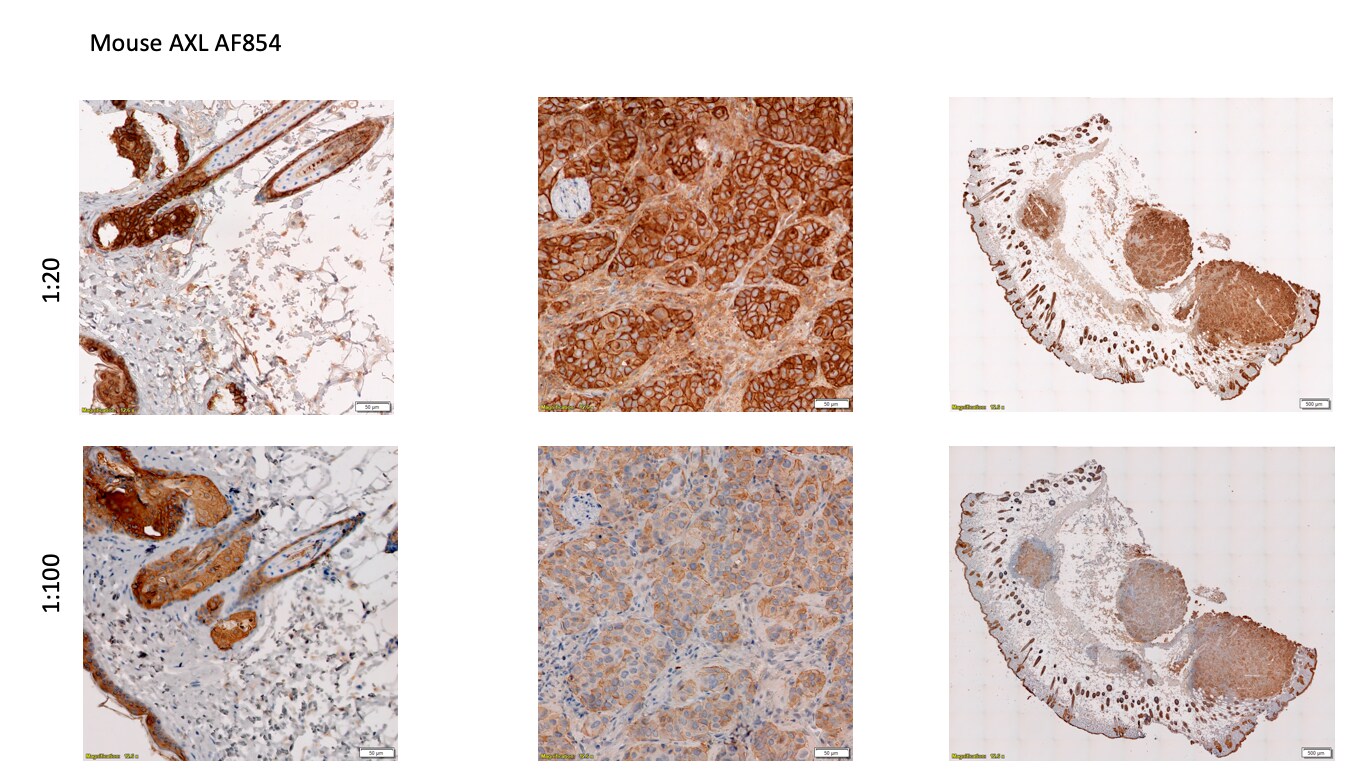
Application: ImmunohistochemistrySample Tested: Cancer TissueSpecies: MouseVerified Customer | Posted 11/19/2019Stained FFPE tissue sections from mouse lung tumors with 1:20 and 1:100 dilutions of AF854, detected with DAB chromagen.

-
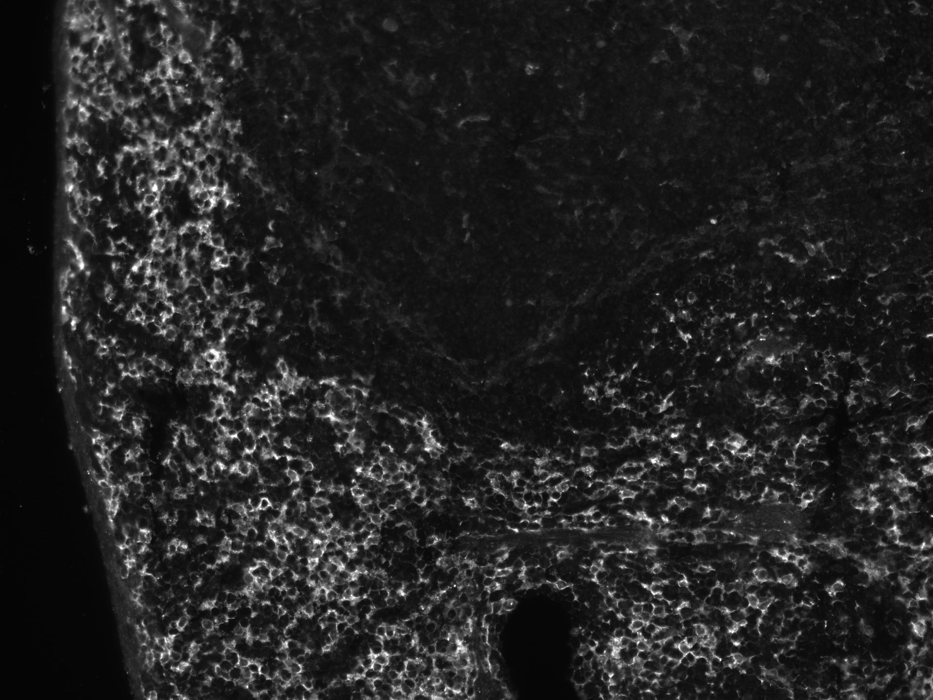
Application: Immunohistochemistry-FrozenSample Tested: Frozen mouse spleenSpecies: MouseVerified Customer | Posted 04/12/2019Immunofluorescence of fresh-frozen mouse spleen stained with Axl 1:100
-
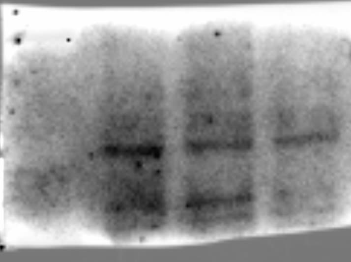
Application: Western BlotSample Tested: B16-F1 mouse melanoma cell lineSpecies: MouseVerified Customer | Posted 02/19/2019The first lane is a wild type line, and the next to are shRNA Axl knockdowns
-
Application: Western BlotSample Tested: Bone marrow cellsSpecies: MouseVerified Customer | Posted 01/04/2019
-
Application: ImmunohistochemistrySample Tested: Brain (cerebral cortex)Species: MouseVerified Customer | Posted 06/22/2017
-
Application: Functional AssaySample Tested: Spinal cord and Brain tissueSpecies: MouseVerified Customer | Posted 06/07/2017
-
Application: in vivoSample Tested: brain and spinal cordSpecies: MouseVerified Customer | Posted 04/03/2017Used in vivo to activate Axl in the CNS.
There are no reviews that match your criteria.
Protocols
Find general support by application which include: protocols, troubleshooting, illustrated assays, videos and webinars.
- Antigen Retrieval Protocol (PIER)
- Antigen Retrieval for Frozen Sections Protocol
- Appropriate Fixation of IHC/ICC Samples
- Cellular Response to Hypoxia Protocols
- Chromogenic IHC Staining of Formalin-Fixed Paraffin-Embedded (FFPE) Tissue Protocol
- Chromogenic Immunohistochemistry Staining of Frozen Tissue
- ClariTSA™ Fluorophore Kits
- Detection & Visualization of Antibody Binding
- Fluorescent IHC Staining of Frozen Tissue Protocol
- Graphic Protocol for Heat-induced Epitope Retrieval
- Graphic Protocol for the Preparation and Fluorescent IHC Staining of Frozen Tissue Sections
- Graphic Protocol for the Preparation and Fluorescent IHC Staining of Paraffin-embedded Tissue Sections
- Graphic Protocol for the Preparation of Gelatin-coated Slides for Histological Tissue Sections
- IHC Sample Preparation (Frozen sections vs Paraffin)
- Immunofluorescent IHC Staining of Formalin-Fixed Paraffin-Embedded (FFPE) Tissue Protocol
- Immunohistochemistry (IHC) and Immunocytochemistry (ICC) Protocols
- Immunohistochemistry Frozen Troubleshooting
- Immunohistochemistry Paraffin Troubleshooting
- Preparing Samples for IHC/ICC Experiments
- Preventing Non-Specific Staining (Non-Specific Binding)
- Primary Antibody Selection & Optimization
- Protocol for Heat-Induced Epitope Retrieval (HIER)
- Protocol for Making a 4% Formaldehyde Solution in PBS
- Protocol for VisUCyte™ HRP Polymer Detection Reagent
- Protocol for the Preparation & Fixation of Cells on Coverslips
- Protocol for the Preparation and Chromogenic IHC Staining of Frozen Tissue Sections
- Protocol for the Preparation and Chromogenic IHC Staining of Frozen Tissue Sections - Graphic
- Protocol for the Preparation and Chromogenic IHC Staining of Paraffin-embedded Tissue Sections
- Protocol for the Preparation and Chromogenic IHC Staining of Paraffin-embedded Tissue Sections - Graphic
- Protocol for the Preparation and Fluorescent IHC Staining of Frozen Tissue Sections
- Protocol for the Preparation and Fluorescent IHC Staining of Paraffin-embedded Tissue Sections
- Protocol for the Preparation of Gelatin-coated Slides for Histological Tissue Sections
- R&D Systems Quality Control Western Blot Protocol
- TUNEL and Active Caspase-3 Detection by IHC/ICC Protocol
- The Importance of IHC/ICC Controls
- Troubleshooting Guide: Immunohistochemistry
- Troubleshooting Guide: Western Blot Figures
- Western Blot Conditions
- Western Blot Protocol
- Western Blot Protocol for Cell Lysates
- Western Blot Troubleshooting
- Western Blot Troubleshooting Guide
- View all Protocols, Troubleshooting, Illustrated assays and Webinars
Loading...