
Hedgehog signaling proteins act as mitogens, morphogens, or inducing factors in many different cell types during embryonic development. They aid in growth, patterning, and morphogenesis in both vertebrates and insects (1). Hip (Hedgehog-interacting protein) is a type I transmembrane protein identified for its ability to bind biologically active Sonic Hedgehog. It is comprised of 700 aa, and includes a hydrophobic signal sequence, two EGF-like domains near the C-terminus, and a 22 amino acid membrane-spanning region at the C-terminal end (2). Hip has only been identified in vertebrates and binds all three mammalian Hedgehogs: sonic (Shh), desert (Dhh), and Indian (Ihh). Like the Hedgehog receptor Patched, Hip is a transcriptional target of Hedgehog signaling (2). Unlike Patched, Hip’s ability to bind hedgehogs is not involved in transducing a signal intracellularly, rather it regulates the availability of Hedgehog ligand extracellularly (3). Transgenic mice overexpressing Hip in proliferating chondrocytes display skeletal defects similar to those observed in Ihh mutant mice. These results indicate that Hip is involved in attenuating Hedgehog signaling (2). The expression pattern of Hip correlates with its ability to interact with all three mammalian Hedgehogs. It is expressed in a variety of organs, adjacent with sites of hedgehog expression. For instance, Shh is expressed in the epithelium of the lung, and Hip is found in the underlying lung mesenchyme (2). In fact, Hip knock-out mice exhibit neonatal lethality with respiratory failure due to defective branching morphogenesis. This phenotype correlates with altered expression of Shh markers suggesting an increase in Shh signaling (3). Interestingly, other developmental mechanisms that rely on normal Shh signaling, such as dorsal-ventral patterning of the neural tube, development of the somites, and organ laterality appeared histologically normal in Hip-/- mice (3). Mouse and human Hip share 94% amino acid identity through the entire protein sequence (4). R&D Systems’ recombinant Hedgehog-interacting protein is a potent antagonist of Shh signaling in vitro.

Scientific images may be resized, cropped, or adjusted for brightness or contrast for presentation purposes. These updates are limited to presentation and do not affect the underlying scientific interpretation of the data. Where available, additional source data may be provided upon request.
Key Product Details
Species Reactivity
Validated:
Cited:
Applications
Validated:
Cited:
Label
Antibody Source
Product Specifications
Immunogen
Lys24-Arg678 (Asn29Ser)
Accession # Q7TN16
Specificity
Clonality
Host
Isotype
Scientific Data Images for Mouse Hip Antibody
Detection of Hip by Immunohistochemistry
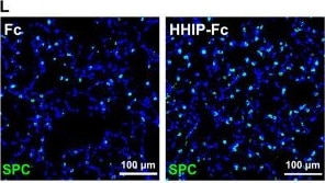
HHIP-Fc administration alleviates emphysema in adult mice with Hhip haploinsufficiency.(A and B) H&E images and MLI quantification of the lungs from adult HhipLacZ/+ and WT mice. (C) IF analysis of PDGFR alpha and SMA in the lungs of adult HhipLacZ/+ and WT mice. Arrow: SMA+ PDGFR alpha + cells. (D and E) Quantification of the number and percentage of myofibroblasts in adult HhipLacZ/+ and WT mice. (F and G) IF analysis of SPC and quantification of AT2s in the lungs of adult HhipLacZ/+ and WT mice. (H and I) H&E images and MLI quantification of the lungs from adult HhipLacZ/+ and WT mice treated with HHIP-Fc or Fc. (J and K) IF analysis and quantification of myofibroblasts in the lungs of adult HhipLacZ/+ and WT mice treated with HHIP-Fc or Fc. Arrow: SMA+ cells. (L and M) IF analysis and quantification of AT2s in the lungs of adult HhipLacZ/+ and WT mice treated with HHIP-Fc or Fc. Each data point represents one mouse [(B), (D), (E), (G), (I), (K), and (M)] of an individual experiment. Data are expressed as mean ± SD. *P < 0.05, **P < 0.005, ***P < 0.0005, and ****P < 0.0001. Image collected and cropped by CiteAb from the following open publication (https://pubmed.ncbi.nlm.nih.gov/40333979), licensed under a CC-BY license. Not internally tested by R&D Systems.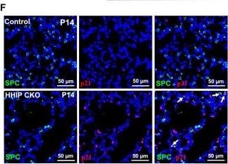
Detection of Hip by Immunohistochemistry
Deletion of Hhip induces cell senescence.(A) Violin plots showing the expression of Cdkn1a in ALMFs and DMFs of Hhip-deleted and control mice. (B) Senescence beta -galactosidase staining of Hhip-deleted and control lungs. (C) IF analysis of SMA, p21, and CDH4 in Hhip-deleted and control lungs. Arrow: p21+ DMFs; arrowhead: p21+ ALMFs. (D and E) Number of p21+ ALMFs (D) and DMFs (E) per unit alveolar area of Hhip-deleted and control mice at P14. (F) IF analysis of SPC and p21 in Hhip-deleted and control lungs. Arrow: p21+ SPC+ cells. (G) Percentage of p21+ cells in AT2s. (H) Activation of senescence pathways in the AT2s of Hhip-deleted mice, relative to control AT2s, and analyzed with IPA. (I) Top 5 upstream regulators in the AT2s of Hhip-deleted mice, relative to control AT2s, analyzed with IPA. (J) AT2 organoids cocultured with lung stromal cells (R26RSmoM2/+) pre-infected with adenovirus-empty and adenovirus-Cre, treated with anti-IGF1 antibody and IgG. (K and L) Quantification of colony-forming efficiency (CFE) and organoid size. (M) IF analysis of SPC and p21 in AT2 organoids. Arrow: p21+ SPC+ cells. (N) Percentage of p21+ cells in AT2s in the organoid assay. All in vitro experiments have been repeated at least one time with consistent results for validation. Each data point represents one mouse [(D), (E), and (G)] of an individual experiment. Data are expressed as Mean ± SD. *P < 0.05, **P < 0.005, ***P < 0.0005, and ****P < 0.0001. Image collected and cropped by CiteAb from the following open publication (https://pubmed.ncbi.nlm.nih.gov/40333979), licensed under a CC-BY license. Not internally tested by R&D Systems.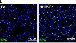
Detection of Hip by Immunohistochemistry
HHIP-Fc administration alleviates emphysema in adult mice with Hhip haploinsufficiency.(A and B) H&E images and MLI quantification of the lungs from adult HhipLacZ/+ and WT mice. (C) IF analysis of PDGFR alpha and SMA in the lungs of adult HhipLacZ/+ and WT mice. Arrow: SMA+ PDGFR alpha + cells. (D and E) Quantification of the number and percentage of myofibroblasts in adult HhipLacZ/+ and WT mice. (F and G) IF analysis of SPC and quantification of AT2s in the lungs of adult HhipLacZ/+ and WT mice. (H and I) H&E images and MLI quantification of the lungs from adult HhipLacZ/+ and WT mice treated with HHIP-Fc or Fc. (J and K) IF analysis and quantification of myofibroblasts in the lungs of adult HhipLacZ/+ and WT mice treated with HHIP-Fc or Fc. Arrow: SMA+ cells. (L and M) IF analysis and quantification of AT2s in the lungs of adult HhipLacZ/+ and WT mice treated with HHIP-Fc or Fc. Each data point represents one mouse [(B), (D), (E), (G), (I), (K), and (M)] of an individual experiment. Data are expressed as mean ± SD. *P < 0.05, **P < 0.005, ***P < 0.0005, and ****P < 0.0001. Image collected and cropped by CiteAb from the following open publication (https://pubmed.ncbi.nlm.nih.gov/40333979), licensed under a CC-BY license. Not internally tested by R&D Systems.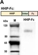
Detection of Hip by Western Blot
HHIP-Fc protein alleviates hyperoxia-induced BPD phenotypes.(A) Design strategy for HHIP-Fc recombinant protein and its analysis by Western blotting. (B) qPCR analysis of Gli1 expression in the lung stromal cells isolated from neonatal mice 3 and 5 days after one dose of HHIP or HHIP-Fc treatment. (C) H&E images of hyperoxia-exposed lungs, treated with HHIP-Fc or Fc fragment control. (D) MLI quantification of hyperoxia-exposed lungs, treated with HHIP-Fc or Fc. (E and F) IF analysis and quantification of myofibroblasts in hyperoxia-exposed lungs, treated with HHIP-Fc or Fc. (G to I) IF analysis and quantification of AT2 number and p21+ AT2 percentage in hyperoxia-exposed lungs, treated with HHIP-Fc or Fc. Arrow: p21+ SPC+ cells. (J) qPCR analysis of Igf1 expression in the lung stromal cells isolated from hyperoxia-exposed mice, treated with HHIP-Fc or Fc. Each data point represents one mouse [(B), (D), (F), and (H) to (J)] of an individual experiment. Data are expressed as mean ± SD. *P < 0.05, **P < 0.005, ***P < 0.0005, and ****P < 0.0001. Image collected and cropped by CiteAb from the following open publication (https://pubmed.ncbi.nlm.nih.gov/40333979), licensed under a CC-BY license. Not internally tested by R&D Systems.Applications for Mouse Hip Antibody
Western Blot
Sample: Recombinant Mouse Hip (Catalog # 1568-HP)
Formulation, Preparation, and Storage
Purification
Reconstitution
Reconstitute at 0.2 mg/mL in sterile PBS. For liquid material, refer to CoA for concentration.
Formulation
Shipping
Stability & Storage
- 12 months from date of receipt, -20 to -70 °C as supplied.
- 1 month, 2 to 8 °C under sterile conditions after reconstitution.
- 6 months, -20 to -70 °C under sterile conditions after reconstitution.
Calculators
Background: Hip
References
- Ingham, P.W. and A.P. McMahon (2001) Genes & Dev 15:3059.
- Chuang, P-T. and A.P. McMahon (1999) Nature 397: 17.
- Chuang, P-T. et al. (2003) Genes & Dev. 17:342.
- Bak, M. et al. (2001) Cytogenet. Cell Genet. 92:300.
Long Name
Alternate Names
Gene Symbol
UniProt
Additional Hip Products
Product Documents for Mouse Hip Antibody
Certificate of Analysis
To download a Certificate of Analysis, please enter a lot or batch number in the search box below.
Note: Certificate of Analysis not available for kit components.
Product Specific Notices for Mouse Hip Antibody
For research use only
Citations for Mouse Hip Antibody
 Powered by Bioz
Customer Reviews for Mouse Hip Antibody
There are currently no reviews for this product. Be the first to review Mouse Hip Antibody and earn rewards!
Have you used Mouse Hip Antibody?
Submit a review and receive an Amazon gift card!
$25/€18/£15/$25CAN/¥2500 for a review with an image
$10/€7/£6/$10CAN/¥1110 for a review without an image
Submit a review
Protocols
Find general support by application which include: protocols, troubleshooting, illustrated assays, videos and webinars.
- Cellular Response to Hypoxia Protocols
- R&D Systems Quality Control Western Blot Protocol
- Troubleshooting Guide: Western Blot Figures
- Western Blot Conditions
- Western Blot Protocol
- Western Blot Protocol for Cell Lysates
- Western Blot Troubleshooting
- Western Blot Troubleshooting Guide
- View all Protocols, Troubleshooting, Illustrated assays and Webinars