
OX40 Ligand (OX40L), also known as gp34, is a type II transmembrane glycoprotein belonging to the TNF superfamily. Murine OX40L cDNA encodes a 198 amino acid (aa) protein comprised of a 28 aa N-terminal cytoplasmic domain, a 20 aa transmembrane segment, and a 150 aa C-terminal extracellular domain (1). Human and mouse OX40L share 46% sequence identity at the amino acid level (1). OX40L is expressed on activated antigen presenting cells such as B cells, macrophages, dendritic cells, and on endothelial cells at the site of inflammation. The receptor for OX40L is OX40 (CD134) which is expressed predominantly on activated CD4+ T cells. Expression of OX40 is transient following engagement of T cell receptors (2). Ligation of OX40L by OX40 stimulates proliferation and differentiation of activated B cells, and increases immunoglobulin secretion (3, 4). The expression of OX40L on B cells is upregulated by CD40 ligation (3). Engagement of the OX40‑OX40L system has co-stimulatory effects on T cells by stimulating the production of cytokines by T helper cells and increasing the survival of memory T cells (2, 5). Blocking of the OX40‑OX40L interaction in vitro inhibits co-stimulation resulting in decreased T cell proliferation and adhesion of T cells to endothelial cells. Inhibition of the OX40‑OX40L interaction in disease models has beneficial effects in acute graft-versus-host disease, inflammatory bowel disease and decreases the development of collagen-induced arthritis and experimental leishmaniasis (6).
Mouse OX40 Ligand/TNFSF4 Antibody (182601)
R&D Systems | Catalog # MAB1236

Scientific images may be resized, cropped, or adjusted for brightness or contrast for presentation purposes. These updates are limited to presentation and do not affect the underlying scientific interpretation of the data. Where available, additional source data may be provided upon request.
Key Product Details
Species Reactivity
Validated:
Cited:
Applications
Validated:
Cited:
Label
Antibody Source
Product Specifications
Immunogen
Gln49-Leu198
Accession # P43488
Specificity
Clonality
Host
Isotype
Endotoxin Level
Scientific Data Images for Mouse OX40 Ligand/TNFSF4 Antibody (182601)
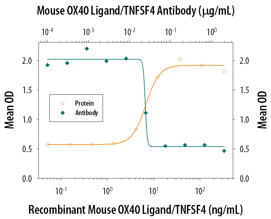
IL‑2 Secretion Induced by OX40 Ligand/TNFSF4 and Neutralization by Mouse OX40 Ligand/TNFSF4 Antibody.
In the presence of a cross-linking antibody, Mouse polyHistidine Monoclonal Antibody (10 µg/mL, Catalog # MAB050) and sub-optimal amounts of Mouse CD3e Monoclonal Antibody (Catalog # MAB484), Recombinant Mouse OX40 Ligand/TNFSF4 (Catalog # 1236-OX) stimulates IL-2 secretion in mouse T cells in a dose-dependent manner (orange line), as measured by the Mouse IL-2 Quantikine ELISA Kit (Catalog # M2000). Under these conditions, IL-2 secretion elicited by Recombinant Mouse OX40 Ligand/TNFSF4 (30 ng/mL) is neutralized (green line) by increasing concentrations of Rat Anti-Mouse OX40 Ligand/TNFSF4 Monoclonal Antibody (Catalog # MAB1236). The ND50 is typically 0.01-0.05 µg/mL.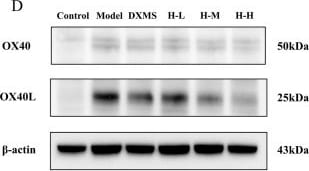
Detection of OX40 Ligand/TNFSF4 by Western Blot
Effects of HLJDD on skin barrier restoration and on OX40/OX40L pathway in AD-like mice (A, B) Representative images of immunohistochemistry labelling and quantification of the expression of FLG (scale bar = 100 μm, n = 3) (C, D) Representative protein bands on the expression of LOR, AQP3, and OX40, OX40L with beta -actin being used as the control (n = 3) (E–H) Quantification of LOR, AQP3, OX40, and OX40L bands (n = 3). Data were the mean ± SEM (n = 3). #p < 0.05, ##p < 0.01 vs control group; *p < 0.05, **p < 0.01 vs model group. Image collected and cropped by CiteAb from the following open publication (https://pubmed.ncbi.nlm.nih.gov/39640480), licensed under a CC-BY license. Not internally tested by R&D Systems.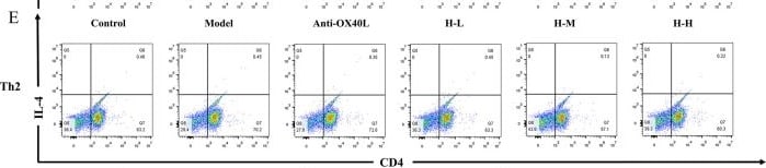
Detection of OX40 Ligand/TNFSF4 by Flow Cytometry
Effect of HLJDD on the differentiation of Th1/Th2 and Th17/Treg in vitro(A–C) Results of Th1, Th2, and Th1/Th2 ratios in each group (n = 3) (D, E) Representative images of Th1/Th2 detected by flow cytometry. (F–H) Results of Th17, Treg, and Th17/Treg ratios in each group (n = 3). (I, J) Representative images of Th17/Treg detected by flow cytometry. Data were the mean ± SEM. ##p < 0.01, ####p < 0.0001 vs control group; *p < 0.05, **p < 0.01, and ***p < 0.001 vs model group. Image collected and cropped by CiteAb from the following open publication (https://pubmed.ncbi.nlm.nih.gov/39640480), licensed under a CC-BY license. Not internally tested by R&D Systems.Applications for Mouse OX40 Ligand/TNFSF4 Antibody (182601)
Western Blot
Sample: Recombinant Mouse OX40 Ligand/TNFSF4 (Catalog # 1236-OX) under non-reducing conditions only
Neutralization
Formulation, Preparation, and Storage
Purification
Reconstitution
Reconstitute at 0.5 mg/mL in sterile PBS. For liquid material, refer to CoA for concentration.
Formulation
Shipping
Stability & Storage
- 12 months from date of receipt, -20 to -70 °C as supplied.
- 1 month, 2 to 8 °C under sterile conditions after reconstitution.
- 6 months, -20 to -70 °C under sterile conditions after reconstitution.
Calculators
Background: OX40 Ligand/TNFSF4
References
- Baum, P.R. et al. (1994) EMBO J. 13:3992.
- Gramaglia, I. et al. (1999) J. Immunol. 161:6510.
- Stuber, E. et al. (1995) Immunity 2:507.
- Malstrom, V. et al. (2001) J. Immunol. 166:6972.
- Maxwell, J.R. et al. (2000) J. Immunol. 164:107.
- Weinberg, A.D. (2002) Trends Immunol. 23:102.
Alternate Names
Gene Symbol
UniProt
Additional OX40 Ligand/TNFSF4 Products
Product Documents for Mouse OX40 Ligand/TNFSF4 Antibody (182601)
Certificate of Analysis
To download a Certificate of Analysis, please enter a lot or batch number in the search box below.
Note: Certificate of Analysis not available for kit components.
Product Specific Notices for Mouse OX40 Ligand/TNFSF4 Antibody (182601)
For research use only
Citations for Mouse OX40 Ligand/TNFSF4 Antibody (182601)
 Powered by Bioz
Customer Reviews for Mouse OX40 Ligand/TNFSF4 Antibody (182601)
There are currently no reviews for this product. Be the first to review Mouse OX40 Ligand/TNFSF4 Antibody (182601) and earn rewards!
Have you used Mouse OX40 Ligand/TNFSF4 Antibody (182601)?
Submit a review and receive an Amazon gift card!
$25/€18/£15/$25CAN/¥2500 for a review with an image
$10/€7/£6/$10CAN/¥1110 for a review without an image
Submit a review
Protocols
Find general support by application which include: protocols, troubleshooting, illustrated assays, videos and webinars.
- Cellular Response to Hypoxia Protocols
- R&D Systems Quality Control Western Blot Protocol
- Troubleshooting Guide: Western Blot Figures
- Western Blot Conditions
- Western Blot Protocol
- Western Blot Protocol for Cell Lysates
- Western Blot Troubleshooting
- Western Blot Troubleshooting Guide
- View all Protocols, Troubleshooting, Illustrated assays and Webinars
Associated Pathways

