Peripherin Antibody - BSA Free
Novus Biologicals | Catalog # NBP1-05423
![Western Blot: Peripherin Antibody [NBP1-05423]](https://resources.rndsystems.com/images/products/Peripherin-Antibody-Western-Blot-NBP1-05423-img0003.jpg "Western Blot: Peripherin Antibody [NBP1-05423]")
Loading...
Key Product Details
Species Reactivity
Validated:
Human, Mouse, Rat, Porcine, Bovine
Cited:
Mouse, Rat
Applications
Validated:
Immunohistochemistry, Immunohistochemistry-Frozen, Western Blot, Immunocytochemistry/ Immunofluorescence
Cited:
Immunohistochemistry-Frozen, Immunocytochemistry/ Immunofluorescence
Label
Unconjugated
Antibody Source
Polyclonal Chicken IgY
Format
BSA Free
Loading...
Product Specifications
Immunogen
Recombinant rat peripherin expressed in and purified from E. coli
Marker
Un-myelinated Neurons Marker
Clonality
Polyclonal
Host
Chicken
Isotype
IgY
Theoretical MW
57 kDa.
Disclaimer note: The observed molecular weight of the protein may vary from the listed predicted molecular weight due to post translational modifications, post translation cleavages, relative charges, and other experimental factors.
Disclaimer note: The observed molecular weight of the protein may vary from the listed predicted molecular weight due to post translational modifications, post translation cleavages, relative charges, and other experimental factors.
Scientific Data Images for Peripherin Antibody - BSA Free
Western Blot: Peripherin Antibody [NBP1-05423]
Western Blot: Peripherin Antibody [NBP1-05423] - Analysis of spinal cord tissue lysates (lanes 2-5) and cell lysates (lanes 6 and 7) using chicken pAb to peripherin, NBP1-05423, dilution 1:10,000 in green: [1] protein standard (red), [2] rat, [3] mouse, [4] pig, [5] cow spinal cord; [6] SH-SY5Y, and [7] PC12 cells. The band at 57kDa corresponds to peripherin protein.![Immunocytochemistry/ Immunofluorescence: Peripherin Antibody [NBP1-05423]](https://resources.rndsystems.com/images/products/Peripherin-Antibody-Immunocytochemistry-Immunofluorescence-NBP1-05423-img0005.jpg "Immunocytochemistry/ Immunofluorescence: Peripherin Antibody [NBP1-05423]")
Immunocytochemistry/ Immunofluorescence: Peripherin Antibody [NBP1-05423]
Immunocytochemistry/Immunofluorescence: Peripherin Antibody [NBP1-05423] - Analysis of mixed fibroblast and PC12 pheochromocytoma cell culture stained with chicken pAb to peripherin, NBP1-05423, dilution 1:5,000 in green, and costained with rabbit pAb to vimentin, RPCA-Vim, dilution 1:2,000, in red. The blue is DAPI staining of nuclear DNA. NBP1-05423 antibody reveals cytoplasmic filamentous staining in the small PC12 cells, while vimentin antibody stains intermediate filaments in the surrounding fibroblastic cells which do not express peripherin.![Immunohistochemistry-Frozen: Peripherin Antibody [NBP1-05423]](https://resources.rndsystems.com/images/products/Peripherin-Antibody-Immunohistochemistry-Frozen-NBP1-05423-img0007.jpg "Immunohistochemistry-Frozen: Peripherin Antibody [NBP1-05423]")
Immunohistochemistry-Frozen: Peripherin Antibody [NBP1-05423]
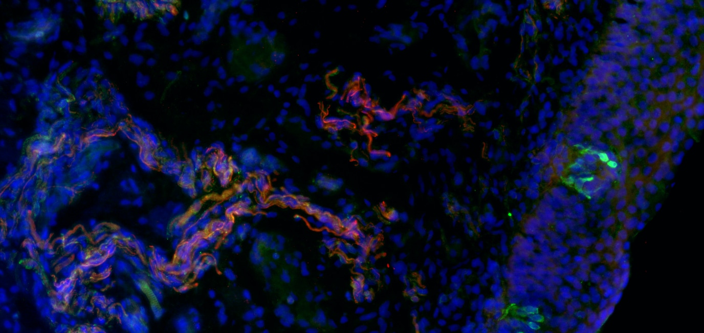
Immunohistochemistry-Frozen: Peripherin Antibody [NBP1-05423] - Peripherin staining (red) in nerve bundles in the human larynx. Image from verified customer review.![Immunocytochemistry/ Immunofluorescence: Peripherin Antibody [NBP1-05423]](https://resources.rndsystems.com/images/products/Peripherin-Antibody-Immunocytochemistry-Immunofluorescence-NBP1-05423-img0001.jpg "Immunocytochemistry/ Immunofluorescence: Peripherin Antibody [NBP1-05423]")
Immunocytochemistry/ Immunofluorescence: Peripherin Antibody [NBP1-05423]
Immunocytochemistry/Immunofluorescence: Peripherin Antibody [NBP1-05423] - shows rat mixed neuron/glial cultures stained with NBP1-05423, our chicken polyclonal antibody to peripherin (green channel) and rabbit antibody to the neurofilament subunit alpha-internexin NBP1-05207 (green channel). These cultures contain mostly neurons which are rich in alpha-internexin, and a subgroup which have a large amount of peripherin also, such as the prominent cell in the middle of the micrograph. Since this cell expresses large amounts of peripherin and alpha-internexin, the green and red signals superimpose to produce a golden cell. Blue is a DNA stain.![Immunohistochemistry-Frozen: Peripherin Antibody [NBP1-05423]](https://resources.rndsystems.com/images/products/Peripherin-Antibody-Immunohistochemistry-Frozen-NBP1-05423-img0006.jpg "Immunohistochemistry-Frozen: Peripherin Antibody [NBP1-05423]")
Immunohistochemistry-Frozen: Peripherin Antibody [NBP1-05423]
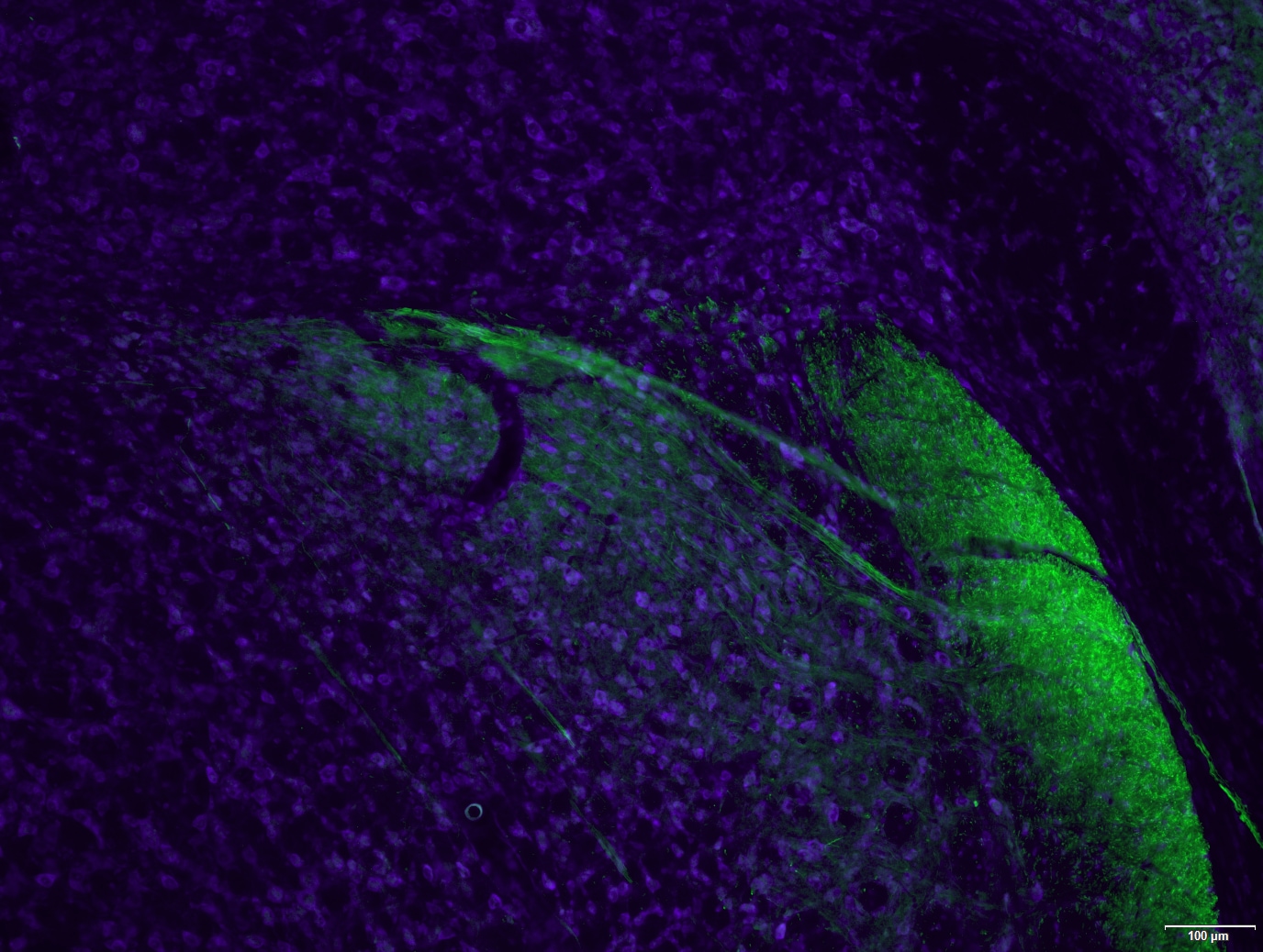
Immunohistochemistry-Frozen: Peripherin Antibody [NBP1-05423] - Peripherin staining (green) in the mouse brainstem including the spinal trigeminal tract (right side of image) and arching fiber bundle to the solitary nuclear tract (center). Nissl Counterstain. Image from verified customer review.Applications for Peripherin Antibody - BSA Free
Application
Recommended Usage
Immunocytochemistry/ Immunofluorescence
1:2000-1:5000
Immunohistochemistry
1:20000
Western Blot
1:20000
Application Notes
This Peripherin antibody is useful for Immunocytochemistry/Immunofluorescence and Western blot, where a band can be seen at ~57 kDa. IHC-Fr reported in verified customer review.
Reviewed Applications
Read 2 reviews rated 4 using NBP1-05423 in the following applications:
Formulation, Preparation, and Storage
Purification
IgY purified
Formulation
Supplied as a concentrated total IgY preparation from egg yolk, dialyzed against PBS with added preservative.
Format
BSA Free
Preservative
0.02% Sodium Azide
Concentration
Please see the vial label for concentration. If unlisted please contact technical services.
Shipping
The product is shipped with polar packs. Upon receipt, store it immediately at the temperature recommended below.
Stability & Storage
Store at 4C short term. Aliquot and store at -20C long term. Avoid freeze-thaw cycles.
Background: Peripherin
Alternate Names
Neurofilament 4, neurofilament 4 (57kD), peripherin, PRPH1NEF4
Entrez Gene IDs
24688 (Rat)
Gene Symbol
PRPH
Additional Peripherin Products
Product Documents for Peripherin Antibody - BSA Free
Certificate of Analysis
To download a Certificate of Analysis, please enter a lot or batch number in the search box below.
Product Specific Notices for Peripherin Antibody - BSA Free
Chicken products cannot be exported to Canada.
This product is for research use only and is not approved for use in humans or in clinical diagnosis. Primary Antibodies are guaranteed for 1 year from date of receipt.
Citations for Peripherin Antibody - BSA Free
 Powered by Bioz
Customer Reviews for Peripherin Antibody - BSA Free (2)
4 out of 5
2 Customer Ratings
Have you used Peripherin Antibody - BSA Free?
Submit a review and receive an Amazon gift card!
$25/€18/£15/$25CAN/¥2500 Yen for a review with an image
$10/€7/£6/$10CAN/¥1110 Yen for a review without an image
Submit a review
Customer Images
Showing
1
-
2 的
2 reviews
Showing All
Filter By:
-
Application: Immunohistochemistry-FrozenSample Tested: larynxSpecies: HumanVerified Customer | Posted 09/17/2018Peripherin staining (red) in nerve bundles in the human larynx.Fixation 4% buffered paraformaldehyde.

-
Application: Immunohistochemistry-FrozenSample Tested: Mouse brainSpecies: MouseVerified Customer | Posted 09/17/2018Peripherin staining (green) in the mouse brainstem including the spinal trigeminal tract (right side of image) and arching fiber bundle to the nuc. solitary tract (center). Nissl Counterstain.Tested on both human and mouse tissues. Works well for staining of small diameter nerve fibers in peripheral tissues. Less effective in staining central terminals of sensory axons (mouse) but still effective (see image).
There are no reviews that match your criteria.
Protocols
Find general support by application which include: protocols, troubleshooting, illustrated assays, videos and webinars.
- Antigen Retrieval Protocol (PIER)
- Antigen Retrieval for Frozen Sections Protocol
- Appropriate Fixation of IHC/ICC Samples
- Cellular Response to Hypoxia Protocols
- Chromogenic IHC Staining of Formalin-Fixed Paraffin-Embedded (FFPE) Tissue Protocol
- Chromogenic Immunohistochemistry Staining of Frozen Tissue
- ClariTSA™ Fluorophore Kits
- Detection & Visualization of Antibody Binding
- Fluorescent IHC Staining of Frozen Tissue Protocol
- Graphic Protocol for Heat-induced Epitope Retrieval
- Graphic Protocol for the Preparation and Fluorescent IHC Staining of Frozen Tissue Sections
- Graphic Protocol for the Preparation and Fluorescent IHC Staining of Paraffin-embedded Tissue Sections
- Graphic Protocol for the Preparation of Gelatin-coated Slides for Histological Tissue Sections
- ICC Cell Smear Protocol for Suspension Cells
- ICC Immunocytochemistry Protocol Videos
- ICC for Adherent Cells
- IHC Sample Preparation (Frozen sections vs Paraffin)
- Immunocytochemistry (ICC) Protocol
- Immunocytochemistry Troubleshooting
- Immunofluorescence of Organoids Embedded in Cultrex Basement Membrane Extract
- Immunofluorescent IHC Staining of Formalin-Fixed Paraffin-Embedded (FFPE) Tissue Protocol
- Immunohistochemistry (IHC) and Immunocytochemistry (ICC) Protocols
- Immunohistochemistry Frozen Troubleshooting
- Immunohistochemistry Paraffin Troubleshooting
- Preparing Samples for IHC/ICC Experiments
- Preventing Non-Specific Staining (Non-Specific Binding)
- Primary Antibody Selection & Optimization
- Protocol for Heat-Induced Epitope Retrieval (HIER)
- Protocol for Making a 4% Formaldehyde Solution in PBS
- Protocol for VisUCyte™ HRP Polymer Detection Reagent
- Protocol for the Fluorescent ICC Staining of Cell Smears - Graphic
- Protocol for the Fluorescent ICC Staining of Cultured Cells on Coverslips - Graphic
- Protocol for the Preparation & Fixation of Cells on Coverslips
- Protocol for the Preparation and Chromogenic IHC Staining of Frozen Tissue Sections
- Protocol for the Preparation and Chromogenic IHC Staining of Frozen Tissue Sections - Graphic
- Protocol for the Preparation and Chromogenic IHC Staining of Paraffin-embedded Tissue Sections
- Protocol for the Preparation and Chromogenic IHC Staining of Paraffin-embedded Tissue Sections - Graphic
- Protocol for the Preparation and Fluorescent ICC Staining of Cells on Coverslips
- Protocol for the Preparation and Fluorescent ICC Staining of Non-adherent Cells
- Protocol for the Preparation and Fluorescent ICC Staining of Stem Cells on Coverslips
- Protocol for the Preparation and Fluorescent IHC Staining of Frozen Tissue Sections
- Protocol for the Preparation and Fluorescent IHC Staining of Paraffin-embedded Tissue Sections
- Protocol for the Preparation of Gelatin-coated Slides for Histological Tissue Sections
- Protocol for the Preparation of a Cell Smear for Non-adherent Cell ICC - Graphic
- R&D Systems Quality Control Western Blot Protocol
- TUNEL and Active Caspase-3 Detection by IHC/ICC Protocol
- The Importance of IHC/ICC Controls
- Troubleshooting Guide: Immunohistochemistry
- Troubleshooting Guide: Western Blot Figures
- Western Blot Conditions
- Western Blot Protocol
- Western Blot Protocol for Cell Lysates
- Western Blot Troubleshooting
- Western Blot Troubleshooting Guide
- View all Protocols, Troubleshooting, Illustrated assays and Webinars
Loading...