SARS-CoV-2 Spike RBD Antibody (1034522)
R&D Systems | Catalog # MAB10540

Key Product Details
Species Reactivity
Validated:
SARS-CoV-2
Cited:
Human, Plant - Nicotiana benthamiana, Primate - Cercopithecus aethiops (African Green Monkey)
Applications
Validated:
Western Blot
Cited:
Western Blot, Bioassay
Label
Unconjugated
Antibody Source
Monoclonal Mouse IgG2B Clone # 1034522
Loading...
Product Specifications
Immunogen
Recombinant SARS-CoV-2 Spike Protein, S1 Subunit, Receptor Binding Domain (RBD)
Arg319-Phe541
Accession # YP_009724390
Arg319-Phe541
Accession # YP_009724390
Specificity
Detects SARS-CoV-2 Spike Protein S1 Receptor Binding Domain (RBD) in direct ELISAs and Western blots. Detects SARS-CoV-2
B.1.1.529 S RBD (Omicron Variant) in direct ELISAs.
Clonality
Monoclonal
Host
Mouse
Isotype
IgG2B
Scientific Data Images for SARS-CoV-2 Spike RBD Antibody (1034522)
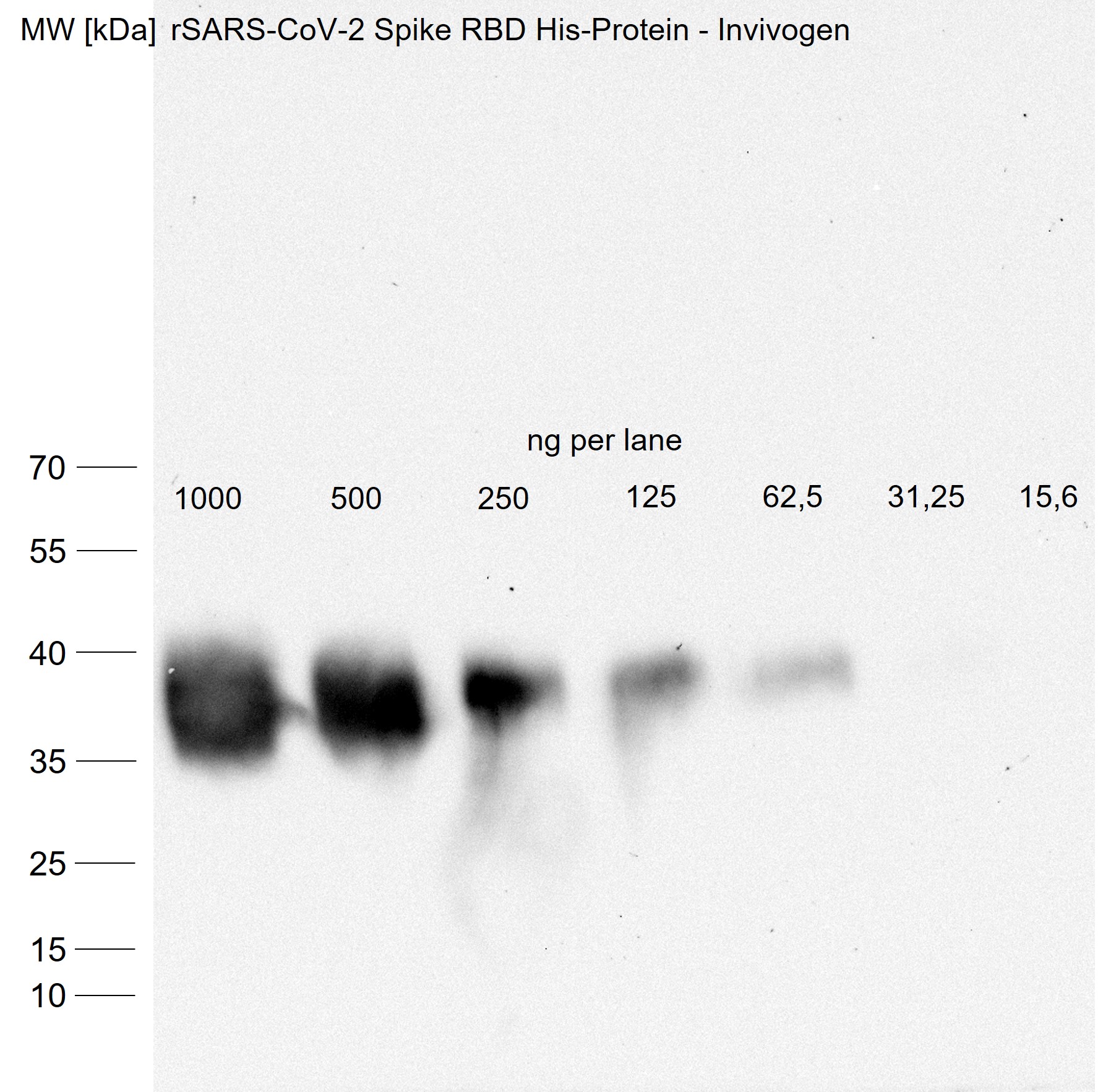
Detection of SARS-CoV-2 Spike RBD by Western Blot.
Western blot shows recombinant SARS-CoV-2 Spike RBD protein. PVDF membrane was probed with 2 µg/mL of Mouse Anti-SARS-CoV-2 Spike RBD Monoclonal Antibody (Catalog # MAB10540) followed by HRP-conjugated Anti-Mouse IgG Secondary Antibody (HAF018). A specific band was detected for SARS-CoV-2 Spike RBD at approximately 35 kDa (as indicated). This experiment was conducted under reducing conditions and using Western Blot Buffer Group 1.
Detection of SARS-CoV-2 Spike RBD by Western Blot
A conjugate vaccine targeting the RBD domain of SARS-CoV-2(A–C) Schematic representation of the RBD (A), CDP (B), and produced CDP-RBD (28.9 kDa) and RBD (22.8 kDa) vaccine proteins (C).(B) CDP is composed of several highly immunogenic clusters originating from three different bacterial proteins (cell division protein ZapB, type-1 fimbrial protein TFP, and small heat shock protein IbpA).(C) The theoretical isoelectric point (pI) and the predicted solubility score (Sol) for each vaccine protein.(D) Coomassie staining after SDS-PAGE (left) and anti-RBD Western blot (right) analysis showing RBD and CDP-RBD.(E) Size-exclusion chromatogram (10/300 GL Superdex 75, Cytiva) showing purified CDP-RBD (black arrow). Image collected and cropped by CiteAb from the following open publication (https://pubmed.ncbi.nlm.nih.gov/35813877), licensed under a CC-BY license. Not internally tested by R&D Systems.
Detection of SARS-CoV-2 Spike RBD by Western Blot
A conjugate vaccine targeting the RBD domain of SARS-CoV-2(A–C) Schematic representation of the RBD (A), CDP (B), and produced CDP-RBD (28.9 kDa) and RBD (22.8 kDa) vaccine proteins (C).(B) CDP is composed of several highly immunogenic clusters originating from three different bacterial proteins (cell division protein ZapB, type-1 fimbrial protein TFP, and small heat shock protein IbpA).(C) The theoretical isoelectric point (pI) and the predicted solubility score (Sol) for each vaccine protein.(D) Coomassie staining after SDS-PAGE (left) and anti-RBD Western blot (right) analysis showing RBD and CDP-RBD.(E) Size-exclusion chromatogram (10/300 GL Superdex 75, Cytiva) showing purified CDP-RBD (black arrow). Image collected and cropped by CiteAb from the following open publication (https://pubmed.ncbi.nlm.nih.gov/35813877), licensed under a CC-BY license. Not internally tested by R&D Systems.Applications for SARS-CoV-2 Spike RBD Antibody (1034522)
Application
Recommended Usage
Western Blot
2 µg/mL
Sample: Recombinant SARS-CoV-2 Spike RBD protein
Sample: Recombinant SARS-CoV-2 Spike RBD protein
Reviewed Applications
Read 1 review rated 4 using MAB10540 in the following applications:
Formulation, Preparation, and Storage
Purification
Protein A or G purified from hybridoma culture supernatant
Reconstitution
Reconstitute at 0.5 mg/mL in sterile PBS. For liquid material, refer to CoA for concentration.
Loading...
Formulation
Lyophilized from a 0.2 μm filtered solution in PBS with Trehalose. *Small pack size (SP) is supplied either lyophilized or as a 0.2 µm filtered solution in PBS.
Shipping
Lyophilized product is shipped at ambient temperature. Liquid small pack size (-SP) is shipped with polar packs. Upon receipt, store immediately at the temperature recommended below.
Stability & Storage
Use a manual defrost freezer and avoid repeated freeze-thaw cycles.
- 12 months from date of receipt, -20 to -70 °C as supplied.
- 1 month, 2 to 8 °C under sterile conditions after reconstitution.
- 6 months, -20 to -70 °C under sterile conditions after reconstitution.
Calculators
Background: Spike RBD
References
- Wu, F. et al. (2020) Nature 579:265.
- Tortorici, M.A. and D. Veesler (2019). Adv. Virus Res. 105:93.
- Bosch, B.J. et al. (2003) J. Virol. 77:8801.
- Belouzard, S. et al. (2009) Proc. Natl. Acad. Sci. 106:5871.
- Millet, J.K. and G. R. Whittaker (2015) Virus Res. 202:120.
- Yuan, Y. et al. (2017) Nat. Commun. 8:15092.
- Walls, A.C. et al. (2010) Cell 180:281.
- Jiang, S. et al. (2020) Trends. Immunol. https://doi.org/10.1016/j.it.2020.03.007.
- Ortega, J.T. et al. (2020) EXCLI J. 19:410.
- Wrapp, D. et al. (2020) Science 367:1260.
- Tai, W. et al. (2020) Cell. Mol. Immunol. https://doi.org/10.1016/j.it.2020.03.007.
- Okba, N. M. A. et al. (2020). Emerg. Infect. Dis. https://doi.org/10.3201/eid2607.200841.
- Wang, X. et al. (2020) https://doi.org/10.1038/s41423-020-0424-9.
- Wang, K. et al. (2020) bioRxiv https://www.biorxiv.org/content/10.1101/2020.03.14.988345v1.
Long Name
Spike Receptor Binding Domain
Gene Symbol
S
UniProt
Additional Spike RBD Products
Product Documents for SARS-CoV-2 Spike RBD Antibody (1034522)
Certificate of Analysis
To download a Certificate of Analysis, please enter a lot or batch number in the search box below.
Note: Certificate of Analysis not available for kit components.
Product Specific Notices for SARS-CoV-2 Spike RBD Antibody (1034522)
For research use only
Related Research Areas
Citations for SARS-CoV-2 Spike RBD Antibody (1034522)
 Powered by Bioz
Customer Reviews for SARS-CoV-2 Spike RBD Antibody (1034522) (1)
4 out of 5
1 Customer Rating
Have you used SARS-CoV-2 Spike RBD Antibody (1034522)?
Submit a review and receive an Amazon gift card!
$25/€18/£15/$25CAN/¥2500 Yen for a review with an image
$10/€7/£6/$10CAN/¥1110 Yen for a review without an image
Submit a review
Customer Images
Showing
1
-
1 的
1 review
Showing All
Filter By:
-
Application: Western BlotSample Tested: Recombinant proteinSpecies: HamsterVerified Customer | Posted 12/14/2021Antibody was diluted in Tris buffered buffer with 0,5% Tween 20 added and incubated overnight at 4°C under constant shaking. Antibody was diluted as instructed and final dilution was 1:1000 equaling 0,5 µg/ml concentration.Samples were boiled for 10 min in 4x Laemmli buffer and subjected to 10% SDS-PAGE.

There are no reviews that match your criteria.
Protocols
Find general support by application which include: protocols, troubleshooting, illustrated assays, videos and webinars.
- Cellular Response to Hypoxia Protocols
- R&D Systems Quality Control Western Blot Protocol
- Troubleshooting Guide: Western Blot Figures
- Western Blot Conditions
- Western Blot Protocol
- Western Blot Protocol for Cell Lysates
- Western Blot Troubleshooting
- Western Blot Troubleshooting Guide
- View all Protocols, Troubleshooting, Illustrated assays and Webinars
Loading...