
Protocol for LipoLIVE™ Deep Red Non-Toxic Dye
In Brief
LipoLIVE™ Deep Red Non-Toxic Dye (Catalog # 9042) is a ready-to-use fluorescent probe for live-cell imaging of lipid droplet and neutral lipids. It does not require cell fixation or affect cell health and maintains the physiological integrity of models, enabling free-wash monitoring of lipid metabolism.
Designed for real-time confocal microscopy, and high-content screening (HCS) workflows, it offers exceptional signal-to-noise ratios and photostability.
LipoLIVE™ is a trademark of Saguaro Biosciences.
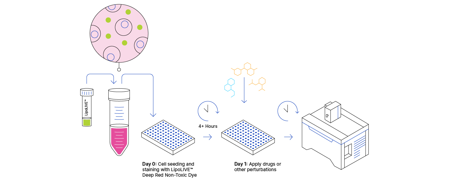
1. Protocol Overview
2. Content and Storage
| Product | Content | Storage | Stability |
|---|---|---|---|
| LipoLIVE™ Deep Red Non-Toxic Dye | Diluted in 50 µl of DMSO | -20° C Delivered at room temperature Protect from light | 1 year |
Table 1. LipoLIVE™ Deep Red Non-Toxic Dye Product Information.
Intended Use: For research use only. Not for use in diagnostics or therapeutic procedures.
3. General Guidelines
LipoLIVE™ Deep Red Non-Toxic Dye Dilution and Preparation
- Warm up the LipoLIVE™ Deep Red Non-Toxic Dye tube to room temperature before use to avoid condensation to form and water to get into the anhydrous dye solution.
- Gently spin the tube before use to collect any dye solution that may remain near the cap.
- Dilute LipoLIVE™ Deep Red Non-Toxic Dye 1,000-fold in preferred culture medium.
- Vortex thoroughly.
NOTE: We recommend 1X as a starting point for optimization. Higher or lower concentrations may be optimal for different imaging systems and cell models.
- Seed cells at desired density (typically to achieve 70-80% confluence) in cell culture medium containing LipoLIVE™ Deep Red Non-Toxic Dye in a black multi-well plate. Return to the incubator at 37°C, 5% CO2 overnight.
- No washing step is required prior to imaging. Keep LipoLIVE™ Deep Red Non-Toxic Dye in solution throughout the assay.
Alternative Cell Culture Indications for LipoLIVE™ Deep Red Non-Toxic Dye
- While we recommend seeding cells in the presence of diluted LipoLIVE™ Deep Red Non-Toxic Dye, the dye can be added after cell seeding, before or following compound addition. Optimisation of seeding density and incubation prior to imaging are required. For reference, LipoLIVE™ Deep Red Non-Toxic Dye staining stabilizes after 4 hours in U2OS cells (at a concentration of 1X).
- A nuclear dye can be added to allow cell segmentation during data analysis, such as NucleoLIVE™ Non-Toxic Dye (Cat. No. 8935) or NucleoLIVE™ Blue Non-Toxic Dye (Cat. No. 9011). We recommend running a preliminary imaging test on cells treated with single dyes to validate the staining kinetics and absence of fluorescence bleed-through between the nuclear dye and LipoLIVE™ Deep Red channels on your system.
4. Technical Specifications & Instrument Compatibility
LipoLIVE™ Deep Red Non-Toxic Dye is a fluorescent dye compatible with GFP, Cy3 or FITC filters.
Instrument compatibility of LipoLIVE™ Deep Red Non-Toxic Dye
| Manufacturer | Instrument | Filters | Mode |
|---|---|---|---|
| Molecular Devices | ImageXpress Confocal ImageXpress Confocal HT.ai | Red / Cy5 / Cy5 | Widefield / Confocal |
| PerkinElmer / Revvity | Opera Phenix Opera Phenix Plus Operetta CLS | 640 / 650-760 615-645 / 655-760 | Widefield only Widefield / Confocal Widefield / Confocal |
| Yokogawa | CQ1 | 640 / 600/37 | Confocal |
| CV8000 | 640 / 676/29 | Confocal |
LipoLIVE™ Deep Red Non-Toxic Dye is compatible with other high-content imagers and confocal microscopes.
5. LipoLIVE™ Deep Red Non-Toxic Dye, Allowing for High Quality Staining of Lipid Droplets and Neutral Lipids without Compromising Cell Health

Figure 2. LipoLIVE™ rapidly stains lipid droplets in live cells. Composite images of MCF-7 and HepG2 cells stained with LipoLIVE™ Deep Red for 24h before overnight incubation with 100 µM Oleic Acid.
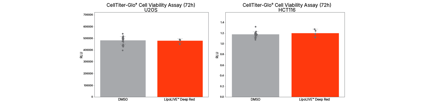
Figure 3. LipoLIVE™ preserves normal cell proliferation and viability, even in sensitive models. U2OS cells and HCT116 cells showed comparable viability to DMSO controls after 72-hour incubation with LipoLIVE™ Deep Red.
6. Recommended Positive Control Compounds for Lipid Studies*
| Compound | Mechanism | Concentrations |
|---|---|---|
| Oleic Acid | Long-chain monounsaturated fatty, stimulating the rapid formation of cytosolic lipid droplets. | 100 nM-10 μM |
| Palmitic Acid | Long-chain saturated fatty acid, inducing lipotoxic stress and the formation of small clustered cytosolic lipid droplets. | 5 μM-500 μM |
Table 2. Compounds and concentration for U2OS cells in 2D.
* Compounds provided as examples only. Validation required for each experimental protocol.
7. Example Protocol (for kinetic, 2D live-cell assay)
U2OS cells are treated with standard compounds for ER stress and apoptosis. MCF7 are cultured in DMEM/F-12 complemented with 10% FBS and 1% Penicillin/Streptomycin.
LipoLIVE™ Deep Red Non-Toxic Dye Dilution and Preparation (Day 0):
- Warm up the LipoLIVE™ Deep Red Non-Toxic Dye tubes to room temperature before use and gently spin to collect any dye solution that may remain near the cap.
- Optional: Dilute 10 μL NucleoLIVE™ Non-Toxic Dye in the same 10 mL culture medium (1000-fold).
- Vortex thoroughly.
Cell Culture Protocol with LipoLIVE™ Deep Red Non-toxic Dye (Day 0):
- Harvest and count MCF7 cells.
- Resuspend cells in prepared culture medium with LipoLIVE™ Deep Red Non-Toxic Dye (and NucleoLIVE™ Non-Toxic Dye, if applicable) at 80,000 cells/mL.
- Seed 96-well plate with 100 μL cell suspension per well to a final density of 8,000 cells per well.
- Incubate overnight at 37°C, 5% CO2.
Standard Compound Preparation and Addition (Day 1):
- Prepare dose-response curves with 10x concentrations, maintaining constant vehicle (0.1% DMSO) solvent concentration.
- Prepare negative controls with 0.1% DMSO in complete media.
- Distribute 12.5 μL of test compounds or controls per well.
Imaging and Data Acquisition (Days 1-3):
- Image 96-well plate at 3h, 6h and 24h after addition of test compounds.
8. Frequently Asked Questions about LipoLIVE™ Deep Red Non-Toxic Dye
What is LipoLIVE™ Deep Red Non-Toxic Dye used for?
LipoLIVE™ Deep Red Non-Toxic Dye is used for the real-time visualization of lipid droplet and neutral lipids in living cells, without affecting cell viability.
Does LipoLIVE™ Deep Red Non-Toxic Dye require cell fixation?
No, LipoLIVE™ Deep Red Non-Toxic Dye does not require cell fixation.
Is a wash step required before imaging?
No washing step is required prior to imaging.
Which imaging systems are compatible with LipoLIVE™ Deep Red Non-Toxic Dye?
LipoLIVE™ Deep Red Non-Toxic Dye is compatible with high-content imagers and confocal microscopes.