ARNT/HIF-1 beta Antibody - BSA Free
Novus Biologicals | Catalog # NB100-110
Key Product Details
Species Reactivity
Validated:
Human, Mouse, Rat, Bovine, Canine, Ferret, Fish, Sheep
Cited:
Human, Mouse, Rat, Bovine, Canine, Fish
Applications
Validated:
Immunohistochemistry, Immunohistochemistry-Paraffin, Western Blot, Immunoblotting, Immunocytochemistry/ Immunofluorescence, Simple Western, Immunoprecipitation, Chromatin Immunoprecipitation (ChIP), Gel Supershift Assay
Cited:
Immunohistochemistry-Paraffin, Western Blot, Immunoblotting, Immunocytochemistry/ Immunofluorescence, Immunoprecipitation, Chromatin Immunoprecipitation, Chromatin Immunoprecipitation (ChIP), Chemotaxis, EMSA, IF/IHC
Label
Unconjugated
Antibody Source
Polyclonal Rabbit IgG
Format
BSA Free
Loading...
Product Specifications
Immunogen
ARNT/HIF-1 beta Antibody was developed against a fusion protein to human HIF-1 beta containing amino acids 496-789. [UniProt# P27540]
Reactivity Notes
ARNT/HIF-1 beta antibody shows Fish reactivity as reported in scientific literature (PMID: 30334616). Canine reactivity reported in scientific literature (PMID:32885302)
Localization
Nuclear
Specificity
ARNT/HIF-1 beta Antibody is specific for HIF-1 beta/ARNT. It is not known if NB100-110 cross-reacts with ARNT2 which is related to HIF-1 beta/ARNT but is the product of a different gene.
Clonality
Polyclonal
Host
Rabbit
Isotype
IgG
Theoretical MW
86.6 kDa.
Disclaimer note: The observed molecular weight of the protein may vary from the listed predicted molecular weight due to post translational modifications, post translation cleavages, relative charges, and other experimental factors.
Disclaimer note: The observed molecular weight of the protein may vary from the listed predicted molecular weight due to post translational modifications, post translation cleavages, relative charges, and other experimental factors.
Scientific Data Images for ARNT/HIF-1 beta Antibody - BSA Free
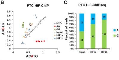
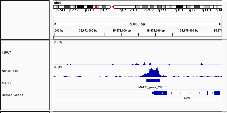
ARNT-HIF-1-beta-Antibody-Chromatin-Immunoprecipitation-NB100-110-img0007.jpg
![Western Blot: ARNT/HIF-1 beta Antibody [NB100-110]](https://resources.rndsystems.com/images/products/ARNT-HIF-1-beta-Antibody-Western-Blot-NB100-110-img0001.jpg "Western Blot: ARNT/HIF-1 beta Antibody [NB100-110]")
Western Blot: ARNT/HIF-1 beta Antibody [NB100-110]
Western Blot: ARNT/HIF-1 beta Antibody [NB100-110] - Analysis of HIF-1 beta using ARNT/HIF-1 beta antibody [NB100-110] in MCF7 whole cell lysate. Theoretical molecular weight is 86.6 kDa. Observed molecular weight ~95 kDa.![Immunohistochemistry: ARNT/HIF-1 beta Antibody [NB100-110]](https://resources.rndsystems.com/images/products/ARNT-HIF-1-beta-Antibody-Immunohistochemistry-NB100-110-img0004.jpg "Immunohistochemistry: ARNT/HIF-1 beta Antibody [NB100-110]")
Immunohistochemistry: ARNT/HIF-1 beta Antibody [NB100-110]
Immunohistochemistry: ARNT/HIF-1 beta Antibody [NB100-110] - Immunohistochemical staining of human skin epidermis with ARNT/HIF-1 beta antibody [NB100-110].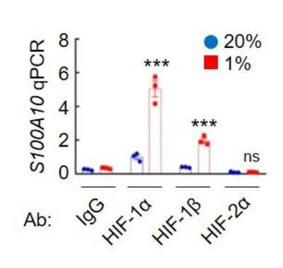
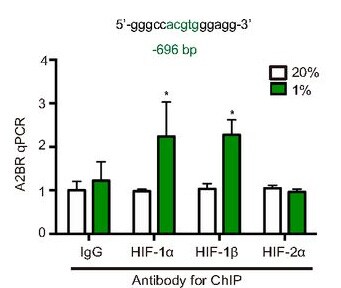
Chromatin Immunoprecipitation: ARNT/HIF-1 beta Antibody [NB100-110] - ChIP analysis with ARNT/HIF-1 beta antibody [NB100-110]. MDA-MB-231 cells were exposed to 20% or 1% O2 for 16 hours, and chromatin immunoprecipitation (ChIP) was performed with the indicated antibody (Ab). Primers flanking the HIF binding site were used for qPCR. ChIP image submitted by a verified customer review.
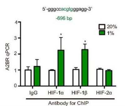
Chromatin Immunoprecipitation (ChIP): ARNT/HIF-1 beta Antibody [NB100-110] - ChIP analysis using ARNT/HIF-1 beta antibody [NB100-110]. MCF7 were exposed to 20% or 1% O2 for 16h, and ChIP assays were performed using IgG or antibodies against HIF-1a, HIF-2a, HIF-1b. ChIP image submitted by a verified customer review.
ARNT-HIF-1-beta-Antibody-Chromatin-Immunoprecipitation-NB100-110-img0008.jpg
![Simple Western: ARNT/HIF-1 beta Antibody [NB100-110]](https://resources.rndsystems.com/images/products/ARNT-HIF-1-beta-Antibody-Simple-Western-NB100-110-img0005.jpg "Simple Western: ARNT/HIF-1 beta Antibody [NB100-110]")
Simple Western: ARNT/HIF-1 beta Antibody [NB100-110]
Simple Western: ARNT/HIF-1 beta Antibody [NB100-110] - Simple Western analysis using ARNT/HIF-1 beta antibody [NB100-110]. Lane view shows a specific band for HIF-1 beta in 0.5 mg/ml of Hypoxic HeLa lysate. This experiment was performed under reducing conditions using the 12-230 kDa separation system. Theoretical molecular weight is 86.6 kDa. Observed molecular weight ~106 kDa.Applications for ARNT/HIF-1 beta Antibody - BSA Free
Application
Recommended Usage
Chromatin Immunoprecipitation (ChIP)
1:10-1:500
Immunohistochemistry
1:150
Immunohistochemistry-Paraffin
1:150
Immunoprecipitation
1:10-1:500
Simple Western
1:1000
Western Blot
1:2000
Reviewed Applications
Read 3 reviews rated 5 using NB100-110 in the following applications:
Formulation, Preparation, and Storage
Purification
Unpurified
Formulation
Whole antisera
Format
BSA Free
Preservative
0.02% Sodium Azide
Concentration
This product is unpurified. The exact concentration of antibody is not quantifiable.
Shipping
The product is shipped with polar packs. Upon receipt, store it immediately at the temperature recommended below.
Stability & Storage
Aliquot and store at -20C or -80C. Avoid freeze-thaw cycles.
Background: ARNT/HIF-1 beta
ARNT has an important role in two specific signaling pathways - the aryl hydrocarbon receptor (AhR) and the hypoxia inducible factor (HIF) pathway (1). In the AhR pathway, AhR in the cytosol is typically inactive and bound to heat shock protein 90 (hsp90) (3). Upon activation and ligand binding by environmental pollutants such as dioxins, AhR is translocated to the nucleus, dissociates from hsp90, and dimerizes with ARNT, leading to binding to response elements and expression of target genes including monooxygenases (1, 3). In the HIF pathway, under hypoxia (low oxygen) conditions prolylhydroxylase domain (PHD) enzymes and factor inhibiting HIF (FIH) are inhibited. HIF-1 alpha (or HIF-2 alpha) accumulates and is transported to the nucleus where it heterodimerizes with ARNT, allowing for binding to target gene's hypoxia response element (HRE), recruitment of coactivators, and transcription (1, 3). HIF-induced gene transcription plays a large role in tumor progression by promoting invasion, metastasis, de-differentiation and altered metabolism, and angiogenesis (1). While HIF-1 alpha's stability is dependent upon oxygen conditions, HIF-1 beta is stable in both normoxia and hypoxia (1-3).
The bHLH-PAS family and ARNT have been linked with a variety of pathologies and diseases including cancer, metabolic diseases, autoimmune diseases, and psychiatric disorders (2). ARNT/AHR is expressed in the skin and its pathway activation enhances skin barrier function and epidermal terminal differentiation, thus AHR agonists are currently being used as therapeutics for atopic dermatitis and psoriasis (4). Accordingly, studies of Arnt-deficient mice show profound abnormalities in skin barrier function and keratinization (4). Additionally, studies suggest that ARNT plays an important role in diabetes and beta-cell function (5). Islets from patients with type 2 diabetes have a significantly decreased ARNT expression compared to glucose-tolerant control donors (5). Modulation and stimulation of the HIF pathway may be a potential therapeutic strategy for treating type 2 diabetes and metabolic syndrome (5).
Alternate names for ARNT/HIF-1 beta include aryl hydrocarbon receptor nuclear translocator, BHLHE2, class E basic helix-loop-helix protein 2, Dixon receptor nuclear translocator, Hypoxia-inducible factor 1-beta, nuclear translocator, and TANGO.
References
1. Mandl, M., & Depping, R. (2014). Hypoxia-inducible aryl hydrocarbon receptor nuclear translocator (ARNT) (HIF-1beta): is it a rare exception?. Molecular medicine (Cambridge, Mass.). https://doi.org/10.2119/molmed.2014.00032
2. Wu, D., & Rastinejad, F. (2017). Structural characterization of mammalian bHLH-PAS transcription factors. Current opinion in structural biology. https://doi.org/10.1016/j.sbi.2016.09.011
3. Esser, C., & Rannug, A. (2015). The aryl hydrocarbon receptor in barrier organ physiology, immunology, and toxicology. Pharmacological reviews.https://doi.org/10.1124/pr.114.009001
4. Furue, M., Hashimoto-Hachiya, A., & Tsuji, G. (2019). Aryl Hydrocarbon Receptor in Atopic Dermatitis and Psoriasis. International journal of molecular sciences. https://doi.org/10.3390/ijms20215424
5. Girgis, C. M., Cheng, K., Scott, C. H., & Gunton, J. E. (2012). Novel links between HIFs, type 2 diabetes, and metabolic syndrome. Trends in endocrinology and metabolism: TEM, https://doi.org/10.1016/j.tem.2012.05.003
Long Name
Aryl Hydrocarbon Receptor Nuclear Translocator
Alternate Names
HIF-1 beta, HIF1 beta, TANGO
Gene Symbol
ARNT
UniProt
Additional ARNT/HIF-1 beta Products
Product Documents for ARNT/HIF-1 beta Antibody - BSA Free
Certificate of Analysis
To download a Certificate of Analysis, please enter a lot or batch number in the search box below.
Product Specific Notices for ARNT/HIF-1 beta Antibody - BSA Free
This product is for research use only and is not approved for use in humans or in clinical diagnosis. Primary Antibodies are guaranteed for 1 year from date of receipt.
Citations for ARNT/HIF-1 beta Antibody - BSA Free
 Powered by Bioz
Customer Reviews for ARNT/HIF-1 beta Antibody - BSA Free (3)
5 out of 5
3 Customer Ratings
Have you used ARNT/HIF-1 beta Antibody - BSA Free?
Submit a review and receive an Amazon gift card!
$25/€18/£15/$25CAN/¥2500 Yen for a review with an image
$10/€7/£6/$10CAN/¥1110 Yen for a review without an image
Submit a review
Customer Images
Showing
1
-
3 的
3 reviews
Showing All
Filter By:
-
Application: Chromatin ImmunoprecipitationSample Tested: Human cancer cell linesSpecies: HumanVerified Customer | Posted 07/05/2019
-
Application: Chromatin ImmunoprecipitationSample Tested: MCF7Species: HumanVerified Customer | Posted 12/02/2018MCF7 were exposed to 20% or 1% O2 for 16h, and ChIP assays were performed using IgG or antibodies against HIF-1a, HIF-2a, HIF-1b.

-
Application: Chromatin ImmunoprecipitationSample Tested: U2OSSpecies: HumanVerified Customer | Posted 07/07/2016
There are no reviews that match your criteria.
Protocols
View specific protocols for ARNT/HIF-1 beta Antibody - BSA Free (NB100-110):
Western Blot Procedure
1. Resolve aliquots (15 mg) of induced * nuclear protein extracts on a SDS/6% polyacrylamide gel.
2. Transfer to nitrocellulose membranes in 20 mM Tris-HCl (pH 8.0)/150 mM glycine/20% (vol/vol) methanol.
3. Block membranes for 1.5 hours with 1X western wash buffer containing 5% non-fat dry milk (NFDM).
4. Incubate membranes for 1.5 hours at room temperature (RT) in NB100-110 diluted 1:2,000 ** in 1X western wash/5% NFDM.
5. Wash with 1X western wash for 35 minutes at RT (1 X 15 minutes, 2 X 10 minutes).
6. Incubate membranes with HRP conjugated anti-Rabbit IgG for 1 hour (RT) in 1X western wash/5% NFDM. Wash with 1X western wash for 35 minutes at RT (1 X 15 minutes, 2 X 10 minutes).
7. Drain membrane and place on saran wrap.
8. Using Amersham ECL Kit, mix equal volumes of two reagents. Pour over membrane (protein side facing up). Let solution sit on membrane for 15-20 seconds.
9. Drain membrane and place on new saran wrap
10. Wrap up membrane and expose to film.
11. Develop accordingly.
Notes: If hypoxia treatment is not hypoxic enough (less than 2% oxygen to get an induction), signal will be absent. Also, if the harvest time is too slow or there are not enough protease inhibitors, etc., the induced protein will be rapidly lost as HIF-1beta has a very short half-life. Whole cell extracts or nuclear extracts of hypoxia induced cell lines (293, Hep3B, COS7, Hepa) are useful as a positive control. Nuclear Extract Preparation Reference: Wang and Semenza. "Purification and Characterization of Hypoxia-Inducible Factor 1". Journal of Biological Chemistry. 270(3): 1230-1237, 1995.
IHC-FFPE sections
I. Deparaffinization:
A. Treat slides with Xylene: 3 changes for 5 minutes each. Drain slides for 10 seconds between changes.
B. Treat slides with 100% Reagent Alcohol: 3 changes for 5 minutes each. Drain slides for 10 seconds between changes.
II. Quench Endogenous Peroxidase:
A. Place slides in peroxidase quenching solution: 15-30 minutes.
To Prepare 200 ml of Quenching Solution:
Add 3 ml of 30% Hydrogen Peroxide to 200 ml of Methanol.
Use within 4 hours of preparation
B. Place slides in distilled water: 2 changes for 2 minutes each.
III. Retrieve Epitopes:
A. Preheat Citrate Buffer. Place 200 ml of Citrate Buffer Working Solution into container, cover and place into steamer. Heat to 90-96 degrees Celcius.
B. Place rack of slides into hot Citrate Buffer for 20 minutes. Cover.
C. Carefully remove container with slides from steamer and cool on bench, uncovered, for 20 minutes.
D. Slowly add distilled water to further cool for 5 minutes.
E. Rinse slides with distilled water. 2 changes for 2 minutes each.
IV. Immunostaining Procedure:
A. Remove each slide from rack and circle tissue section with a hydrophobic barrier pen (e.g. Liquid Blocker-Super Pap Pen).
B. Flood slide with Wash Solution. Do not allow tissue sections to dry for the rest of the procedure.
C. Drain wash solution and apply 4 drops of Blocking Reagent to each slide and incubate for 15 minutes.
D. Drain Blocking Reagent (do not wash off the Blocking Reagent), apply 200 ul of Primary Antibody solution to each slide, and incubate for 1 hour.
E. Wash slides with Wash Solution: 3 changes for 5 minutes each.
F. Drain wash solution, apply 4 drops of Secondary antibody to each slide and incubate for 1 hour.
G. Wash slides with Wash Solution: 3 changes for 5 minutes each.
H. Drain wash solution, apply 4 drops of DAB Substrate to each slide and develop for 5-10 minutes. Check development with microscope.
I. Wash slides with Wash Solution: 3 changes for 5 minutes each.
J. Drain wash solution, apply 4 drops of Hematoxylin to each slide and stain for 1-3 minutes. Increase time if darker counterstaining is desired.
K. Wash slides with Wash Solution: 2-3 changes for 2 minutes each.
L. Drain wash solution and apply 4 drops of Bluing Solution to each slide for 1-2 minutes.
M. Rinse slides in distilled water.
N. Soak slides in 70% reagent alcohol: 3 minutes with intermittent agitation.
O. Soak slides in 95% reagent alcohol: 2 changes for 3 minutes each with intermittent agitation.
P. Soak slides in 100% reagent alcohol: 3 changes for 3 minutes each with intermittent agitation. Drain slides for 10 seconds between each change.
Q. Soak slides in Xylene: 3 changes for 3 minutes each with intermittent agitation. Drain slides for 10 seconds between each change.
R. Apply 2-3 drops of non-aqueous mounting media to each slide and mount coverslip.
S. Lay slides on a flat surface to dry prior to viewing under microscope.
NOTES:
-Use treated slides (e.g. HistoBond) to assure adherence of FFPE sections to slide.
-Prior to deparaffinization, heat slides overnight in a 60 degrees Celcius oven.
-All steps in which Xylene is used should be performed in a fume hood.
-For Epitope Retrieval, a microwave or pressure cooker may be substituted for the steamer method. Adjust times as necessary depending on conditions.
-For the initial IHC run with a new primary antibody, test tissues with and without Epitope Retrieval. In some instances, Epitope Retrieval may not be necessary.
-200 ul is the recommended maximum volume to apply to a slide for full coverage. Using more than 200 ul may allow solutions to wick off the slide and create drying artifacts. For small tissue sections less than 200 ul may be used.
-5 minutes of development with DAB Substrate should be sufficient. Do not develop for more than 10 minutes. If 5 minutes of development causes background staining, further dilution of the primary antibody may be necessary.
-Hematoxylin should produce a light nuclear counterstain so as not to obscure the DAB staining. Counterstain for 1-1 1/2 minutes for nuclear antigens. Counterstain for 2-3 minutes for cytoplasmic and membranous antigens. If darker counterstaining is desired increase time (up to 10 minutes).
Find general support by application which include: protocols, troubleshooting, illustrated assays, videos and webinars.
- Antigen Retrieval Protocol (PIER)
- Antigen Retrieval for Frozen Sections Protocol
- Appropriate Fixation of IHC/ICC Samples
- Cellular Response to Hypoxia Protocols
- ChIP Protocol Video
- Chromatin Immunoprecipitation (ChIP) Protocol
- Chromatin Immunoprecipitation Protocol
- Chromogenic IHC Staining of Formalin-Fixed Paraffin-Embedded (FFPE) Tissue Protocol
- Chromogenic Immunohistochemistry Staining of Frozen Tissue
- ClariTSA™ Fluorophore Kits
- Detection & Visualization of Antibody Binding
- Fluorescent IHC Staining of Frozen Tissue Protocol
- Graphic Protocol for Heat-induced Epitope Retrieval
- Graphic Protocol for the Preparation and Fluorescent IHC Staining of Frozen Tissue Sections
- Graphic Protocol for the Preparation and Fluorescent IHC Staining of Paraffin-embedded Tissue Sections
- Graphic Protocol for the Preparation of Gelatin-coated Slides for Histological Tissue Sections
- ICC Cell Smear Protocol for Suspension Cells
- ICC Immunocytochemistry Protocol Videos
- ICC for Adherent Cells
- IHC Sample Preparation (Frozen sections vs Paraffin)
- Immunocytochemistry (ICC) Protocol
- Immunocytochemistry Troubleshooting
- Immunofluorescence of Organoids Embedded in Cultrex Basement Membrane Extract
- Immunofluorescent IHC Staining of Formalin-Fixed Paraffin-Embedded (FFPE) Tissue Protocol
- Immunohistochemistry (IHC) and Immunocytochemistry (ICC) Protocols
- Immunohistochemistry Frozen Troubleshooting
- Immunohistochemistry Paraffin Troubleshooting
- Immunoprecipitation Protocol
- Preparing Samples for IHC/ICC Experiments
- Preventing Non-Specific Staining (Non-Specific Binding)
- Primary Antibody Selection & Optimization
- Protocol for Heat-Induced Epitope Retrieval (HIER)
- Protocol for Making a 4% Formaldehyde Solution in PBS
- Protocol for VisUCyte™ HRP Polymer Detection Reagent
- Protocol for the Fluorescent ICC Staining of Cell Smears - Graphic
- Protocol for the Fluorescent ICC Staining of Cultured Cells on Coverslips - Graphic
- Protocol for the Preparation & Fixation of Cells on Coverslips
- Protocol for the Preparation and Chromogenic IHC Staining of Frozen Tissue Sections
- Protocol for the Preparation and Chromogenic IHC Staining of Frozen Tissue Sections - Graphic
- Protocol for the Preparation and Chromogenic IHC Staining of Paraffin-embedded Tissue Sections
- Protocol for the Preparation and Chromogenic IHC Staining of Paraffin-embedded Tissue Sections - Graphic
- Protocol for the Preparation and Fluorescent ICC Staining of Cells on Coverslips
- Protocol for the Preparation and Fluorescent ICC Staining of Non-adherent Cells
- Protocol for the Preparation and Fluorescent ICC Staining of Stem Cells on Coverslips
- Protocol for the Preparation and Fluorescent IHC Staining of Frozen Tissue Sections
- Protocol for the Preparation and Fluorescent IHC Staining of Paraffin-embedded Tissue Sections
- Protocol for the Preparation of Gelatin-coated Slides for Histological Tissue Sections
- Protocol for the Preparation of a Cell Smear for Non-adherent Cell ICC - Graphic
- R&D Systems Quality Control Western Blot Protocol
- TUNEL and Active Caspase-3 Detection by IHC/ICC Protocol
- The Importance of IHC/ICC Controls
- Troubleshooting Guide: Immunohistochemistry
- Troubleshooting Guide: Western Blot Figures
- Western Blot Conditions
- Western Blot Protocol
- Western Blot Protocol for Cell Lysates
- Western Blot Troubleshooting
- Western Blot Troubleshooting Guide
- View all Protocols, Troubleshooting, Illustrated assays and Webinars
FAQs for ARNT/HIF-1 beta Antibody - BSA Free
Showing
1
-
5 的
5 FAQs
Showing All
-
Q: Do you have a concentration for NB100-110? what dilution do you recommend for a ChIP
A: NB100-110 is actually unpurified product, so there is no concentration listed. The recommended dilution for chromatin Immunoprecipitation for this product is 1:10-1:500.
-
Q: For use in Western Blot with HIF-1 beta antibodies, what molecular weight of the band should I expect to see?
A: The theoretical molecular weight determined by our technical team for ARNT/HIF-1 beta antibodies is 86.6 kDa.
-
Q: If this product is used in an application or species as a part of a customer review, will that validate this product in the application/species?
A: If any of our primary antibodes are used in an untested application or species and it is shown to work through images from customer reviews or through publications, this validates the application/species for this product, allowing the tested application/species to fall under our 100% guarantee. Please check out our Innovator's Reward Program if you decide to test a primary antibody with a species or application that is not currently listed. Please note that the Innovator's Reward Program only applies to our primary antibodies.
-
Q: Is this target appropriate for use in Chromatin Research?
A: Yes; here is a list of research areas that we have deemed appropriate for the target "ARNT/HIF-1 beta": Angiogenesis, Autophagy, Cancer, Cellular Markers, Chromatin Research, HIF Target Genes, Hypoxia, Transcription Factors and Regulators, Cardiovascular Biology, Lipid and Metabolism.
-
Q: So this NB100-110 does not have a concentration. How much would you recommend to use per chip reaction where I normally use 5 to 10 ug of antibody?
A: Our HIF-1 beta product NB100-110 is provided as whole antisera, so we have not determined the protein concentration. We recommend using a 1:10-1:500 dilution for ChIP. You will need to determine the optimal amount for your experiment.
-
Q: Do you have a concentration for NB100-110? what dilution do you recommend for a ChIP
A: NB100-110 is actually unpurified product, so there is no concentration listed. The recommended dilution for chromatin Immunoprecipitation for this product is 1:10-1:500.
-
Q: For use in Western Blot with HIF-1 beta antibodies, what molecular weight of the band should I expect to see?
A: The theoretical molecular weight determined by our technical team for ARNT/HIF-1 beta antibodies is 86.6 kDa.
-
Q: If this product is used in an application or species as a part of a customer review, will that validate this product in the application/species?
A: If any of our primary antibodes are used in an untested application or species and it is shown to work through images from customer reviews or through publications, this validates the application/species for this product, allowing the tested application/species to fall under our 100% guarantee. Please check out our Innovator's Reward Program if you decide to test a primary antibody with a species or application that is not currently listed. Please note that the Innovator's Reward Program only applies to our primary antibodies.
-
Q: Is this target appropriate for use in Chromatin Research?
A: Yes; here is a list of research areas that we have deemed appropriate for the target "ARNT/HIF-1 beta": Angiogenesis, Autophagy, Cancer, Cellular Markers, Chromatin Research, HIF Target Genes, Hypoxia, Transcription Factors and Regulators, Cardiovascular Biology, Lipid and Metabolism.
-
Q: So this NB100-110 does not have a concentration. How much would you recommend to use per chip reaction where I normally use 5 to 10 ug of antibody?
A: Our HIF-1 beta product NB100-110 is provided as whole antisera, so we have not determined the protein concentration. We recommend using a 1:10-1:500 dilution for ChIP. You will need to determine the optimal amount for your experiment.
Loading...
Associated Pathways

