GW182 Antibody - BSA Free
Novus Biologicals | Catalog # NBP1-28751
![Western Blot: GW182 Antibody [NBP1-28751]](https://resources.rndsystems.com/images/products/GW182-Antibody-Knockdown-Validated-NBP1-28751-img0005.jpg "Western Blot: GW182 Antibody [NBP1-28751]")
![Western Blot: GW182 Antibody [NBP1-28751]](https://resources.rndsystems.com/images/products/GW182-Antibody-Western-Blot-NBP1-28751-img0003.jpg "Western Blot: GW182 Antibody [NBP1-28751]")
Key Product Details
Validated by
Species Reactivity
Validated:
Cited:
Applications
Validated:
Cited:
Label
Antibody Source
Format
Product Specifications
Immunogen
Reactivity Notes
Marker
Clonality
Host
Isotype
Scientific Data Images for GW182 Antibody - BSA Free
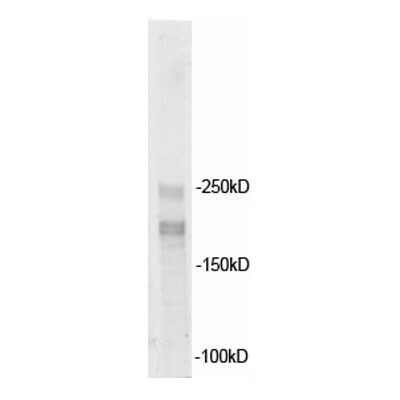
Western Blot: GW182 Antibody [NBP1-28751]
Western Blot: GW182 Antibody [NBP1-28751] - Detection of Human GW182 by Western Blot. Samples: Whole cell lysate (50 ug) from HeLa, 293T, and Jurkat cells. Antibodies: Affinity purified rabbit anti-GW182 antibody NBP1-28751 used for WB at 0.1 ug/ml. Detection: Chemiluminescence with an exposure time of 30 seconds.
Western Blot: Rabbit Polyclonal GW182 Antibody [NBP1-28751] -
Western Blot: Rabbit Polyclonal GW182 Antibody [NBP1-28751] - Detection of human GW182 by western blot of immunoprecipitates. Samples: Whole cell lysate (50 ug) from HeLa cells. Antibodies: Affinity purified rabbit anti-GW182 antibodyNBP1-28751 (lot NBP1-28751-2) used for IP at 6 ug/mg lysate. GW182 was also immunoprecipitated by a previous lot (lot NBP1-28751-1) of this antibody. For blotting immunoprecipitated GW182, NBP1-28751 was used at 1 ug/ml. Detection: Chemiluminescence with an exposure time of 3 seconds.
Western Blot: GW182 Antibody [NBP1-28751] -
Western Blot: GW182 Antibody [NBP1-28751] - TNRC6A interacts with eIF4E2. (A) Plasmids expressing proteins indicated were transfected into HEK293T cells. At 48 h posttransfection, cells were lysed & the lysates were immunoprecipitated (IP) in the presence of RNase A. The precipitates were resolved on SDS-PAGE followed by Western blotting. (B) A plasmid expressing Flag-tagged eIF4E1 or eIF4E2 was transfected into HEK293T cells. At 48 h posttransfection, cells were lysed & the lysates were immunoprecipitated with the anti-TNRC6A antibody or control IgG in the presence of RNase A. The precipitates were resolved on SDS-PAGE followed by Western blotting. (C) Upper: schematic representation of TNRC6A truncation mutants. Lower: A plasmid expressing the TNRC6A mutant indicated & a plasmid expressing myc-tagged eIF4E2 were transiently transfected into HEK293T cells. At 48 h posttransfection, cells were lysed & the lysates were immunoprecipitated with anti-Flag antibody in the presence of RNase A followed by Western blotting. (D) Bacterially expressed Flag-tagged eIF4E2 or eIF4E1 was incubated with Glutathione Sepharose 4B bound fusion protein of GST & the C-terminal domain of TNRC6A (6A-C). The precipitates were washed & resolved on SDS-PAGE followed by commassie brilliant blue staining (lower) & Western blotting (upper) Image collected & cropped by CiteAb from the following publication (https://academic.oup.com/proteincell/article/8/10/750/6765124), licensed under a CC-BY license. Not internally tested by Novus Biologicals.
Western Blot: GW182 Antibody [NBP1-28751] -
Western Blot: GW182 Antibody [NBP1-28751] - Cellular expression of TBG nanocapsule uptake & RNAi-CK2 oligomer processing markers in xenograft tumors(A) Expression levels for key nanocapsule entry & oligomer processing proteins were detected by immunoblot in PC3-LN4 & 22Rv1 cytosolic tumor lysates from the dose response studies. The signals for four mice per group are shown, the proteins detected are indicated on the right, & the size markers are indicated on the left. Two exposures are provided for GW182 in order to show detectable signals in linear range in all lanes for both PC3-LN4 & 22Rv1 tumor lysates. T1, T2, T3, & T4 labels indicate different tumors within the treatment & xenograft model groups. Antibody sourcing information is listed in Materials & Methods. Actin signal was used as the loading control. (B) Indirect immunofluorescence detection of GW182 proteins & GW bodies in PC3-LN4 tumors. Results from 3 mice treated with TNG-RNAi-CK2 & 3 mice treated with TBG-RNAi-F7 are shown. T1, T2, & T3 labels indicate different tumors. Antibody sourcing information is listed in Materials & Methods. Nuclei were counterstained with Sytox® Green. Scale bar represents 20 μm. Image collected & cropped by CiteAb from the following publication (https://www.oncotarget.com/lookup/doi/10.18632/oncotarget.11442), licensed under a CC-BY license. Not internally tested by Novus Biologicals.
Western Blot: GW182 Antibody [NBP1-28751] -
Western Blot: GW182 Antibody [NBP1-28751] - TNRC6A interacts with eIF4E2. (A) Plasmids expressing proteins indicated were transfected into HEK293T cells. At 48 h posttransfection, cells were lysed & the lysates were immunoprecipitated (IP) in the presence of RNase A. The precipitates were resolved on SDS-PAGE followed by Western blotting. (B) A plasmid expressing Flag-tagged eIF4E1 or eIF4E2 was transfected into HEK293T cells. At 48 h posttransfection, cells were lysed & the lysates were immunoprecipitated with the anti-TNRC6A antibody or control IgG in the presence of RNase A. The precipitates were resolved on SDS-PAGE followed by Western blotting. (C) Upper: schematic representation of TNRC6A truncation mutants. Lower: A plasmid expressing the TNRC6A mutant indicated & a plasmid expressing myc-tagged eIF4E2 were transiently transfected into HEK293T cells. At 48 h posttransfection, cells were lysed & the lysates were immunoprecipitated with anti-Flag antibody in the presence of RNase A followed by Western blotting. (D) Bacterially expressed Flag-tagged eIF4E2 or eIF4E1 was incubated with Glutathione Sepharose 4B bound fusion protein of GST & the C-terminal domain of TNRC6A (6A-C). The precipitates were washed & resolved on SDS-PAGE followed by commassie brilliant blue staining (lower) & Western blotting (upper) Image collected & cropped by CiteAb from the following publication (https://academic.oup.com/proteincell/article/8/10/750/6765124), licensed under a CC-BY license. Not internally tested by Novus Biologicals.
Western Blot: GW182 Antibody [NBP1-28751] -
Western Blot: GW182 Antibody [NBP1-28751] - Downregulation of eIF4E2 increases the protein levels of endogenous IMP1. HeLa cells were transfected with siRNAs indicated. At 48 h posttransfection, cells were lysed. (A) A fraction of the lysate was subjected to SDS-PAGE followed by Western blotting. (B) The rest cell lysate was used to extract RNA, followed by RT-qPCR measurement of the RNA levels. Relative IMP1 protein levels were quantified with the Image J software & normalized with the beta -actin levels. Translational efficiency was calculated as relative protein level divided by mRNA level. Fold repression was calculated as the value in the presence of the control siRNA divided by that in the presence of the targeting siRNA. Data presented are means ± SD of three independent experiments. The P value is determined by two-tailed Student’s t test. ns, nonsignificant. *P < 0.05; **P < 0.01; ***P < 0.005. Ctrli, control siRNA; TNi, siRNAs targeting TNRC6A & TNRC6B; 4E2i, siRNA targeting eIF4E2; 4Ei, siRNA targeting eIF4E1 Image collected & cropped by CiteAb from the following publication (https://academic.oup.com/proteincell/article/8/10/750/6765124), licensed under a CC-BY license. Not internally tested by Novus Biologicals.Applications for GW182 Antibody - BSA Free
Immunoprecipitation
Western Blot
Reviewed Applications
Read 1 review rated 5 using NBP1-28751 in the following applications:
Formulation, Preparation, and Storage
Purification
Formulation
Format
Preservative
Concentration
Shipping
Stability & Storage
Background: GW182
Alternate Names
Entrez Gene IDs
Gene Symbol
UniProt
Additional GW182 Products
Product Documents for GW182 Antibody - BSA Free
Certificate of Analysis
To download a Certificate of Analysis, please enter a lot or batch number in the search box below.
Product Specific Notices for GW182 Antibody - BSA Free
This product is for research use only and is not approved for use in humans or in clinical diagnosis. Primary Antibodies are guaranteed for 1 year from date of receipt.
Citations for GW182 Antibody - BSA Free
 Powered by Bioz
Customer Reviews for GW182 Antibody - BSA Free (1)
Have you used GW182 Antibody - BSA Free?
Submit a review and receive an Amazon gift card!
$25/€18/£15/$25CAN/¥2500 Yen for a review with an image
$10/€7/£6/$10CAN/¥1110 Yen for a review without an image
Submit a review
Customer Images
-
Application: Western BlotSample Tested: Hela whole cell lysateSpecies: HumanVerified Customer | Posted 06/20/2012

There are no reviews that match your criteria.
Protocols
Find general support by application which include: protocols, troubleshooting, illustrated assays, videos and webinars.
- Cellular Response to Hypoxia Protocols
- Immunoprecipitation Protocol
- R&D Systems Quality Control Western Blot Protocol
- Troubleshooting Guide: Western Blot Figures
- Western Blot Conditions
- Western Blot Protocol
- Western Blot Protocol for Cell Lysates
- Western Blot Troubleshooting
- Western Blot Troubleshooting Guide
- View all Protocols, Troubleshooting, Illustrated assays and Webinars
FAQs for GW182 Antibody - BSA Free
-
Q: Can I use 3-8% gradient?
A: Yes, for this protein that should be fine.
-
Q: I have recently tried the NBP1-28751 anti TNRC6A antibody and it works wonders (especially it is long known that it is very hard to get decent antibody against TNRC6 proteins). However there are 3 member of TNRC6 protein, TNRC6A, TNRC6B and TNRC6C which are highly similar. I just wonder if this antibody cross react with other member of TNRC6 protein family? I understand the immunogen information is proprietary, but if you can let me know whether it cross react with TNRC6B and C it will be most helpful!!
A: We tested this antibody via IP with two different antibodies for TNRC6A followed by western blotting with NBP1-28751, confirming the specificity. We have not tested for cross-reactivity with other TNRC6 proteins, however, based on amino acid sequence homology we do not expect NBP1-28751 to cross-react with either TNRC6B or TNRC6C.
-
Q: What is the % of the gel shown here?
A: For this image we used a 4-12% gradient gel.
-
Q: Can I use 3-8% gradient?
A: Yes, for this protein that should be fine.
-
Q: I have recently tried the NBP1-28751 anti TNRC6A antibody and it works wonders (especially it is long known that it is very hard to get decent antibody against TNRC6 proteins). However there are 3 member of TNRC6 protein, TNRC6A, TNRC6B and TNRC6C which are highly similar. I just wonder if this antibody cross react with other member of TNRC6 protein family? I understand the immunogen information is proprietary, but if you can let me know whether it cross react with TNRC6B and C it will be most helpful!!
A: We tested this antibody via IP with two different antibodies for TNRC6A followed by western blotting with NBP1-28751, confirming the specificity. We have not tested for cross-reactivity with other TNRC6 proteins, however, based on amino acid sequence homology we do not expect NBP1-28751 to cross-react with either TNRC6B or TNRC6C.
-
Q: What is the % of the gel shown here?
A: For this image we used a 4-12% gradient gel.