
Histone H3 [Dimethyl Lys79] Antibody - BSA Free
Novus Biologicals | Catalog # NBP2-59194

Scientific images may be resized, cropped, or adjusted for brightness or contrast for presentation purposes. These updates are limited to presentation and do not affect the underlying scientific interpretation of the data. Where available, additional source data may be provided upon request.
Loading...
Key Product Details
Species Reactivity
Human
Applications
Western Blot, ELISA, Chromatin Immunoprecipitation (ChIP), Chromatin Immunoprecipitation Sequencing, Dot Blot, Single Cell Western
Label
Unconjugated
Antibody Source
Polyclonal Rabbit IgG
Format
BSA Free
Loading...
Product Specifications
Immunogen
The exact sequence of the immunogen to this Histone H3 [Dimethyl Lys79] antibody is proprietary.
Modification
Dimethyl Lys79
Clonality
Polyclonal
Host
Rabbit
Isotype
IgG
Theoretical MW
15 kDa.
Disclaimer note: The observed molecular weight of the protein may vary from the listed predicted molecular weight due to post translational modifications, post translation cleavages, relative charges, and other experimental factors.
Disclaimer note: The observed molecular weight of the protein may vary from the listed predicted molecular weight due to post translational modifications, post translation cleavages, relative charges, and other experimental factors.
Scientific Data Images for Histone H3 [Dimethyl Lys79] Antibody - BSA Free
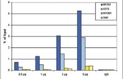
Chromatin Immunoprecipitation: Histone H3 [Dimethyl Lys79] Antibody [NBP2-59194] - ChIP assays were performed using human HeLa cells, the antibody against H3K79me2 and optimized PCR primer pairs for qPCR. ChIP was performed using sheared chromatin from 1 million cells. A titration consisting of 0.5, 1, 2 and 5 ug of antibody per ChIP experiment was analyzed. IgG (1 ug/IP) was used as a negative IP control. Quantitative PCR was performed with primers specific for the coding regions of the active EIF2S3 and CCT5 genes, used as positive controls, and for the inactive MYOD1) gene and the Sat2 satellite repeat, used as negative controls. Figure shows the recovery, expressed as a % of input (the relative amount of immunoprecipitated DNA compared to input DNA after qPCR analysis).
![Western Blot: Histone H3 [Dimethyl Lys79] Antibody [NBP2-59194]](https://resources.rndsystems.com/images/products/Histone-H3-[Dimethyl-Lys79]-Antibody-Western-Blot-NBP2-59194-img0001.jpg "Western Blot: Histone H3 [Dimethyl Lys79] Antibody [NBP2-59194]")
Western Blot: Histone H3 [Dimethyl Lys79] Antibody [NBP2-59194]
Western Blot: Histone H3 [Dimethyl Lys79] Antibody [NBP2-59194] - Western blot was performed on whole cell (25 ug, lane 1) and histone extracts (15 ug, lane 2) from HeLa cells, and on 1 ug of recombinant histone H2A, H2B, H3 and H4 (lane 3, 4, 5 and 6, respectively) using the antibody against H3K79me2 diluted 1:200 in TBS-Tween containing 5% skimmed milk. Observed molecular weight is ~17 kDa.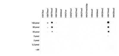
Dot Blot: Histone H3 [Dimethyl Lys79] Antibody [NBP2-59194] - Analysis was performed to test the cross reactivity of the Diagenode antibody against H3K79me2 with peptides containing other modifications and unmodified sequences of histone H3. One hundred to 0.2 pmol of peptide containing the respective histone modification were spotted on a membrane. The antibody was used at a dilution of 1:5,000. Figure 4 shows a high specificity of the antibody for the modification of interest.
![ELISA: Histone H3 [Dimethyl Lys79] Antibody [NBP2-59194]](https://resources.rndsystems.com/images/products/Histone-H3-[Dimethyl-Lys79]-Antibody-ELISA-NBP2-59194-img0002.jpg "ELISA: Histone H3 [Dimethyl Lys79] Antibody [NBP2-59194]")
ELISA: Histone H3 [Dimethyl Lys79] Antibody [NBP2-59194]
ELISA: Histone H3 [Dimethyl Lys79] Antibody [NBP2-59194] - To determine the titer of the antibody, an ELISA was performed using a serial dilution of the antibody directed against H3K79me2. The antigen used was a peptide containing the histone modification of interest. By plotting the absorbance against the antibody dilution, the titer of the antibody was estimated to be 1:6,600.Applications for Histone H3 [Dimethyl Lys79] Antibody - BSA Free
Application
Recommended Usage
Chromatin Immunoprecipitation (ChIP)
1-2 ug/IP
Dot Blot
1:5000
ELISA
1:500
Single Cell Western
100 ug/ml
Western Blot
1:200
Formulation, Preparation, and Storage
Purification
Peptide affinity purified
Formulation
PBS
Format
BSA Free
Preservative
0.05% Sodium Azide and 0.05% ProClin 300
Concentration
Please see the vial label for concentration. If unlisted please contact technical services.
Shipping
The product is shipped with polar packs. Upon receipt, store it immediately at the temperature recommended below.
Stability & Storage
Store at -20C. Avoid freeze-thaw cycles.
Background: Histone H3
Histones are nuclear proteins responsible for the nucleosome structure of the chromosomal fiber in eukaryotes. Changes in chromatin structure play a large role in the regulation of transcription. The chromatin fibers are compacted through the interaction of a linker histone, H1, with the DNA between the nucleosomes to form higher order chromatin structures.
Common histone modifications include methylation of lysine and arginine, acetylation of lysine, phosphorylation of threonine and serine, and sumoylation, biotinylation, and ubiquitylation of lysine. Posttranslational modifications such as acetylation of core histones regulates gene expression, thus altering protein function and regulation (1). Histone H3 is primarily acetylated at lysines 9, 14, 18, and 23 and have a theoretical molecular weight of 15 kDa. Acetylation at lysine 9 and 14 appears to control histone deposition, chromatin assembly and active transcription. Methylation of arginine residues within histone H3 has also been linked to transcription regulation. Histone H3 has been linked to various types of cancer as a biomarker through the aberrant expression of histone deacetylase (HDAC) enzymes and changes to chromatins (2-4).
References
1. Zhang, Y. X., Akumuo, R. C., Espana, R. A., Yan, C. X., Gao, W. J., & Li, Y. C. (2018). The histone demethylase KDM6B in the medial prefrontal cortex epigenetically regulates cocaine reward memory. Neuropharmacology, 141, 113-125. doi:10.1016/j.neuropharm.2018.08.030
2. Nandakumar, V., Hansen, N., Glenn, H. L., Han, J. H., Helland, S., Hernandez, K,...Meldrum, D. R. (2016). Vorinostat differentially alters 3D nuclear structure of cancer and non-cancerous esophageal cells. Sci Rep, 6, 30593. doi:10.1038/srep30593
3. Zhou, M., Li, Y., Lin, S., Chen, Y., Qian, Y., Zhao, Z., & Fan, H. (2019). H3K9me3, H3K36me3, and H4K20me3 Expression Correlates with Patient Outcome in Esophageal Squamous Cell Carcinoma as Epigenetic Markers. Dig Dis Sci, 64(8), 2147-2157. doi:10.1007/s10620-019-05529-2
4. Li, Y., Guo, D., Sun, R., Chen, P., Qian, Q., & Fan, H. (2019). Methylation Patterns of Lys9 and Lys27 on Histone H3 Correlate with Patient Outcome in Gastric Cancer. Dig Dis Sci, 64(2), 439-446. doi:10.1007/s10620-018-5341-8
Alternate Names
H3F3A, H3K79Me2
Gene Symbol
H3C14
Additional Histone H3 Products
Product Documents for Histone H3 [Dimethyl Lys79] Antibody - BSA Free
Certificate of Analysis
To download a Certificate of Analysis, please enter a lot or batch number in the search box below.
Product Specific Notices for Histone H3 [Dimethyl Lys79] Antibody - BSA Free
This product is for research use only and is not approved for use in humans or in clinical diagnosis. Primary Antibodies are guaranteed for 1 year from date of receipt.
Related Research Areas
Customer Reviews for Histone H3 [Dimethyl Lys79] Antibody - BSA Free
There are currently no reviews for this product. Be the first to review Histone H3 [Dimethyl Lys79] Antibody - BSA Free and earn rewards!
Have you used Histone H3 [Dimethyl Lys79] Antibody - BSA Free?
Submit a review and receive an Amazon gift card!
$25/€18/£15/$25CAN/¥2500 for a review with an image
$10/€7/£6/$10CAN/¥1110 for a review without an image
Submit a review
Protocols
Find general support by application which include: protocols, troubleshooting, illustrated assays, videos and webinars.
- Cellular Response to Hypoxia Protocols
- ChIP Protocol Video
- Chromatin Immunoprecipitation (ChIP) Protocol
- Chromatin Immunoprecipitation Protocol
- ELISA Sample Preparation & Collection Guide
- ELISA Troubleshooting Guide
- How to Run an R&D Systems DuoSet ELISA
- How to Run an R&D Systems Quantikine ELISA
- How to Run an R&D Systems Quantikine™ QuicKit™ ELISA
- Quantikine HS ELISA Kit Assay Principle, Alkaline Phosphatase
- Quantikine HS ELISA Kit Principle, Streptavidin-HRP Polymer
- R&D Systems Quality Control Western Blot Protocol
- Sandwich ELISA (Colorimetric) – Biotin/Streptavidin Detection Protocol
- Sandwich ELISA (Colorimetric) – Direct Detection Protocol
- Troubleshooting Guide: ELISA
- Troubleshooting Guide: Western Blot Figures
- Western Blot Conditions
- Western Blot Protocol
- Western Blot Protocol for Cell Lysates
- Western Blot Troubleshooting
- Western Blot Troubleshooting Guide
- View all Protocols, Troubleshooting, Illustrated assays and Webinars
Loading...