

Scientific images may be resized, cropped, or adjusted for brightness or contrast for presentation purposes. These updates are limited to presentation and do not affect the underlying scientific interpretation of the data. Where available, additional source data may be provided upon request.
Key Product Details
Species Reactivity
Validated:
Cited:
Applications
Validated:
Cited:
Label
Antibody Source
Product Specifications
Immunogen
Asp24-Leu197
Accession # Q04900
Specificity
Clonality
Host
Isotype
Scientific Data Images for Human CD164 Antibody
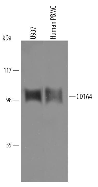
Detection of Human CD164 by Western Blot.
Western blot shows lysates of U937 human histiocytic lymphoma cell line and human peripheral blood mononuclear cells (PBMC). PVDF Membrane was probed with 1 µg/mL of Sheep Anti-Human CD164 Antigen Affinity-purified Polyclonal Antibody (Catalog # AF5790) followed by HRP-conjugated Anti-Sheep IgG Secondary Antibody (Catalog # HAF016). A specific band was detected for CD164 at approximately 100 kDa (as indicated). This experiment was conducted under reducing conditions and using Immunoblot Buffer Group 2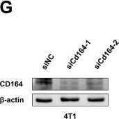
Detection of CD164 by Western Blot
The downregulation of CD164 impedes breast cancer progression in vivo. (A) Schematic representation of the tumor implantation and therapeutic protocol. (B) Measurement of tumor volume and (C) assessment of tumor growth rates comparing the si-CD164 treatment group to the control group throughout the treatment period in nude mice. (D) Monitoring alterations in body weight of nude mice during the treatment duration. (E) Immunohistochemical staining of tumor tissues for Ki67, a marker of proliferation. (F) Histological analysis of primary internal organs to evaluate potential toxicity (scale bars = 50 μm). (G) Western blot analysis evaluating the knockdown efficiency of siRNA-Cd164 in the 4T1 cell line. (H) Measurement of tumor volume and (I) assessment of tumor growth rates comparing the si-Cd164 treatment group to the control group throughout the treatment period in Balb/c mice. (J) Immunohistochemical staining of tumor tissues for Ki67 and CD164 (scale bars = 50 μm). (K) Monitoring alterations in body weight of Balb/c mice during the treatment duration. Data are expressed as mean ± SD. Statistical significance was assessed utilizing two-way ANOVA and t-tests. Image collected and cropped by CiteAb from the following open publication (https://www.frontiersin.org/articles/10.3389/fimmu.2025.1601547/full), licensed under a CC-BY license. Not internally tested by R&D Systems.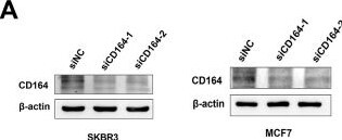
Detection of CD164 by Western Blot
CD164 knockdown suppresses proliferation and induces apoptosis in breast cancer cells. (A) Western blot analysis evaluating the knockdown efficacy of siRNA-CD164 in MCF7 and SKBR3 cell lines. (B) Cell viability assessed via CCK-8 assay subsequent to CD164 knockdown. (C) EDU incorporation assay and (D) clonogenic assay employed to evaluate cell proliferation. (E, F) Flow cytometry analysis of cell cycle distribution subsequent to CD164 inhibition. Annexin V/PI staining (G, H) to assess apoptosis levels. For flow cytometry analysis of apoptosis, quadrants were defined as follows: LL (Annexin V-/PI-) = viable cells; LR (Annexin V+/PI-) = early apoptotic cells; UR (Annexin V+/PI+) = late apoptotic/necrotic cells; UL (Annexin V-/PI+) = necrotic/mechanically damaged cells. Quadrant boundaries were established using unstained and single-stained controls. Data are expressed as mean ± standard deviation (SD). Statistical significance was evaluated through one-way ANOVA. Image collected and cropped by CiteAb from the following open publication (https://www.frontiersin.org/articles/10.3389/fimmu.2025.1601547/full), licensed under a CC-BY license. Not internally tested by R&D Systems.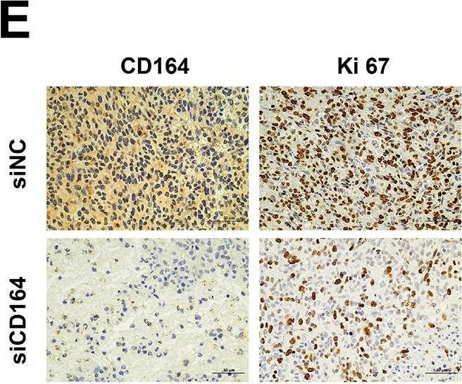
Detection of CD164 by Immunohistochemistry
The downregulation of CD164 impedes breast cancer progression in vivo. (A) Schematic representation of the tumor implantation and therapeutic protocol. (B) Measurement of tumor volume and (C) assessment of tumor growth rates comparing the si-CD164 treatment group to the control group throughout the treatment period in nude mice. (D) Monitoring alterations in body weight of nude mice during the treatment duration. (E) Immunohistochemical staining of tumor tissues for Ki67, a marker of proliferation. (F) Histological analysis of primary internal organs to evaluate potential toxicity (scale bars = 50 μm). (G) Western blot analysis evaluating the knockdown efficiency of siRNA-Cd164 in the 4T1 cell line. (H) Measurement of tumor volume and (I) assessment of tumor growth rates comparing the si-Cd164 treatment group to the control group throughout the treatment period in Balb/c mice. (J) Immunohistochemical staining of tumor tissues for Ki67 and CD164 (scale bars = 50 μm). (K) Monitoring alterations in body weight of Balb/c mice during the treatment duration. Data are expressed as mean ± SD. Statistical significance was assessed utilizing two-way ANOVA and t-tests. Image collected and cropped by CiteAb from the following open publication (https://www.frontiersin.org/articles/10.3389/fimmu.2025.1601547/full), licensed under a CC-BY license. Not internally tested by R&D Systems.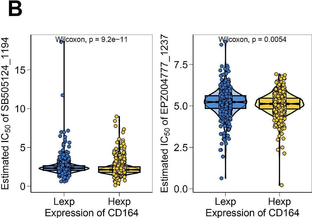
Detection of CD164 by Flow Cytometry
Chemosensitivity and functional enrichment analyses of CD164 in breast carcinoma. (A) Chemosensitivity analysis of breast cancer specimens utilizing drug response data from the GDSC database. The pharmacological sensitivity profiles of four targeted agents—YK-4-279 (RNA helicase A inhibitor), BMS-345541 (IKK inhibitor), AZ960 (JAK2/3 inhibitor), and Talazoparib (PARP inhibitor)—were stratified by CD164 expression status (Low Expression [Lexp] vs. High Expression [Hexp]). Violin plot distributions revealed consistently lower IC50 values (indicating greater sensitivity) in CD164-low tumors across above compounds. (B) The pharmacological sensitivity profiles of two targeted agents—SB505124 (TGF-beta receptor inhibitor) and EPZ004777 (DOT1L inhibitor)—were also stratified by CD164 expression status (Lexp vs. Hexp). Violin plot distributions revealed consistently lower IC50 values (indicating greater sensitivity) in CD164-high tumors across above compounds. (C) GSEA analysis revealed CD164’s involvement in regulating the AMPK signaling pathway, Butanoate metabolism, and Circadian rhythm pathways. (D) GSVA analysis was performed to investigate the molecular mechanisms through which CD164 facilitates tumor progression. The gene sets were ranked based on the GSVA scores. Data was acquired from the Molecular Signatures Database. Image collected and cropped by CiteAb from the following open publication (https://www.frontiersin.org/articles/10.3389/fimmu.2025.1601547/full), licensed under a CC-BY license. Not internally tested by R&D Systems.Applications for Human CD164 Antibody
Western Blot
Sample: U937 human histiocytic lymphoma cell line and human peripheral blood mononuclear cells (PBMC)
Formulation, Preparation, and Storage
Purification
Reconstitution
Reconstitute at 0.2 mg/mL in sterile PBS. For liquid material, refer to CoA for concentration.
Formulation
Shipping
Stability & Storage
- 12 months from date of receipt, -20 to -70 °C as supplied.
- 1 month, 2 to 8 °C under sterile conditions after reconstitution.
- 6 months, -20 to -70 °C under sterile conditions after reconstitution.
Calculators
Background: CD164
Alternate Names
Gene Symbol
UniProt
Additional CD164 Products
Product Documents for Human CD164 Antibody
Certificate of Analysis
To download a Certificate of Analysis, please enter a lot or batch number in the search box below.
Note: Certificate of Analysis not available for kit components.
Product Specific Notices for Human CD164 Antibody
For research use only
Related Research Areas
Citations for Human CD164 Antibody
 Powered by Bioz
Customer Reviews for Human CD164 Antibody
There are currently no reviews for this product. Be the first to review Human CD164 Antibody and earn rewards!
Have you used Human CD164 Antibody?
Submit a review and receive an Amazon gift card!
$25/€18/£15/$25CAN/¥2500 for a review with an image
$10/€7/£6/$10CAN/¥1110 for a review without an image
Submit a review
Protocols
Find general support by application which include: protocols, troubleshooting, illustrated assays, videos and webinars.
- Cellular Response to Hypoxia Protocols
- R&D Systems Quality Control Western Blot Protocol
- Troubleshooting Guide: Western Blot Figures
- Western Blot Conditions
- Western Blot Protocol
- Western Blot Protocol for Cell Lysates
- Western Blot Troubleshooting
- Western Blot Troubleshooting Guide
- View all Protocols, Troubleshooting, Illustrated assays and Webinars
Associated Pathways
