
Crossveinless-2 (CV-2), also known as bone morphogenetic protein-binding endothelial cell precursor-derived regulator (BMPER), is a secreted Chordin-like protein that modulates the BMP signaling pathway (1‑3). Human CV-2 is synthesized as a 685 amino acid (aa) precursor protein with a putative 39 aa signal peptide, five tandem chordin-like cysteine-rich (CR) domains, a partial von Willebrand factor type D domain (vWD), and a carboxyl trypsin inhibitor-like cysteine-rich domain (TIL) (1, 4). Secreted CV-2 is reported to be proteolytically cleaved to generate two fragments that are disulfide-linked (1, 2). The cleavage site of R&D Systems’ recombinant CV-2 is found to be between Asp369 and Pro370 in the GDPH sequence within the vWD domain. This cleavage is likely due to an autocatalytic mechanism triggered by low pH comparable to that of the late secretory pathway (5). The GDPH sequence is conserved in CV-2 from other species. It is also found in multiple proteins that undergo a similar type of cleavage (5). Human CV-2 message is detected in many tissues, with the highest expression detected in adult brain and adult and fetal lung (1). It is also expressed in Flk-1+ endothelial cell precursors and in primary chondrocytes (2). During embryonic development, CV-2 is expressed in regions of high BMP signaling, such as the posterior primitive streak and the ventral tail bud (4). Human CV-2 shares 92% and 34% aa sequence identity with the mouse and Drosophila homologs, respectively (1, 4). Results from biochemical experiments using recombinant CV-2 show that CV-2 directly interacts with BMP-2, -4, and -6 to antagonize BMP signaling, which can regulate a wide range of differentiation processes (1, 2). In contrast, genetic data from Drosophila suggest that CV-2 potentiates BMP-signaling (6). It is possible that like TSG, CV-2 can positively and negatively modulate BMP signal transduction depending on the cell context (7).
Human Crossveinless-2/CV-2 Antibody (355304)
R&D Systems | Catalog # MAB1956

Scientific images may be resized, cropped, or adjusted for brightness or contrast for presentation purposes. These updates are limited to presentation and do not affect the underlying scientific interpretation of the data. Where available, additional source data may be provided upon request.
Key Product Details
Species Reactivity
Validated:
Cited:
Applications
Validated:
Cited:
Label
Antibody Source
Product Specifications
Immunogen
Val34-Arg685
Accession # Q8N8U9
Specificity
Clonality
Host
Isotype
Scientific Data Images for Human Crossveinless-2/CV-2 Antibody (355304)
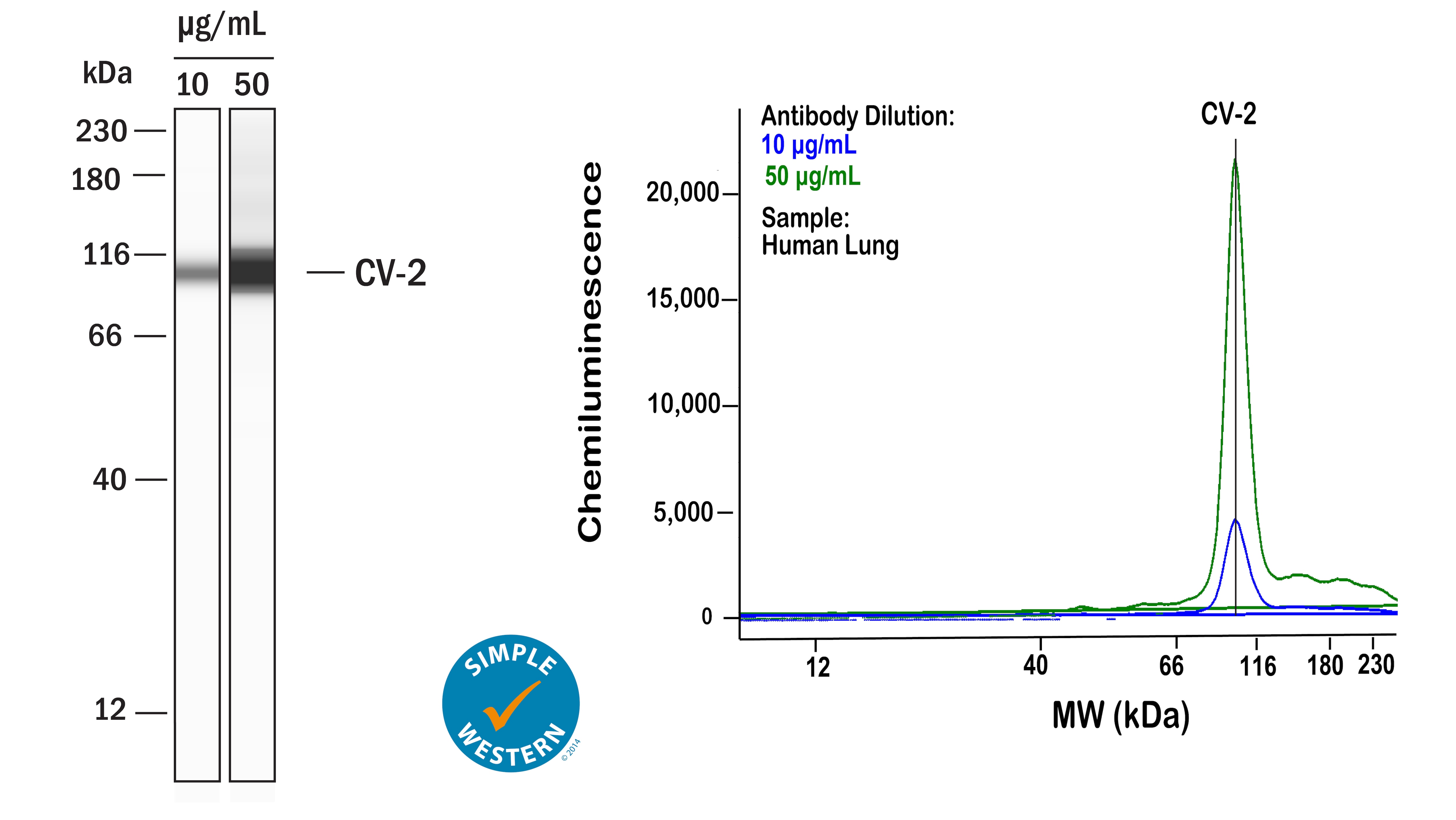
Detection of Human Crossveinless‑2/CV‑2 by Simple WesternTM.
Left: Simple Western lane view shows lysates of human lung, loaded at 1 mg/ml. A specific band was detected for Crossveinless‑2/CV‑2 at approximately 103 kDa (as indicated) using both 10 µg/ml and 50 µg/ml of Rat Anti-Human Crossveinless‑2/CV‑2 Monoclonal Antibody (Catalog # MAB1956) followed by 1:50 dilution of HRP-conjugated Anti-Rat IgG Secondary Antibody (Catalog # HAF005) in Milk-free Antibody Diluent (Catalog # 043-524). This experiment was conducted under reducing conditions and using the 12-230kDa separation system. Right: Simple Western electropherogram showing the same Rat Anti-Human Crossveinless‑2/CV‑2 Monoclonal Antibody (Catalog # MAB1956) tested at 10 µg/ml (blue line) and 50 µg/ml (green line) in the human lung.Applications for Human Crossveinless-2/CV-2 Antibody (355304)
Simple Western
Sample: Human lung
Western Blot
Sample: Recombinant Human Crossveinless‑2/CV‑2 (Catalog # 1956-CV)
Formulation, Preparation, and Storage
Purification
Reconstitution
Reconstitute at 0.5 mg/mL in sterile PBS. For liquid material, refer to CoA for concentration.
Formulation
Shipping
Stability & Storage
- 12 months from date of receipt, -20 to -70 °C as supplied.
- 1 month, 2 to 8 °C under sterile conditions after reconstitution.
- 6 months, -20 to -70 °C under sterile conditions after reconstitution.
Calculators
Background: Crossveinless-2/CV-2
References
- Binnerts, M.E. et al. (2004) Biochem Biophys Res Commun. 315:272.
- Moser, M. et al. (2003) Mol Cell Biol. 23:5664.
- Garcia-Abreu, J. et al. (2002) Gene, 287:39.
- Coffinier, C. et al. (2002) Mech Dev. 119:S179.
- Lidell, M.E. et al. (2003) J. Biol. Chem. 278:13944.
- Conley, C.A. et al. (2000) Development 127:3947.
- Kamimura, M. et al. (2004) Developmental Dynamics 230:434.
Long Name
Alternate Names
Gene Symbol
UniProt
Additional Crossveinless-2/CV-2 Products
Product Documents for Human Crossveinless-2/CV-2 Antibody (355304)
Certificate of Analysis
To download a Certificate of Analysis, please enter a lot or batch number in the search box below.
Note: Certificate of Analysis not available for kit components.
Product Specific Notices for Human Crossveinless-2/CV-2 Antibody (355304)
For research use only
Related Research Areas
Citations for Human Crossveinless-2/CV-2 Antibody (355304)
 Powered by Bioz
Customer Reviews for Human Crossveinless-2/CV-2 Antibody (355304)
There are currently no reviews for this product. Be the first to review Human Crossveinless-2/CV-2 Antibody (355304) and earn rewards!
Have you used Human Crossveinless-2/CV-2 Antibody (355304)?
Submit a review and receive an Amazon gift card!
$25/€18/£15/$25CAN/¥2500 for a review with an image
$10/€7/£6/$10CAN/¥1110 for a review without an image
Submit a review
Protocols
Find general support by application which include: protocols, troubleshooting, illustrated assays, videos and webinars.
- Cellular Response to Hypoxia Protocols
- R&D Systems Quality Control Western Blot Protocol
- Troubleshooting Guide: Western Blot Figures
- Western Blot Conditions
- Western Blot Protocol
- Western Blot Protocol for Cell Lysates
- Western Blot Troubleshooting
- Western Blot Troubleshooting Guide
- View all Protocols, Troubleshooting, Illustrated assays and Webinars