
phospho-Akt (S473) Pan Specific Antibody (545007)
R&D Systems | Catalog # MAB887

Scientific images may be resized, cropped, or adjusted for brightness or contrast for presentation purposes. These updates are limited to presentation and do not affect the underlying scientific interpretation of the data. Where available, additional source data may be provided upon request.
Key Product Details
Validated by
Species Reactivity
Validated:
Cited:
Applications
Validated:
Cited:
Label
Antibody Source
Product Specifications
Immunogen
Specificity
Clonality
Host
Isotype
Scientific Data Images for phospho-Akt (S473) Pan Specific Antibody (545007)
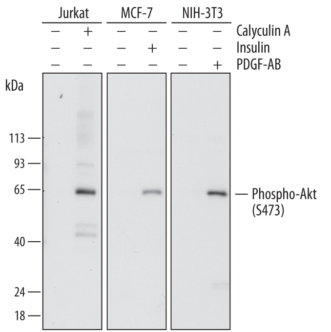
Detection of Human and Mouse Phospho-Akt (S473) by Western Blot.
Western blot shows lysates of Jurkat human acute T cell leukemia cell line, MCF-7 human breast cancer cell line, and NIH-3T3 mouse embryonic fibroblast cell line untreated (-) or treated (+) with 100 nm Calyculin for 30 minutes, 1 µg/mL insulin for 5 minutes, or 100 ng/mL Recombinant Human PDGF-AB (Catalog # 222-AB) for 20 minutes. PVDF membrane was probed with 0.2 µg/mL of Mouse Anti-Human Phospho-Akt (S473) Pan Specific Monoclonal Antibody (Catalog # MAB887) followed by HRP-conjugated Anti-Mouse IgG Secondary Antibody (Catalog # HAF007). A specific band was detected for Phospho-Akt (S473) at approximately 65 kDa (as indicated). This experiment was conducted under reducing conditions and using Immunoblot Buffer Group 1.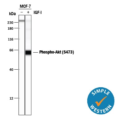
Detection of Human Phospho-Akt (S473) by Simple WesternTM.
Simple Western lane view shows lysates of MCF-7 human breast cancer cell line untreated (-) or treated (+) with 100 ng/mL Recombinant Human IGF-I (Catalog # 291-G1) for 20 minutes, loaded at 0.2 mg/mL. A specific band was detected for Phospho-Akt (S473) at approximately 65 kDa (as indicated) using 2 µg/mL of Mouse Anti-Human/Mouse Phospho-Akt (S473) Pan Specific Monoclonal Antibody (Catalog # MAB887). This experiment was conducted under reducing conditions and using the 12-230 kDa separation system.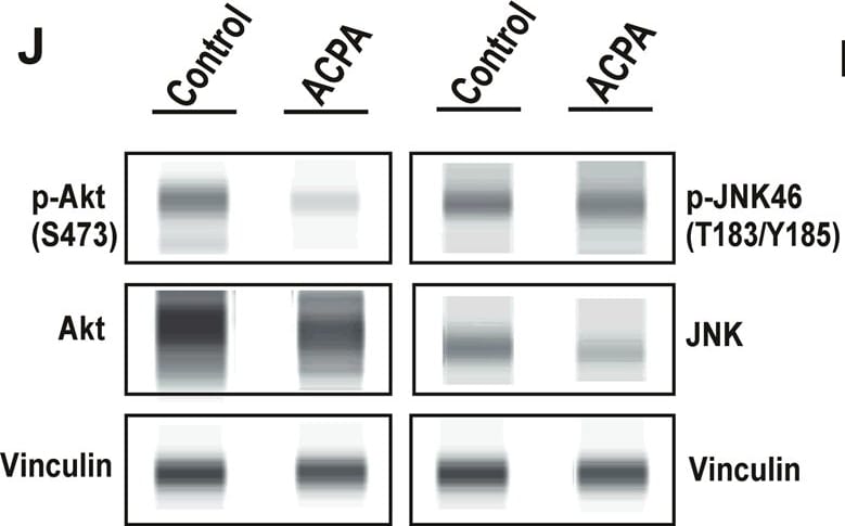
Detection of Human AKT by Simple Western
ACPA inhibits p-Akt, induces p-JNK and affects levels of specific metabolites in NSCLC lines.a Principal component analysis (PCA) score plot: Metabolomics profiling of control and ACPA-treated A549, H1299, H358, and H838 cells. b Changes in variable importance in projection (VIP) values for 19 metabolites in A549 cells. c, d, e Changes in VIP values for 20 metabolites in H1299, H358, and H838 cells. Significantly changed metabolites (*p < 0.05, indicated by arrows) were matched to apoptotic pathways. f, g, h, i Increase and decrease in several metabolites of ACPA-treated A549, H1299, H358, and H838 cells (*p < 0.05). j Simple Western showing total Akt, p-Akt (S473), total JNK46 and JNK54 and p-JNK46 and p-JNK54 (T183/Y185) in A549 cells at 24 hours after treatment with IC50 dose of ACPA. k Relative expression levels of Akt and p-Akt for control and ACPA-treated A549 cells after normalization by total vinculin protein. l Relative expression levels of JNK (46 and 54 kDa) and p-JNK for control and ACPA-treated A549 cells after normalization by total vinculin protein. *p < 0.05, Student’s t-test. All tests were done in quadruplicates. Image collected and cropped by CiteAb from the following publication (https://pubmed.ncbi.nlm.nih.gov/33431819), licensed under a CC-BY license. Not internally tested by R&D Systems.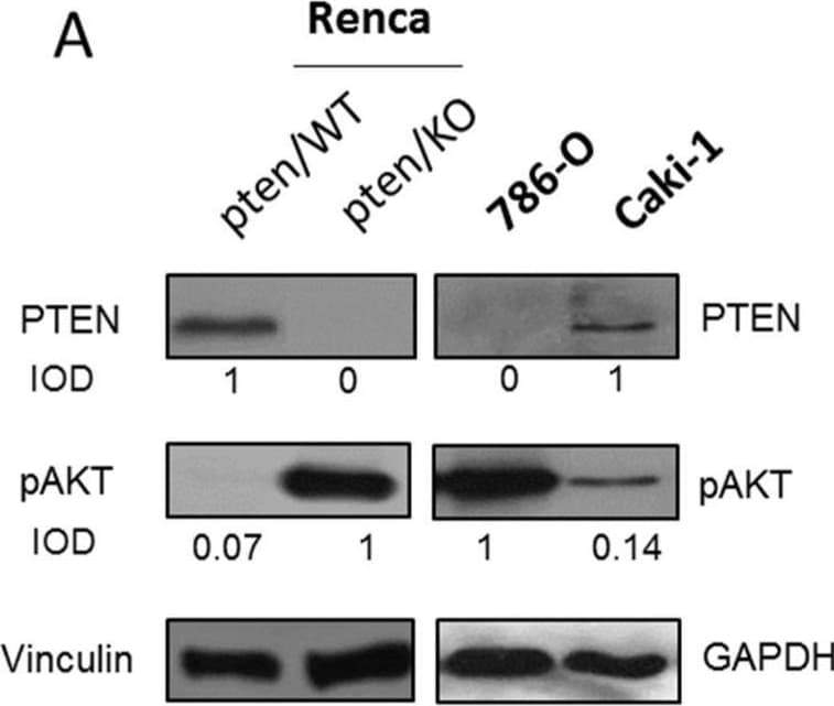
Detection of Phospho-Akt (S473) Pan Specific by Western Blot
PTEN status in tested models (A) PTEN and pAKT levels in kidney cancer cell lines, IOD calculated relative to Vinculin (B) Relative to GusB expression of Pten in RNA isolates from kidney cancer and healthy kidney tissues (C) graphical workflow. Image collected and cropped by CiteAb from the following open publication (https://pubmed.ncbi.nlm.nih.gov/35625614), licensed under a CC-BY license. Not internally tested by R&D Systems.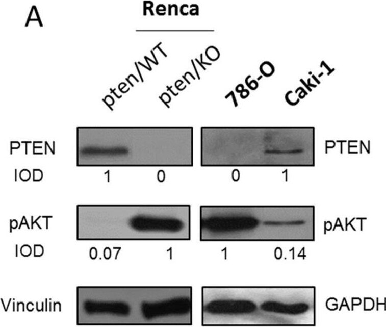
Detection of Phospho-Akt (S473) Pan Specific by Western Blot
PTEN status in tested models (A) PTEN and pAKT levels in kidney cancer cell lines, IOD calculated relative to Vinculin (B) Relative to GusB expression of Pten in RNA isolates from kidney cancer and healthy kidney tissues (C) graphical workflow. Image collected and cropped by CiteAb from the following open publication (https://pubmed.ncbi.nlm.nih.gov/35625614), licensed under a CC-BY license. Not internally tested by R&D Systems.Applications for phospho-Akt (S473) Pan Specific Antibody (545007)
Simple Western
Sample: MCF‑7 human breast cancer cell line treated with Recombinant Human IGF‑I (Catalog # 291-G1)
Western Blot
Sample: Jurkat human acute T cell leukemia cell line, MCF‑7 human breast cancer cell line, and NIH‑3T3 mouse embryonic fibroblast cell line treated with calyculin, insulin, or Recombinant Human PDGF-AB (Catalog # 222-AB)
Reviewed Applications
Read 2 reviews rated 5 using MAB887 in the following applications:
Formulation, Preparation, and Storage
Purification
Reconstitution
Sterile PBS to a final concentration of 0.5 mg/mL. For liquid material, refer to CoA for concentration.
Formulation
Shipping
Stability & Storage
- 12 months from date of receipt, -20 to -70 °C as supplied.
- 1 month, 2 to 8 °C under sterile conditions after reconstitution.
- 6 months, -20 to -70 °C under sterile conditions after reconstitution.
Calculators
Background: Akt
Long Name
Alternate Names
Additional Akt Products
Product Documents for phospho-Akt (S473) Pan Specific Antibody (545007)
Certificate of Analysis
To download a Certificate of Analysis, please enter a lot or batch number in the search box below.
Note: Certificate of Analysis not available for kit components.
Product Specific Notices for phospho-Akt (S473) Pan Specific Antibody (545007)
For research use only
Citations for phospho-Akt (S473) Pan Specific Antibody (545007)
 Powered by Bioz
Customer Reviews for phospho-Akt (S473) Pan Specific Antibody (545007) (2)
Have you used phospho-Akt (S473) Pan Specific Antibody (545007)?
Submit a review and receive an Amazon gift card!
$25/€18/£15/$25CAN/¥2500 for a review with an image
$10/€7/£6/$10CAN/¥1110 for a review without an image
Submit a review
Customer Images
-
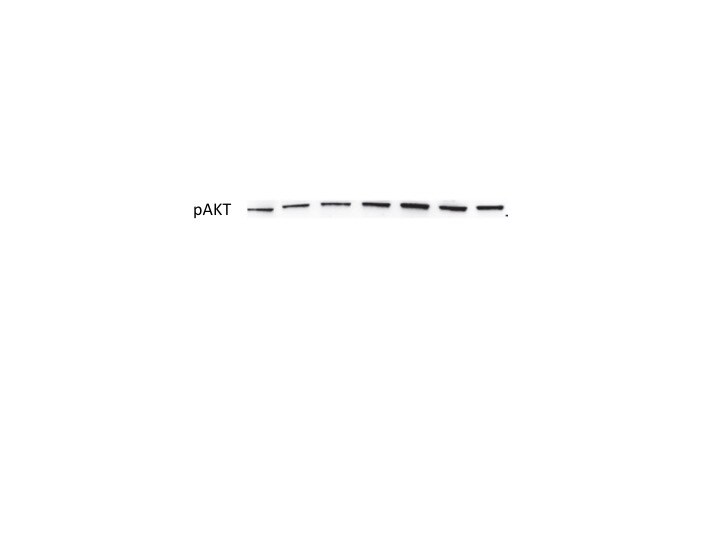
Application: Western BlotSample Tested: HEK293 human embryonic kidney cell lineSpecies: HumanVerified Customer | Posted 07/21/2018

-
Application: Functional AssaySample Tested: Purified StandardSpecies: HumanVerified Customer | Posted 12/01/2017
There are no reviews that match your criteria.
Protocols
Find general support by application which include: protocols, troubleshooting, illustrated assays, videos and webinars.
- Cellular Response to Hypoxia Protocols
- R&D Systems Quality Control Western Blot Protocol
- Troubleshooting Guide: Western Blot Figures
- Western Blot Conditions
- Western Blot Protocol
- Western Blot Protocol for Cell Lysates
- Western Blot Troubleshooting
- Western Blot Troubleshooting Guide
- View all Protocols, Troubleshooting, Illustrated assays and Webinars