
Activin A betaA subunit Antibody (69403)
R&D Systems | Catalog # MAB3381

Scientific images may be resized, cropped, or adjusted for brightness or contrast for presentation purposes. These updates are limited to presentation and do not affect the underlying scientific interpretation of the data. Where available, additional source data may be provided upon request.
Key Product Details
Species Reactivity
Validated:
Cited:
Applications
Validated:
Cited:
Label
Antibody Source
Product Specifications
Immunogen
Specificity
Clonality
Host
Isotype
Endotoxin Level
Scientific Data Images for Activin A betaA subunit Antibody (69403)
Hemoglobin Expression Induced by Activin A and Neutralization by Human/ Mouse/Rat Activin A Antibody.
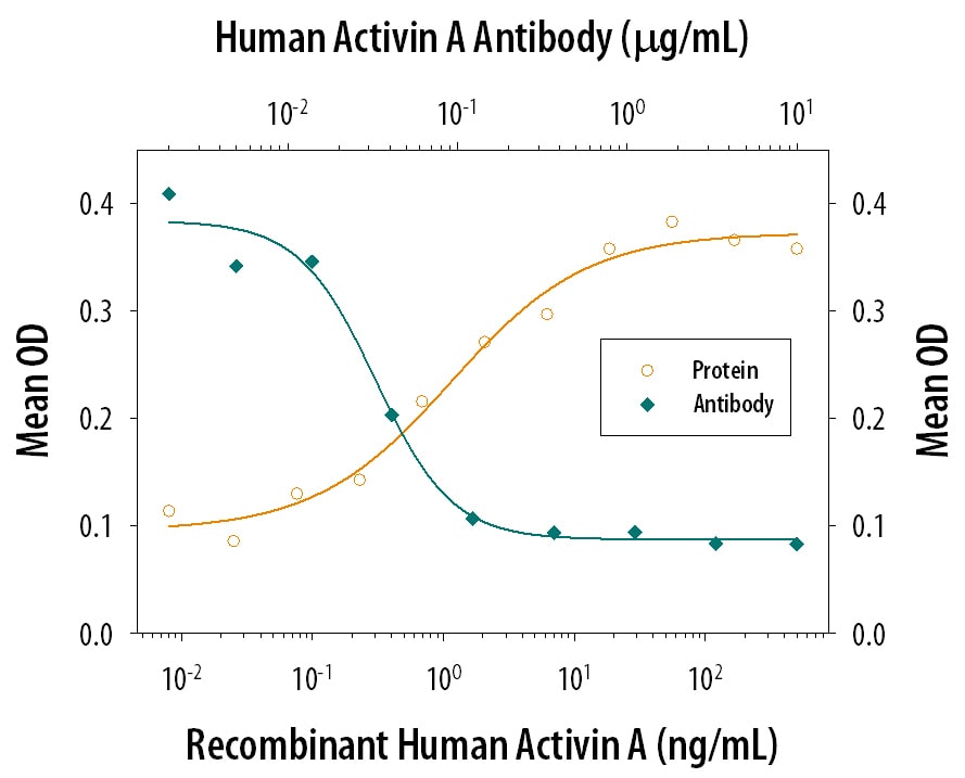
Recombinant Human/Mouse/ Rat Activin A (Catalog # 338-AC) increases hemoglobin expression in the K562 human chronic myelogenous leukemia cell line in a dose-dependent manner (orange line), as measured by the psuedoperoxidase assay. Hemoglobin Expression elicited by Recombinant Human/Mouse/Rat Activin A (7.5 ng/mL) is neutralized (green line) by increasing concentrations of Goat Anti-Human/Mouse/Rat Activin A beta A subunit Monoclonal Antibody (Catalog # MAB3381). The ND50 is typically 0.02-0.06 µg/mL.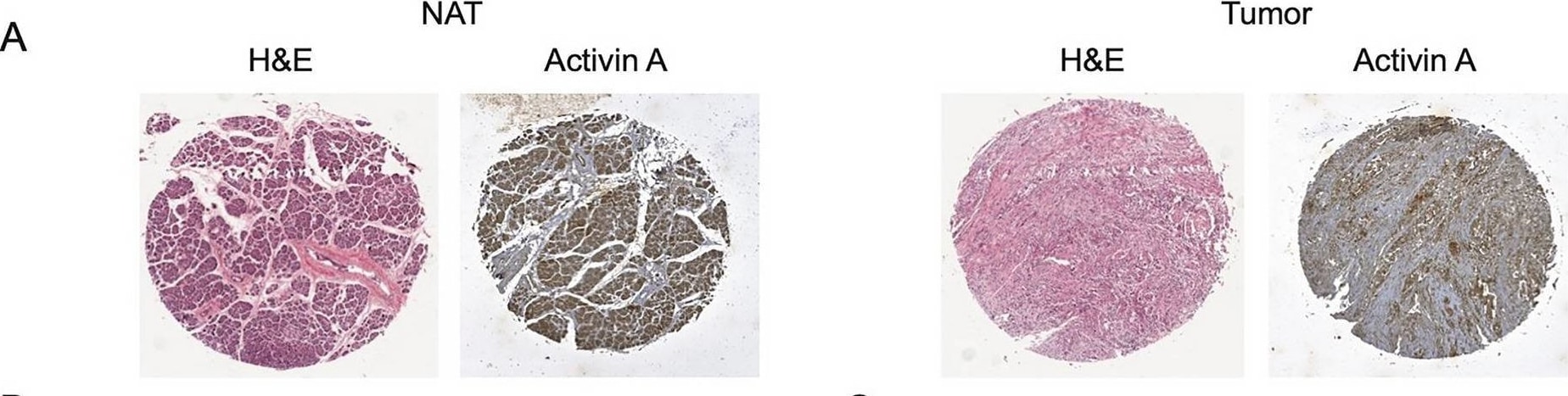
Detection of Activin A beta A subunit by Immunohistochemistry
Activin A levels are significantly higher in tumor tissue and correlate to worse prognosis in PDAC. (A) Representative cores of pancreatic tumors from a human pancreatic tissue microarray stained with H&E to assess the percentage of epithelial versus stromal cells in each core. Adjacent sections were immunostained for activin A expression. (B) Quantification of the percentage of epithelial cells (left panel) or stromal cells (right panel) in normal tissue (white bar) versus tumor tissue (black bar). Statistical analysis is unpaired t-test. (C) Quantification of activin A in TMA (n = 63). An average of the percentage of epithelial and stromal fraction was calculated and scored in each TMA. Comparing epithelial (left panel) and stromal fraction (right panel) in normal tissue (white bar) versus tumor tissue (black bar). Statistical analysis 2-way ANOVA; mean ± SEM, ***(p < 0.001). (D) Kaplan–Meier plots of patient overall survival versus expression of activin A in either epithelial cells (left panel) or stromal cells (right panel). Statistical analysis Log-rank (Mantel-Cox) test, ** (p < 0.01), ***(p < 0.001). Image collected and cropped by CiteAb from the following open publication (https://pubmed.ncbi.nlm.nih.gov/33846512), licensed under a CC-BY license. Not internally tested by R&D Systems.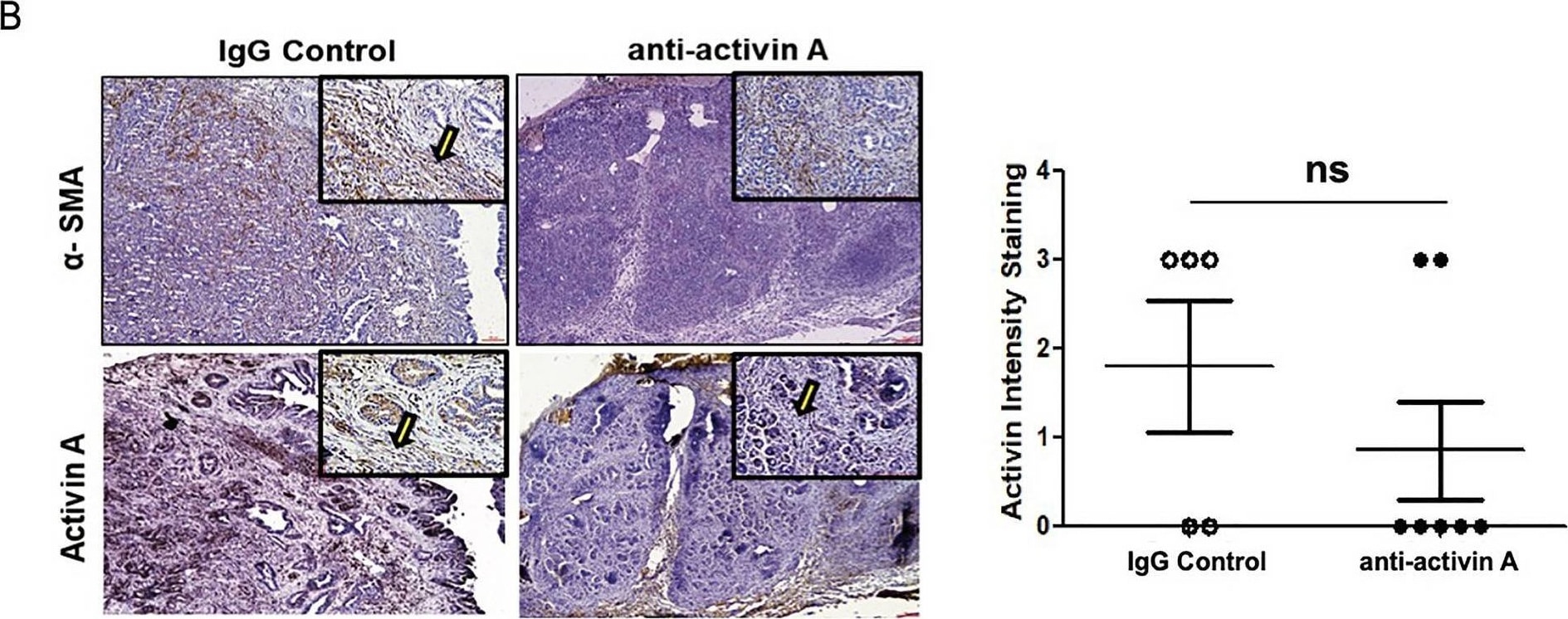
Detection of Activin A beta A subunit by Immunohistochemistry
Inhibition of activin A in KPC mice significantly reduces metastasis. (A) Time course of anti-activin A neutralizing antibody injected in mice weekly (2 mg/kg). Control animals were injected with the same concentration of IgG isotype. Remaining animals were collected at the end of week 13 of treatment. (B) Representative image (×4 magnification) of H&E staining of pancreas from control treated (IgG) and anti-activin A treated KPC mice IHC-stained for either alpha -SMA (to indicate stromal cells) or activin A. Representative stromal cells marked by arrows. (C) Representative image (×4 magnification) of H&E staining of pancreas used for quantification of CN (yellow arrow) cystic lesions. Quantification of lesions is documented in both IgG control and anti-activin A antibody treated mice. (D) Left panel: Representative images (×10 and ×40 magnification) of liver tissue from control treated versus anti-activin A treated KPC mice stained with H&E (upper panel) or ductal marker protein CK19 (Lower panel). Right panel: Quantification of distant metastatic formation in the liver tissue of control (n = 5) and treated (n = 7) mice. Statistical analysis unpaired Student t-test (ns: not significant). Image collected and cropped by CiteAb from the following open publication (https://pubmed.ncbi.nlm.nih.gov/33846512), licensed under a CC-BY license. Not internally tested by R&D Systems.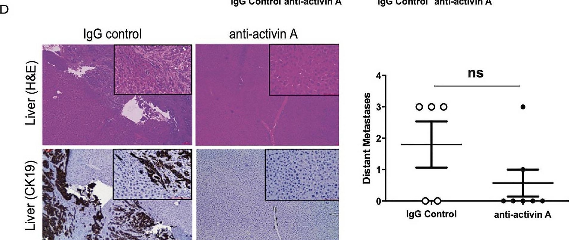
Detection of Activin A beta A subunit by Immunohistochemistry
Inhibition of activin A in KPC mice significantly reduces metastasis. (A) Time course of anti-activin A neutralizing antibody injected in mice weekly (2 mg/kg). Control animals were injected with the same concentration of IgG isotype. Remaining animals were collected at the end of week 13 of treatment. (B) Representative image (×4 magnification) of H&E staining of pancreas from control treated (IgG) and anti-activin A treated KPC mice IHC-stained for either alpha -SMA (to indicate stromal cells) or activin A. Representative stromal cells marked by arrows. (C) Representative image (×4 magnification) of H&E staining of pancreas used for quantification of CN (yellow arrow) cystic lesions. Quantification of lesions is documented in both IgG control and anti-activin A antibody treated mice. (D) Left panel: Representative images (×10 and ×40 magnification) of liver tissue from control treated versus anti-activin A treated KPC mice stained with H&E (upper panel) or ductal marker protein CK19 (Lower panel). Right panel: Quantification of distant metastatic formation in the liver tissue of control (n = 5) and treated (n = 7) mice. Statistical analysis unpaired Student t-test (ns: not significant). Image collected and cropped by CiteAb from the following open publication (https://pubmed.ncbi.nlm.nih.gov/33846512), licensed under a CC-BY license. Not internally tested by R&D Systems.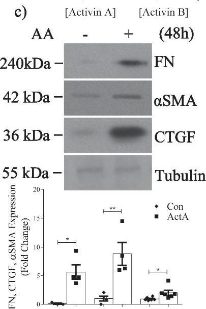
Detection of Activin A beta A subunit by Western Blot
Specific activin A inhibition attenuates TGF beta 1-induced Smad3 activation and profibrotic responses in MC. a ELISA demonstrates that TGF beta 1 (24 h) increases actA and actB secretion (n = 3) to 19.5 ng/ml and 2.5 ng/ml, representing an 8.9- and 1.06-fold induction respectively. b TGF beta 1 increases actA in whole cell lysate by 1.8-fold (n = 3). c ActA (20 ng/ml) upregulates FN (n = 3–4), CTGF (n = 4) and alpha SMA (n = 6) at 48 h. d TGF beta 1 and actA both increase Smad3 transcriptional activity; no synergistic effect is seen (n = 6–12). e An actA neutralizing antibody attenuates TGF beta 1-induced FN (n = 5–6), alpha SMA (n = 5), CTGF (n = 5–6), and Smad3 activation (n = 10–12). f ActA neutralization decreases TGF beta 1-induced Smad3 transcriptional activity at 24 h (n = 9–15), but this is not decreased by actB neutralization (n = 6) (g). h MC were stimulated with TGF beta 1 or actA for 1 h, then treated with their type I receptor inhibitor SB431542 (50 µM). Restimulation with the same ligand shows that cells become refractory to TGF beta 1, but not actA (n = 4). *, **, ***, ****P < 0.05, 0.01, 0.001, 0.0001; one-way ANOVA with Tukey’s multiple comparisons post hoc test Image collected and cropped by CiteAb from the following open publication (https://pubmed.ncbi.nlm.nih.gov/36717814), licensed under a CC-BY license. Not internally tested by R&D Systems.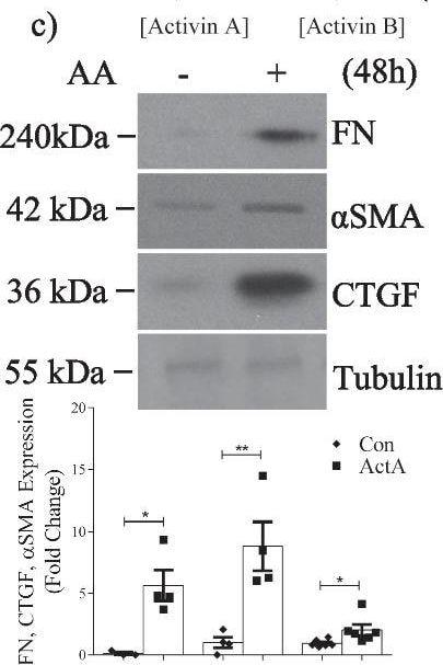
Detection of Activin A beta A subunit by Western Blot
Specific activin A inhibition attenuates TGF beta 1-induced Smad3 activation and profibrotic responses in MC. a ELISA demonstrates that TGF beta 1 (24 h) increases actA and actB secretion (n = 3) to 19.5 ng/ml and 2.5 ng/ml, representing an 8.9- and 1.06-fold induction respectively. b TGF beta 1 increases actA in whole cell lysate by 1.8-fold (n = 3). c ActA (20 ng/ml) upregulates FN (n = 3–4), CTGF (n = 4) and alpha SMA (n = 6) at 48 h. d TGF beta 1 and actA both increase Smad3 transcriptional activity; no synergistic effect is seen (n = 6–12). e An actA neutralizing antibody attenuates TGF beta 1-induced FN (n = 5–6), alpha SMA (n = 5), CTGF (n = 5–6), and Smad3 activation (n = 10–12). f ActA neutralization decreases TGF beta 1-induced Smad3 transcriptional activity at 24 h (n = 9–15), but this is not decreased by actB neutralization (n = 6) (g). h MC were stimulated with TGF beta 1 or actA for 1 h, then treated with their type I receptor inhibitor SB431542 (50 µM). Restimulation with the same ligand shows that cells become refractory to TGF beta 1, but not actA (n = 4). *, **, ***, ****P < 0.05, 0.01, 0.001, 0.0001; one-way ANOVA with Tukey’s multiple comparisons post hoc test Image collected and cropped by CiteAb from the following open publication (https://pubmed.ncbi.nlm.nih.gov/36717814), licensed under a CC-BY license. Not internally tested by R&D Systems.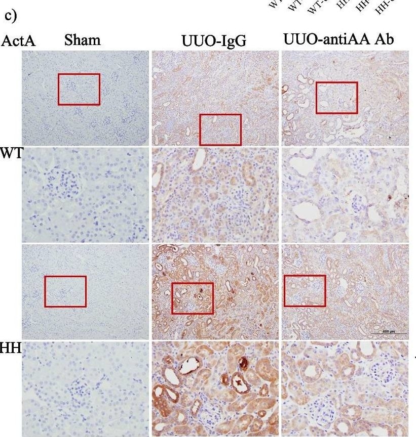
Detection of Activin A beta A subunit by Immunohistochemistry
Activin A neutralization inhibits renal fibrosis in TGF beta 1-overexpressing mice. a TGF beta 1 transcript is increased in mice genetically engineered to overexpress TGF beta 1 (HH) compared with wild-type mice (WT) (n = 6–7, *p ≤ 0.05). b Serum actA is elevated in wild-type and HH mice after UUO. This is decreased by treatment with a neutralizing actA antibody (anti-actA) in HH mice. c Renal actA is increased after UUO, with a greater induction in HH mice. Both are attenuated by actA neutralization. Boxed areas are shown at higher magnification immediately below. ActA increases are seen particularly in tubular epithelial cells. d Renal alpha SMA, fibronectin (FN), pSmad3 and MRTF-A are increased after UUO and this is augmented in HH kidneys. Expression of all is attenuated by actA neutralization in both WT and HH kidneys. (n = 6–9) *, **, ***, ****P < 0.05, 0.01, 0.001, 0.0001; one-way ANOVA with Tukey’s multiple comparisons post hoc test where there are > 2 groups; t-test for 2 groups Image collected and cropped by CiteAb from the following open publication (https://pubmed.ncbi.nlm.nih.gov/36717814), licensed under a CC-BY license. Not internally tested by R&D Systems.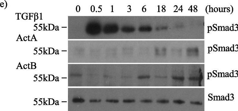
Detection of Activin A beta A subunit by Western Blot
Activin inhibition attenuates TGF beta 1-induced fibrotic responses and Smad3 activation in MC. Activin inhibition with follistatin (FST) decreases TGF beta 1-induced: a FN, alpha SMA and CTGF upregulation at 48 h (n = 5), b Smad3 phosphorylation (pSmad3) at 24 h (n = 5), c Smad3 nuclear translocation as assessed using eGFP-Smad3 (n = 3; 25–30 cells quantified per treatment group) at 24 h, and d Smad3 transcriptional activity at 24 h (n = 8). e Time course experiments show increases in pSmad3 occur earlier (30–60 min) with TGF beta 1 (n = 4) compared with actA (n = 4) or actB (n = 3) (18–48 h). *, **, ***, ****P < 0.05, 0.01, 0.001, 0.0001; one-way ANOVA with Tukey’s multiple comparisons post hoc test Image collected and cropped by CiteAb from the following open publication (https://pubmed.ncbi.nlm.nih.gov/36717814), licensed under a CC-BY license. Not internally tested by R&D Systems.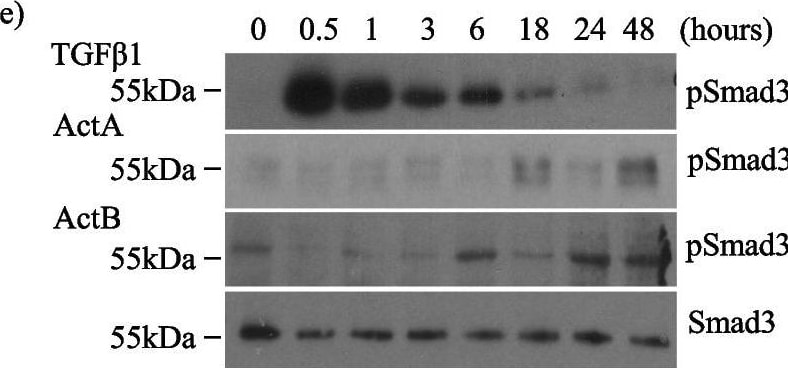
Detection of Activin A beta A subunit by Western Blot
Activin inhibition attenuates TGF beta 1-induced fibrotic responses and Smad3 activation in MC. Activin inhibition with follistatin (FST) decreases TGF beta 1-induced: a FN, alpha SMA and CTGF upregulation at 48 h (n = 5), b Smad3 phosphorylation (pSmad3) at 24 h (n = 5), c Smad3 nuclear translocation as assessed using eGFP-Smad3 (n = 3; 25–30 cells quantified per treatment group) at 24 h, and d Smad3 transcriptional activity at 24 h (n = 8). e Time course experiments show increases in pSmad3 occur earlier (30–60 min) with TGF beta 1 (n = 4) compared with actA (n = 4) or actB (n = 3) (18–48 h). *, **, ***, ****P < 0.05, 0.01, 0.001, 0.0001; one-way ANOVA with Tukey’s multiple comparisons post hoc test Image collected and cropped by CiteAb from the following open publication (https://pubmed.ncbi.nlm.nih.gov/36717814), licensed under a CC-BY license. Not internally tested by R&D Systems.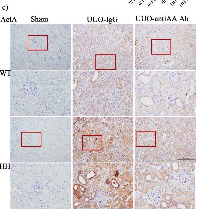
Detection of Activin A beta A subunit by Immunohistochemistry
Activin A neutralization inhibits renal fibrosis in TGF beta 1-overexpressing mice. a TGF beta 1 transcript is increased in mice genetically engineered to overexpress TGF beta 1 (HH) compared with wild-type mice (WT) (n = 6–7, *p ≤ 0.05). b Serum actA is elevated in wild-type and HH mice after UUO. This is decreased by treatment with a neutralizing actA antibody (anti-actA) in HH mice. c Renal actA is increased after UUO, with a greater induction in HH mice. Both are attenuated by actA neutralization. Boxed areas are shown at higher magnification immediately below. ActA increases are seen particularly in tubular epithelial cells. d Renal alpha SMA, fibronectin (FN), pSmad3 and MRTF-A are increased after UUO and this is augmented in HH kidneys. Expression of all is attenuated by actA neutralization in both WT and HH kidneys. (n = 6–9) *, **, ***, ****P < 0.05, 0.01, 0.001, 0.0001; one-way ANOVA with Tukey’s multiple comparisons post hoc test where there are > 2 groups; t-test for 2 groups Image collected and cropped by CiteAb from the following open publication (https://pubmed.ncbi.nlm.nih.gov/36717814), licensed under a CC-BY license. Not internally tested by R&D Systems.
Human Activin A ELISA Standard Curve
Human Activin A was serially diluted and captured by Mouse Anti-Human/Mouse/Rat Activin A beta A subunit Monoclonal Antibody (Catalog # MAB3381) coated on a Clear Polystyrene Microplate (Catalog # DY990). Mouse Anti-Human/Mouse/Rat Activin A beta A subunit Monoclonal Antibody (Catalog # MAB3381) was biotinylated and incubated with the protein captured on the plate. Detection of the standard curve was achieved by incubating Streptavidin-HRP (Catalog # DY998)Applications for Activin A betaA subunit Antibody (69403)
Neutralization
Human/Mouse/Rat Activin A Sandwich Immunoassay
Reviewed Applications
Read 2 reviews rated 5 using MAB3381 in the following applications:
Formulation, Preparation, and Storage
Purification
Reconstitution
Reconstitute at 0.5 mg/mL in sterile PBS. For liquid material, refer to CoA for concentration.
Formulation
Shipping
Stability & Storage
- 12 months from date of receipt, -20 to -70 °C as supplied.
- 1 month, 2 to 8 °C under sterile conditions after reconstitution.
- 6 months, -20 to -70 °C under sterile conditions after reconstitution.
Calculators
Background: Activin A betaA subunit
References
- Kumanov, P. et al. (2005) Reprod. Biomed. Online 10:786.
- Maeshima, A. et al. (2008) Endocr. J. 55:1.
- Rodgarkia-Dara, C. et al. (2006) Mutat. Res. 613:123.
- Werner, S. and C. Alzheimer (2006) Cytokine Growth Factor Rev. 17:157.
- Xu, P. and A.K. Hall (2006) Dev. Biol. 299:303.
- Shav-Tal, Y. and D. Zipori (2002) Stem Cells 20:493.
- Chen, Y.G. et al. (2006) Exp. Biol. Med. 231:534.
- Gray, A.M. and A.J. Mason (1990) Science 247:1328.
- Mason, A.J. et al. (1996) Mol. Endocrinol. 10:1055.
- Thompson, T.B. et al. (2004) Mol. Cell. Endocrinol. 225:9.
- Harrison, C.A. et al. (2005) Trends Endocrinol. Metab. 16:73.
- Onichtchouk, D. et al. (1999) Nature 401:480.
- Gray, P.C. et al. (2002) Mol. Cell. Endocrinol. 188:254.
- Kelber, J.A. et al. (2008) J. Biol. Chem. 283:4490.
- Phillips, D.J. et al. (1997) J. Endocrinol. 155:65.
- Schneyer, A. et al. (2003) Endocrinology 144:1671.
Gene Symbol
Additional Activin A betaA subunit Products
Product Documents for Activin A betaA subunit Antibody (69403)
Certificate of Analysis
To download a Certificate of Analysis, please enter a lot or batch number in the search box below.
Note: Certificate of Analysis not available for kit components.
Product Specific Notices for Activin A betaA subunit Antibody (69403)
For research use only
Citations for Activin A betaA subunit Antibody (69403)
 Powered by Bioz
Customer Reviews for Activin A betaA subunit Antibody (69403) (2)
Have you used Activin A betaA subunit Antibody (69403)?
Submit a review and receive an Amazon gift card!
$25/€18/£15/$25CAN/¥2500 for a review with an image
$10/€7/£6/$10CAN/¥1110 for a review without an image
Submit a review
Customer Images
-
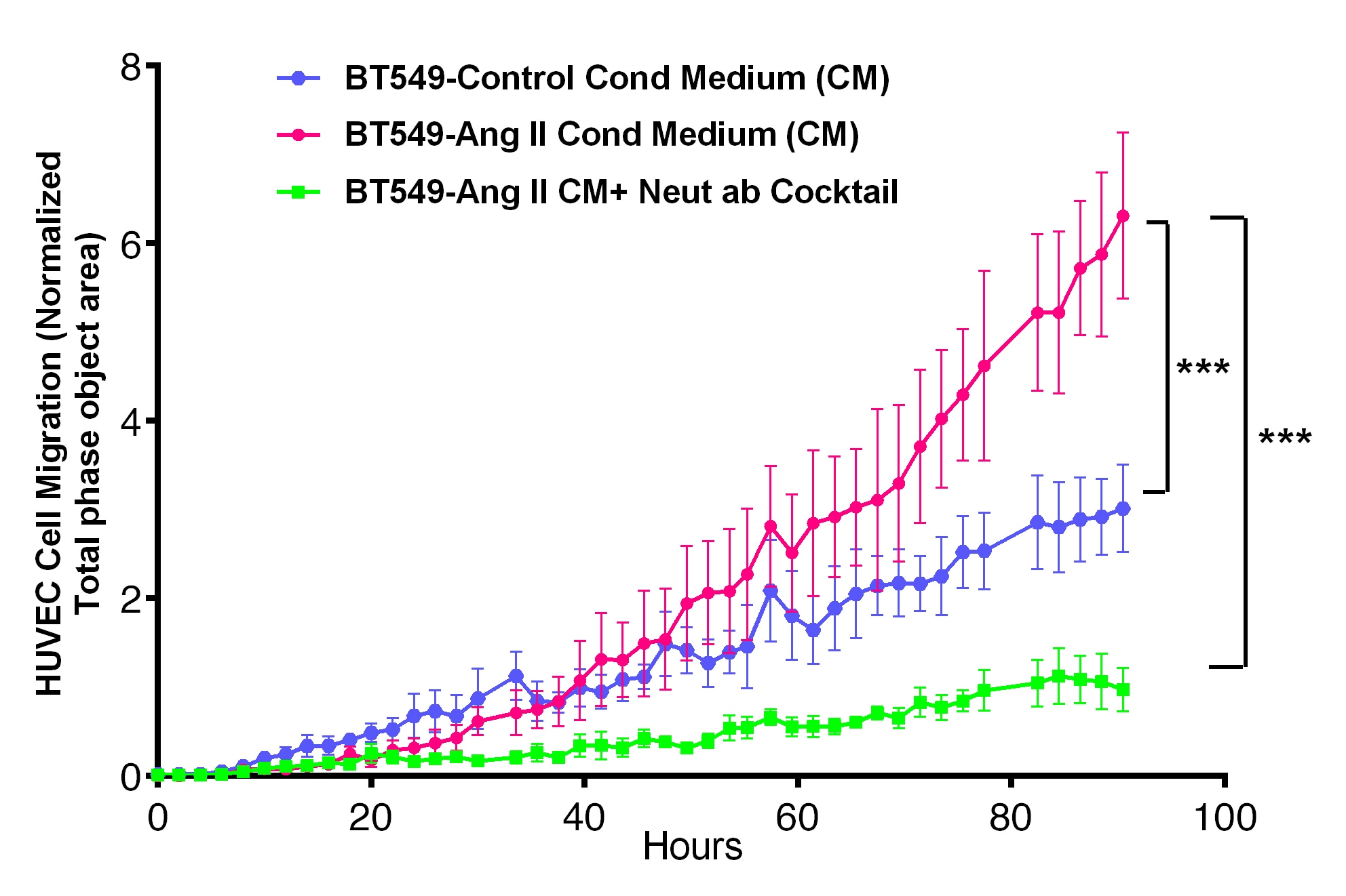
Application: Block/NeutralizeSample Tested: Serum-free Cell Culture SupernatesSpecies: HumanVerified Customer | Posted 07/07/2020We used this INHBA (100ng/mL) neutralizing antibody as part of an antibody cocktail containing several other antibodies to neutralize INHBA present in the Conditioned Medium (CM) from BT549 Cells treated with Angiotensin II. The effect of cytokines in the CM (+/-Neutralizing ab Cocktail) on Endothelial (HUVEC) Cell migration was measured using Incucyte Zoom Chemotaxis Assay.

-
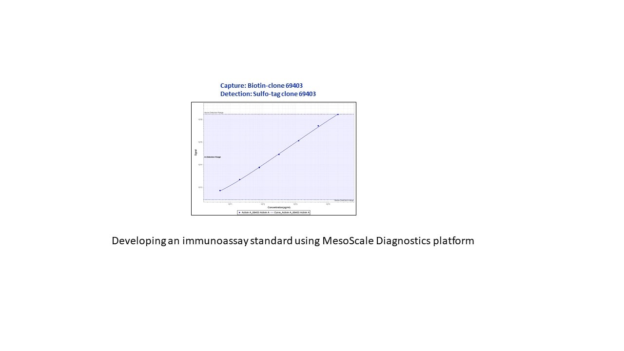
Application: ELISASample Tested: Recombinant proteinSpecies: HumanVerified Customer | Posted 03/28/2019I added a sulfo tag to this antibody and used it as a detection antibody when developing an immunoassay for Activin A.
There are no reviews that match your criteria.