
HSP90 is a composite name for a large group of Heat Shock Proteins whose molecular weights average 90 kDa. HSPs functions primarily as a molecular chaperone, facilitating the folding of other cellular proteins, preventing protein aggregation, or targeting improperly folded proteins to specific degradative pathways. HSP90 is a central component in a number of basic cellular processes including hormone signaling and cell cycle control. HSP90 is ubiquitously expressed, highly conserved and accounts for 1‑2% of the total cellular protein. Recently introduced, standardized nomenclature has divided the 17 identified HSP90 genes into three related and one unrelated classes, HSP90AA, HSP90AB, HSP90BB, and TRAP, respectively. Eukaryotic cells have two principal isoforms of HSP90. The antibody described here reacts with the 724 amino acid constitutively expressed HSP90AB1-isoform 1 (also known as HSP90 beta, HSP90B, HSPCB, HSPC2, and HSP89 beta ) and also with the 737 amino acid inducible form, HSP90AA1 (also known as HSP90 alpha, HSP90A, HSPC1, HSPCA, HSP89 alpha and LAP2). HSP90AB1-1 and HSP90AA share 90% identity.
HSP90 Antibody (341320)
R&D Systems | Catalog # MAB3286


Scientific images may be resized, cropped, or adjusted for brightness or contrast for presentation purposes. These updates are limited to presentation and do not affect the underlying scientific interpretation of the data. Where available, additional source data may be provided upon request.
Key Product Details
Species Reactivity
Validated:
Cited:
Applications
Validated:
Cited:
Label
Antibody Source
Product Specifications
Immunogen
Met1-Asp724
Accession # P08238
Specificity
Clonality
Host
Isotype
Scientific Data Images for HSP90 Antibody (341320)
Detection of Human/Mouse/Rat HSP90 by Western Blot.
Western blot shows lysates of Raji human Burkitt's lymphoma cell line and HeLa human cervical epithelial carcinoma cell line, CH-1 mouse B cell lymphoma cell line, A20 mouse B cell lymphoma cell line, and L6 rat myoblast cell line. PVDF membrane was probed with 0.25 µg/mL Human/Mouse/Rat HSP90 Monoclonal Antibody (Catalog # MAB3286) followed by HRP-conjugated Anti-Mouse IgG Secondary Antibody (Catalog # HAF007). For additional reference, recombinant human HSP90a and HSP90 beta (5 ng/lane) were included. A specific band for HSP90 was detected at approximately 100 kDa (as indicated). This experiment was conducted under reducing conditions and using Immunoblot Buffer Group 2.
Detection of Human and Rat HSP90 by Simple WesternTM.
Simple Western lane view shows lysates of Raji human Burkitt's lymphoma cell line and L6 rat myoblast cell line, loaded at 0.2 mg/mL. A specific band was detected for HSP90 at approximately 95 kDa (as indicated) using 2.5 µg/mL of Mouse Anti-Human/Mouse/Rat HSP90 Monoclonal Antibody (Catalog # MAB3286). This experiment was conducted under reducing conditions and using the 12-230 kDa separation system.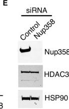
Detection of HSP90 by Western Blot
Nup358 is required for HDAC3 function and nuclear translocation.(A) Upstream transcriptional regulators (P < 0.05) predicted to be altered in Nup358 knockout LSK cells belonging to cluster 4 and cluster 9 ranked by P value. (B) The intracellular localization of HDAC3, E2F1, RB1, p53, RBL2, DMTF1, and GTF2I was analyzed by immunofluorescence in 293T cells transfected with control or Nup358-specific siRNAs. Scale bars, 25 μm. (C) HDAC3 intracellular localization was analyzed by immunofluorescence in 293T cells transfected with control or Nup358-specific siRNAs. Cells were costained with antibodies against nucleoporins Nup358 and Nup98. Scale bars, 25 μm. (D) Quantification of HDAC3 nuclear/cytoplasmic signal ratio in control or Nup358 knockdown cells. Data are expressed as mean ± SD. ****P ≤ 0.0001. (E) Protein levels of HDAC3 and Nup358 in cells transfected with scramble control or Nup358-specific siRNAs were analyzed by Western blot. HSP90 was used as loading control. Image collected and cropped by CiteAb from the following open publication (https://pubmed.ncbi.nlm.nih.gov/38838144), licensed under a CC-BY license. Not internally tested by R&D Systems.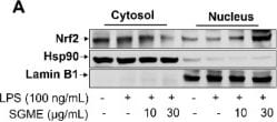
Detection of HSP90 by Western Blot
Modulatory effects of SGME on the Nrf2/HO-1 signaling pathway. (A–D) Raw 264.7 macrophages were pretreated with SGME for 3 h and then stimulated with LPS for 1 h. (A and B) Protein levels of Nrf2 in nuclear and cytoplasmic lysates were examined by western blot. (C and D) Nrf2 was labeled by immunostaining. Representative images showed the distribution of Nrf2 (green) in relation to nuclei (red); the bar graph depicted relative nuclear fluorescence intensity of Nrf2 calculated by ImageJ; scale bar: 20 μm. (E–G) After treatment with SGME for 3 h, Raw 264.7 macrophages were treated with LPS for 3 h. Protein and mRNA levels of HO-1 were measured by western blot and RT-qPCR. * denotes p < 0.05; n = 3.– Image collected and cropped by CiteAb from the following open publication (https://pubmed.ncbi.nlm.nih.gov/42031984), licensed under a CC-BY license. Not internally tested by R&D Systems.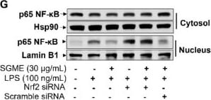
Detection of HSP90 by Western Blot
Roles of Nrf2/HO-1 in modulation of inflammatory responses by SGME. (A) Raw 264.7 cells were sequentially treated with ML385 (10 µM) or SnPP (10 µM) (pharmacological inhibitors of Nrf2 and HO-1, respectively) for 1 h, SGME (30 µg/mL) for 3 h, and LPS (100 ng/mL) for 24 h. Nitrite/nitrate levels were measured in culture medium. (B–H) Cells were transfected with an siRNA targeting Nrf2 (20 nM) or a scramble siRNA for 24 h. Then, cells were treated with SGME (30 µg/mL) for 3 h, followed by LPS (100 ng/mL) for 24 h (B), 6 h (C–F), or 1 h (G–H). (B) NO production was determined by measuring nitrite/nitrate release into culture medium. (C–F) The mRNA levels of genes encoding iNOS (C), IL-6 (D), COX-2 (E), and TNF-alpha (F) were measured using RT-qPCR. (G and H) The expression of p65 NF-kappa B in cytoplasmic and nuclear fractions was examined by western blot. (I and J) BMDMs were treated with ML385 (10 µM) or SnPP (10 µM) for 1 h and then with SGME (30 µg/mL) for an additional 3 h. BMDMs were next incubated with LPS/IFN-gamma (100/10 ng/mL) for 24 h to induce M1 polarization. The expression of CD86 (I) and CD80 (J) on the cell surface was analyzed using flow cytometry. * denotes p < 0.05; ns: statistically non-significant; n = 3. Image collected and cropped by CiteAb from the following open publication (https://pubmed.ncbi.nlm.nih.gov/42031984), licensed under a CC-BY license. Not internally tested by R&D Systems.Applications for HSP90 Antibody (341320)
Simple Western
Sample: Raji human Burkitt's lymphoma cell line and L6 rat myoblast cell line
Western Blot
Sample: Raji human Burkitt's lymphoma cell line and HeLa human cervical epithelial carcinoma cell line, CH-1 mouse B cell lymphoma cell line, A20 mouse B cell lymphoma cell line, and L6 rat myoblast cell line
Formulation, Preparation, and Storage
Purification
Reconstitution
Reconstitute at 0.5 mg/mL in sterile PBS. For liquid material, refer to CoA for concentration.
Formulation
Shipping
Stability & Storage
- 12 months from date of receipt, -20 to -70 °C as supplied.
- 1 month, 2 to 8 °C under sterile conditions after reconstitution.
- 6 months, -20 to -70 °C under sterile conditions after reconstitution.
Calculators
Background: HSP90
Long Name
Alternate Names
UniProt
Additional HSP90 Products
Product Documents for HSP90 Antibody (341320)
Certificate of Analysis
To download a Certificate of Analysis, please enter a lot or batch number in the search box below.
Note: Certificate of Analysis not available for kit components.
Product Specific Notices for HSP90 Antibody (341320)
For research use only
Related Research Areas
Citations for HSP90 Antibody (341320)
 Powered by Bioz
Customer Reviews for HSP90 Antibody (341320)
There are currently no reviews for this product. Be the first to review HSP90 Antibody (341320) and earn rewards!
Have you used HSP90 Antibody (341320)?
Submit a review and receive an Amazon gift card!
$25/€18/£15/$25CAN/¥2500 for a review with an image
$10/€7/£6/$10CAN/¥1110 for a review without an image
Submit a review
Protocols
Find general support by application which include: protocols, troubleshooting, illustrated assays, videos and webinars.
- Cellular Response to Hypoxia Protocols
- R&D Systems Quality Control Western Blot Protocol
- Troubleshooting Guide: Western Blot Figures
- Western Blot Conditions
- Western Blot Protocol
- Western Blot Protocol for Cell Lysates
- Western Blot Troubleshooting
- Western Blot Troubleshooting Guide
- View all Protocols, Troubleshooting, Illustrated assays and Webinars
Associated Pathways
