
Ninjurin-1 (nerve injury-induced protein 1) is a 20-22 kDa member of the Ninjurin family of transmembrane (TM) proteins. It is expressed by Schwann cells, neurons and hepatocytes and participates in intercellular homophilic binding during nerve regeneration. Human Ninjurin-1 is 152 amino acids in length. It has an unusual membrane orientation. There is an 80 amino acid (aa) N-terminal extracellular domain (ECD) (aa 1-80), followed by a TM segment, a cytoplasmic region, a second TM segment and a C-terminal ECD (aa 142-152). Homophilic binding is divalent-cation dependent and occurs between Pro26 and Asn37. Over aa 1-80, human Ninjurin-1 shares 84% aa sequence identity with mouse Ninjurin-1. Human Ninjurin-1 shares only 50% aa sequence identity with human Ninjurin-2.
Human Ninjurin-1 Antibody (758943)
R&D Systems | Catalog # MAB5105


Scientific images may be resized, cropped, or adjusted for brightness or contrast for presentation purposes. These updates are limited to presentation and do not affect the underlying scientific interpretation of the data. Where available, additional source data may be provided upon request.
Key Product Details
Species Reactivity
Validated:
Cited:
Applications
Validated:
Cited:
Label
Antibody Source
Product Specifications
Immunogen
Asp2-Val81
Accession # Q92982
Specificity
Clonality
Host
Isotype
Scientific Data Images for Human Ninjurin-1 Antibody (758943)
Detection of Human Ninjurin‑1 by Western Blot.
Western blot shows lysates of human liver tissue. PVDF membrane was probed with 2 µg/mL of Mouse Anti-Human Ninjurin-1 Monoclonal Antibody (Catalog # MAB5105) followed by HRP-conjugated Anti-Mouse IgG Secondary Antibody (Catalog # HAF007). A specific band was detected for Ninjurin-1 at approximately 20 kDa (as indicated). This experiment was conducted under reducing conditions and using Immunoblot Buffer Group 1.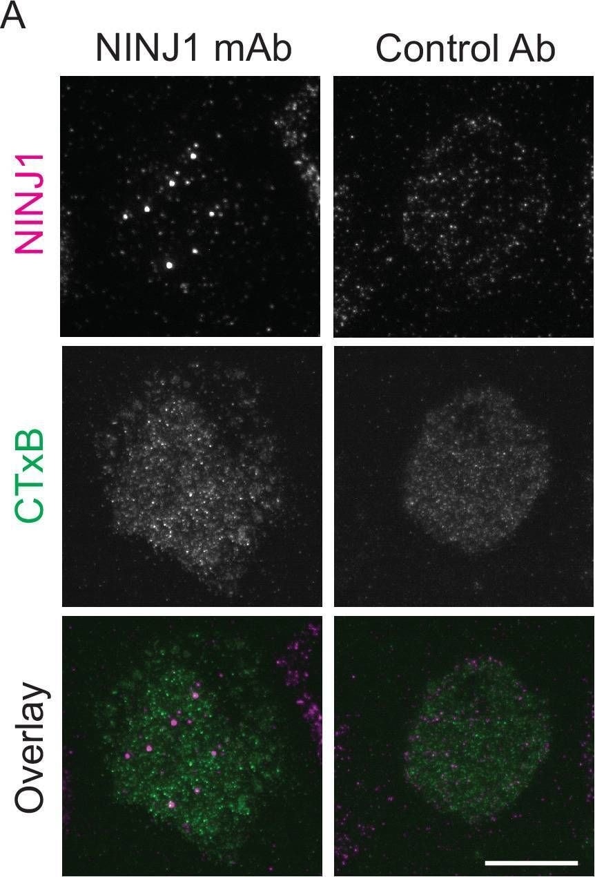
Detection of Ninjurin-1 by Immunocytochemistry/ Immunofluorescence
Mouse anti-human NINJ1 monoclonal antibody validation.LPS and nigericin treated human monocyte-derived macrophages (hMDMs) were fixed and stained with the indicated antibodies as described in the main text prior to imaging by total internal reflection fluorescence (TIRF) microscopy. (A) Cells were stained with mouse anti-human NINJ1 (IgG2b) Ab or a control Ab (mouse IgG2a) followed by fluorescent rabbit anti-mouse Ab. The cell plasma membrane was stained using fluorescent cholera toxin subunit B (CTxB). (B) In a separate experiment, cells were stained with mouse anti-human NINJ1 (IgG2b) Ab or no primary Ab, followed by fluorescent rabbit anti-mouse Ab. Scale bars 20 μm. Image collected and cropped by CiteAb from the following open publication (https://pubmed.ncbi.nlm.nih.gov/36468682), licensed under a CC-BY license. Not internally tested by R&D Systems.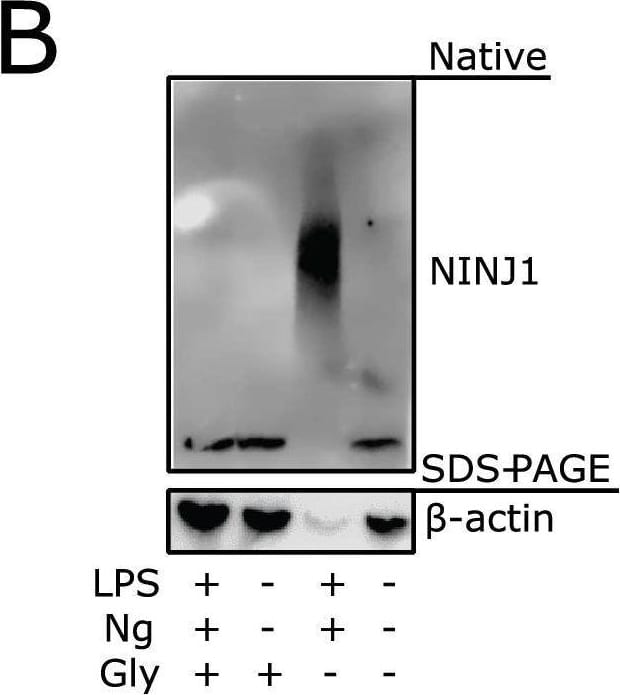
Detection of Ninjurin-1 by Western Blot
Glycine prevents NINJ1 aggregation and pyroptotic lysis in induced pluripotent stem-cell (iPSC) derived human macrophages (iPSDMs) without affecting IL-1 beta secretion.(A) Quantification of NINJ1 native-PAGE signal from unstimulated human monocyte-derived macrophage (hMDM) lysates confirm NINJ1 silencing in siNINJ1-treated cells compared to siCtrl-treated cells. (B–D) iPSDMs were LPS-primed and stimulated to undergo pyroptosis with nigericin in the presence or absence of 50 mM glycine. (B) Native-PAGE analysis of endogenous NINJ1 in iPSDMs display a shift to higher molecular weight when cells are stimulated to undergo pyroptosis, which is abrogated by glycine pretreatment. (C) LDH release was measured in the supernatants to assess cell rupture and was reduced with glycine co-treatment. Data are expressed as % of LDH in supernatant from LPS and nigericin-treated cells in n=3 independent experiments. Data points from each experiment is shown along with their mean and SD. (D) IL-1 beta release from pyroptotic cells was not altered by the presence of glycine. Data points from three technical replicates from one experiment are shown along with their mean and SD.Figure 4—figure supplement 2—source data 1.Numerical values for experimental data plotted in Figure 4—figure supplement 2.Numerical values for experimental data plotted in Figure 4—figure supplement 2. Image collected and cropped by CiteAb from the following open publication (https://pubmed.ncbi.nlm.nih.gov/36468682), licensed under a CC-BY license. Not internally tested by R&D Systems.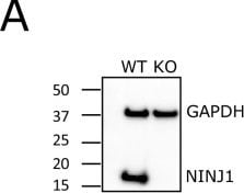
Detection of Ninjurin-1 by Western Blot
Glycine cytoprotection phenocopies NINJ1 knockout and prevents NINJ1 aggregation in a mouse macrophage cell line.Using a CRISPR-Cas9 system, Ninj1 was knocked out of a RAW mouse macrophage cell line engineered to constitutively express ASC (RAW-ASC) and therefore have the capacity to undergo pyroptosis. (A) Western blot demonstrating NINJ1 knockout compared with the parental line. GAPDH is shown as a loading control. (B) Wildtype and NINJ1 knockout cells were induced to undergo pyroptosis with LPS + nigericin treatment with or without glycine. Supernatant LDH was evaluated as a marker of cytotoxicity by colorimetric assay. Cytotoxicity was decreased in glycine-treated (5 mM) wildtype cells comparably to NINJ1 knockout. Glycine (5 mM) treatment of NINJ1 KO cells provided no additional protection to knockout cells without glycine. * p<0.05 by two-sided ANOVA with Tukey’s multiple comparison correction. (C) LPS-primed wildtype RAW-ASC induced to undergo pyroptosis in the presence of glycine demonstrate similar plasma membrane ballooning to NINJ1 knockout RAW-ASC cells induced to undergo pyroptosis. Membrane ballooning is shown in live cells labeled with the plasma membrane dye FM4-64. Scale bar 20 µm. (D) Native-PAGE analysis of endogenous NINJ1 in RAW-ASC macrophages demonstrates a shift to high molecular weight aggregate upon pyroptosis stimulation, which is abrogated by glycine treatment.Figure 1—figure supplement 2—source data 1.Numerical values for experimental data plotted in Figure 1—figure supplement 2.Numerical values for experimental data plotted in Figure 1—figure supplement 2. Image collected and cropped by CiteAb from the following open publication (https://pubmed.ncbi.nlm.nih.gov/36468682), licensed under a CC-BY license. Not internally tested by R&D Systems.Applications for Human Ninjurin-1 Antibody (758943)
Western Blot
Sample: Human liver tissue
Formulation, Preparation, and Storage
Purification
Reconstitution
Sterile PBS to a final concentration of 0.5 mg/mL. For liquid material, refer to CoA for concentration.
Formulation
Shipping
Stability & Storage
- 12 months from date of receipt, -20 to -70 °C as supplied.
- 1 month, 2 to 8 °C under sterile conditions after reconstitution.
- 6 months, -20 to -70 °C under sterile conditions after reconstitution.
Calculators
Background: Ninjurin-1
Alternate Names
Gene Symbol
UniProt
Additional Ninjurin-1 Products
Product Documents for Human Ninjurin-1 Antibody (758943)
Certificate of Analysis
To download a Certificate of Analysis, please enter a lot or batch number in the search box below.
Note: Certificate of Analysis not available for kit components.
Product Specific Notices for Human Ninjurin-1 Antibody (758943)
For research use only
Related Research Areas
Citations for Human Ninjurin-1 Antibody (758943)
 Powered by Bioz
Customer Reviews for Human Ninjurin-1 Antibody (758943)
There are currently no reviews for this product. Be the first to review Human Ninjurin-1 Antibody (758943) and earn rewards!
Have you used Human Ninjurin-1 Antibody (758943)?
Submit a review and receive an Amazon gift card!
$25/€18/£15/$25CAN/¥2500 for a review with an image
$10/€7/£6/$10CAN/¥1110 for a review without an image
Submit a review
Protocols
Find general support by application which include: protocols, troubleshooting, illustrated assays, videos and webinars.
- Cellular Response to Hypoxia Protocols
- R&D Systems Quality Control Western Blot Protocol
- Troubleshooting Guide: Western Blot Figures
- Western Blot Conditions
- Western Blot Protocol
- Western Blot Protocol for Cell Lysates
- Western Blot Troubleshooting
- Western Blot Troubleshooting Guide
- View all Protocols, Troubleshooting, Illustrated assays and Webinars