PERK, a type 1 ER membrane kinase, mediates eIF2 alpha phosphorylation at Ser51 during the UPR (unfolded protein response). Protein synthesis is inhibited, thereby reducing the burden of protein substrate for the ER folding and degradation mechanism. Phosphorylation of eIF2 alpha also selectively promotes the expression of UPR target genes such as Chop and BiP. PERK may also play a role in tumor cell adaptation to hypoxic stress by regulating the translation of angiogenic factors necessary for the development of functional microvessels. Mutations in PERK are responsible for the rare autosomal-recessive disorder, WRS (Wolcott-Rallison syndrome).

Key Product Details
Validated by
Knockout/Knockdown, Biological Validation
Species Reactivity
Validated:
Human
Cited:
Human, Mouse
Applications
Validated:
Knockout Validated, Western Blot
Cited:
Western Blot
Label
Unconjugated
Antibody Source
Polyclonal Goat IgG
Loading...
Product Specifications
Immunogen
E. coli-derived recombinant human PERK
Ala29-Gln230
Accession # Q9NZJ5
Ala29-Gln230
Accession # Q9NZJ5
Specificity
Detects human PERK in Western blots.
Clonality
Polyclonal
Host
Goat
Isotype
IgG
Scientific Data Images for Human PERK Antibody
Detection of Human PERK by Western Blot.
Western blot shows lysates of HepG2 human hepatocellular carcinoma cell line and NTera-2 human testicular embryonic carcinoma cell line. PVDF membrane was probed with 1 µg/mL of Human PERK Antigen Affinity-purified Polyclonal Antibody (Catalog # AF3999) followed by HRP-conjugated Anti-Goat IgG Secondary Antibody (Catalog # HAF017). A specific band was detected for PERK at approximately 130 kDa (as indicated). This experiment was conducted using Immunoblot Buffer Group 1.
Western Blot Shows Human PERK Specificity by Using Knockout Cell Line.
Western blot shows lysates of HeLa human cervical epithelial carcinoma parental cell line and PERK knockout HeLa cell line (KO). PVDF membrane was probed with 1 µg/mL of Goat Anti-Human PERK Antigen Affinity-purified Polyclonal Antibody (Catalog # AF3999) followed by HRP-conjugated Anti-Goat IgG Secondary Antibody (Catalog # HAF017). A specific band was detected for PERK at approximately 150 kDa (as indicated) in the parental HeLa cell line, but is not detectable in knockout HeLa cell line. GAPDH (Catalog # AF5718) is shown as a loading control. This experiment was conducted under reducing conditions and using Immunoblot Buffer Group 1.
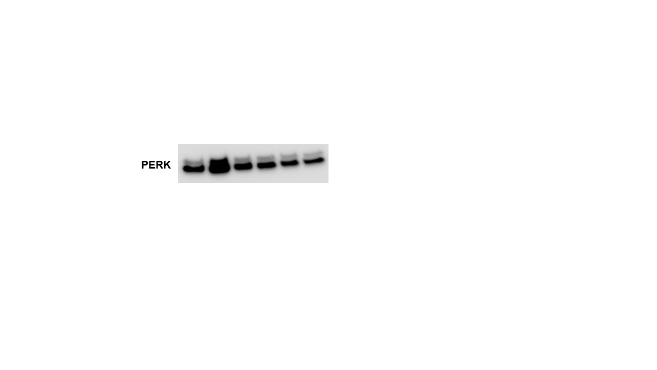
Detection of Human PERK by Western Blot
Activation of the ER stress signaling pathway leads to the LX-2 cell apoptosis induced by VP-16.(a) Western blotting analysis of ER stress-associated proteins in LX-2 cells after treatment with VP-16 for 72 h. (b) After exposure to VP-16 for 72 h, the protein phosphorylation levels of PERK and eIF2 alpha were analyzed by western blotting. (c) After exposure to VP-16 for 72 h, the proteins of the IRE1 alpha /ASK1/JNK signaling pathway were analyzed by western blotting. (d,e) LX-2 cells were pretreated with a JNK inhibitor (SP600125, 40 μM) for 1 h, and then treated with 4 μM VP-16 for 72 h. (d) Cell viability was measured using the CCK-8 assay. The percentage of apoptosis cells was analyzed by flow cytometry. The data are presented as the mean ± SD of at least three independent experiments performed in triplicates. **P < 0.01, compared with the group treated with VP-16 alone. (e) Western blotting analysis of the cell lysates was performed using the indicated antibodies. In all of the western blotting analyses, beta -actin was used as a loading control. *P < 0.05, compared with the control group. #P < 0.05, compared with the group treated with VP-16 alone. Image collected and cropped by CiteAb from the following publication (https://pubmed.ncbi.nlm.nih.gov/27680712), licensed under a CC-BY license. Not internally tested by R&D Systems.Applications for Human PERK Antibody
Application
Recommended Usage
Knockout Validated
PERK
is specifically detected in HeLa human cervical epithelial carcinoma parental cell line but is not detectable in
PERK knockout HeLa cell line.
Western Blot
1 µg/mL
Sample: HepG2 human hepatocellular carcinoma cell line and NTera-2 human testicular embryonic carcinoma cell line
Sample: HepG2 human hepatocellular carcinoma cell line and NTera-2 human testicular embryonic carcinoma cell line
Reviewed Applications
Read 1 review rated 5 using AF3999 in the following applications:
Formulation, Preparation, and Storage
Purification
Antigen Affinity-purified
Reconstitution
Reconstitute at 0.2 mg/mL in sterile PBS. For liquid material, refer to CoA for concentration.
Loading...
Formulation
Lyophilized from a 0.2 μm filtered solution in PBS with Trehalose. *Small pack size (SP) is supplied either lyophilized or as a 0.2 µm filtered solution in PBS.
Shipping
Lyophilized product is shipped at ambient temperature. Liquid small pack size (-SP) is shipped with polar packs. Upon receipt, store immediately at the temperature recommended below.
Stability & Storage
Use a manual defrost freezer and avoid repeated freeze-thaw cycles.
- 12 months from date of receipt, -20 to -70 °C as supplied.
- 1 month, 2 to 8 °C under sterile conditions after reconstitution.
- 6 months, -20 to -70 °C under sterile conditions after reconstitution.
Calculators
Background: PERK
Long Name
Eukaryotic Translation Initiation Factor 4B
Alternate Names
EIF2AK3, PEK, WRS
Gene Symbol
EIF2AK3
UniProt
Additional PERK Products
Product Documents for Human PERK Antibody
Certificate of Analysis
To download a Certificate of Analysis, please enter a lot or batch number in the search box below.
Note: Certificate of Analysis not available for kit components.
Product Specific Notices for Human PERK Antibody
For research use only
Citations for Human PERK Antibody
 Powered by Bioz
Customer Reviews for Human PERK Antibody (1)
5 out of 5
1 Customer Rating
Have you used Human PERK Antibody?
Submit a review and receive an Amazon gift card!
$25/€18/£15/$25CAN/¥2500 Yen for a review with an image
$10/€7/£6/$10CAN/¥1110 Yen for a review without an image
Submit a review
Customer Images
Showing
1
-
1 的
1 review
Showing All
Filter By:
-
Application: Western BlotSample Tested: Pancreatic cancer cellsSpecies: HumanVerified Customer | Posted 12/27/2017

There are no reviews that match your criteria.
Protocols
Find general support by application which include: protocols, troubleshooting, illustrated assays, videos and webinars.
- Cellular Response to Hypoxia Protocols
- R&D Systems Quality Control Western Blot Protocol
- Troubleshooting Guide: Western Blot Figures
- Western Blot Conditions
- Western Blot Protocol
- Western Blot Protocol for Cell Lysates
- Western Blot Troubleshooting
- Western Blot Troubleshooting Guide
- View all Protocols, Troubleshooting, Illustrated assays and Webinars
Loading...