Human Repulsive Guidance molecule (RGM) is a 33 kDa GPI-linked member of an expanding RGM-related family of neuronal and muscle-expressed membrane proteins (1). It is synthesized as a 450 amino acid (aa) preproprotein that contains a 47 aa signal sequence, a 121 aa N-terminal prosegment, a 256 mature region and a 26 aa C-terminal prosegment (2). The N-terminal prosegment contains an RGD tripeptide and the molecule’s only two potential N-linked glycosylation sites. The mature segment shows an abbreviated von Willebrand factor domain. Proteolytic processing occurs at an aspartic acid-proline bond, creating a predicted 32 kDa mature region (2). The mature region of human RGM-A has 88% and 93% aa identity to the chick and mouse mature region of RGM-A, respectively. When compared to human RGMb and c, the mature region of human RGM-A shows 58% and 54% aa identity, respectively. Recombinant chick RGM-A has been reported to induce collapse of temporal but not nasal growth cones, and to repel temporal retinal axons in vitro. This suggests a role in the development of the retina-superior colliculus connection. In mammals, however, this activity is not so evident, and thus its function in this system is uncertain (3). Alternatively, in mice, RGM-A is said to be needed for neural tube closure, and may play a role in entorhinal-hippocampal connections (3, 4). The receptor for RGM-A is reported to be neogenin (5, 6). RGM-A has also been shown to be a bone morphogenic protein co-receptor, able to bind both BMP-2 and BMP-4 (7).

Key Product Details
Species Reactivity
Validated:
Human
Cited:
Human, Mouse
Applications
Validated:
Immunohistochemistry, Western Blot
Cited:
Immunohistochemistry, Western Blot
Label
Unconjugated
Antibody Source
Polyclonal Goat IgG
Loading...
Product Specifications
Immunogen
Mouse myeloma cell line NS0-derived recombinant human RGM-A
Cys48-Gly422
Accession # Q96B86
Cys48-Gly422
Accession # Q96B86
Specificity
Detects human RGM-A in direct ELISAs and Western blots.
Clonality
Polyclonal
Host
Goat
Isotype
IgG
Scientific Data Images for Human RGM-A Antibody
RGM‑A in Human Brain.
RGM-A was detected in immersion fixed paraffin-embedded sections of human brain (spinal cord) using Goat Anti-Human RGM-A Antigen Affinity-purified Polyclonal Antibody (Catalog # AF2459) at 15 µg/mL overnight at 4 °C. Before incubation with the primary antibody, tissue was subjected to heat-induced epitope retrieval using Antigen Retrieval Reagent-Basic (Catalog # CTS013). Tissue was stained using the Anti-Goat HRP-DAB Cell & Tissue Staining Kit (brown; Catalog # CTS008) and counterstained with hematoxylin (blue). Specific staining was localized to the dorsal horn. View our protocol for Chromogenic IHC Staining of Paraffin-embedded Tissue Sections.
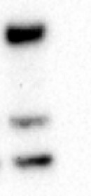
Detection of RGM-A by Western Blot
HFE2 administration significantly improves EAE disease outcome. A Western Blot analysis shows a strong HFE2 reduction in EAE animal serum when compared to control. ELISA quantification showing HFE2 decrease and RGMa increase in EAE animal serum (mean ± s.d.; EAE n = 3 and RGMa n = 3). b Body conditioning assessment (‘0’ no paralysis to ‘4’ forelimb weakness) of EAE-induced mice with HFE2 + /- RGMa treatment every 3 days (d3, 6, 9, 12 and 15; mean ± s.e.m.; unpaired two-tail t-tests; PBS n = 8, HFE2 n = 6, RGMa+ HFE2 n = 8). c Body conditioning assessment of EAE-induced Hfe2fl/fl and Hfe2 delta Alb-cre mice (mean ± s.e.m.; unpaired two-tail t-tests; Hfe2fl/fln = 8 and Hfe2 delta Alb-cren = 8). d Representative confocal images of CD3 +, B220+ and CD11b+ immune staining in spinal cord of EAE-induced mice 3 weeks after induction and quantification (mean ± s.e.m.; unpaired two-tail t-test; PBS n = 7, HFE2 n = 6 mice; technical replicates shown). e Schematic of activated immune cell adoptive transfer. Representative confocal images of CD4-positive labelled transferred cell (CellVue) in PBS-treated or HFE2-treated recipient WT mice and quantification (mean ± s.e.m.; unpaired two-tail t-test; PBS n = 6, HFE2 n = 6). Ptx; Pertussis Toxin. f Representative confocal images of fibrinogen in the spinal cord of HFE2-treated EAE-mice and quantification (mean ± s.e.m.; unpaired two-tail t-test; PBS-treated n = 6 and HFE2 n = 6). *P < 0.05, **P < 0.01, ***P < 0.001. Source data are provided as a Source Data file (includes exact p-values). Image collected and cropped by CiteAb from the following open publication (https://www.nature.com/articles/s41467-024-45303-1), licensed under a CC-BY license. Not internally tested by R&D Systems.Applications for Human RGM-A Antibody
Application
Recommended Usage
Immunohistochemistry
10-25 µg/mL
Sample: Formalin fixed paraffin-embedded sections of human spinal cord subjected to Antigen Retrieval Reagent-Basic (Catalog # CTS013)
Sample: Formalin fixed paraffin-embedded sections of human spinal cord subjected to Antigen Retrieval Reagent-Basic (Catalog # CTS013)
Western Blot
0.1 µg/mL
Sample: Recombinant Human Repulsive Guidance Molecule A/RGM-A (Catalog # 2459-RM)
Sample: Recombinant Human Repulsive Guidance Molecule A/RGM-A (Catalog # 2459-RM)
Reviewed Applications
Read 1 review rated 5 using AF2459 in the following applications:
Formulation, Preparation, and Storage
Purification
Antigen Affinity-purified
Reconstitution
Reconstitute at 0.2 mg/mL in sterile PBS. For liquid material, refer to CoA for concentration.
Loading...
Formulation
Lyophilized from a 0.2 μm filtered solution in PBS with Trehalose. *Small pack size (SP) is supplied either lyophilized or as a 0.2 µm filtered solution in PBS.
Shipping
Lyophilized product is shipped at ambient temperature. Liquid small pack size (-SP) is shipped with polar packs. Upon receipt, store immediately at the temperature recommended below.
Stability & Storage
Use a manual defrost freezer and avoid repeated freeze-thaw cycles.
- 12 months from date of receipt, -20 to -70 °C as supplied.
- 1 month, 2 to 8 °C under sterile conditions after reconstitution.
- 6 months, -20 to -70 °C under sterile conditions after reconstitution.
Calculators
Background: RGM-A
References
- Samad, T.A. et al. (2004) J. Neurosci. 24:2027.
- Monnier P. et al. (2002) Nature 419:392.
- Niederkofler V. et al. (2004) J. Neurosci. 24:808.
- Brinks, H. et al. (2004) J. Neurosci. 24:3862.
- Rajagopalan S. et al. (2004) Nat. Cell Biol. 6:756.
- Matsunaga E. et al. (2004) Nat. Cell Biol. 6:749.
- Babitt J.L. et al. (2005) J. Biol. Chem. 280(33):29820.
Long Name
Repulsive Guidance Molecule A
Alternate Names
RGMA
Gene Symbol
RGMA
UniProt
Additional RGM-A Products
Product Documents for Human RGM-A Antibody
Certificate of Analysis
To download a Certificate of Analysis, please enter a lot or batch number in the search box below.
Note: Certificate of Analysis not available for kit components.
Product Specific Notices for Human RGM-A Antibody
For research use only
Related Research Areas
Citations for Human RGM-A Antibody
 Powered by Bioz
Customer Reviews for Human RGM-A Antibody (1)
5 out of 5
1 Customer Rating
Have you used Human RGM-A Antibody?
Submit a review and receive an Amazon gift card!
$25/€18/£15/$25CAN/¥2500 Yen for a review with an image
$10/€7/£6/$10CAN/¥1110 Yen for a review without an image
Submit a review
Customer Images
Showing
1
-
1 的
1 review
Showing All
Filter By:
-
Application: Functional AssaySample Tested: Recombinant proteinSpecies: HumanVerified Customer | Posted 02/04/2025Worked in detecting 2459-RM in in vitro assay

There are no reviews that match your criteria.
Protocols
Find general support by application which include: protocols, troubleshooting, illustrated assays, videos and webinars.
- Antigen Retrieval Protocol (PIER)
- Antigen Retrieval for Frozen Sections Protocol
- Appropriate Fixation of IHC/ICC Samples
- Cellular Response to Hypoxia Protocols
- Chromogenic IHC Staining of Formalin-Fixed Paraffin-Embedded (FFPE) Tissue Protocol
- Chromogenic Immunohistochemistry Staining of Frozen Tissue
- ClariTSA™ Fluorophore Kits
- Detection & Visualization of Antibody Binding
- Fluorescent IHC Staining of Frozen Tissue Protocol
- Graphic Protocol for Heat-induced Epitope Retrieval
- Graphic Protocol for the Preparation and Fluorescent IHC Staining of Frozen Tissue Sections
- Graphic Protocol for the Preparation and Fluorescent IHC Staining of Paraffin-embedded Tissue Sections
- Graphic Protocol for the Preparation of Gelatin-coated Slides for Histological Tissue Sections
- IHC Sample Preparation (Frozen sections vs Paraffin)
- Immunofluorescent IHC Staining of Formalin-Fixed Paraffin-Embedded (FFPE) Tissue Protocol
- Immunohistochemistry (IHC) and Immunocytochemistry (ICC) Protocols
- Immunohistochemistry Frozen Troubleshooting
- Immunohistochemistry Paraffin Troubleshooting
- Preparing Samples for IHC/ICC Experiments
- Preventing Non-Specific Staining (Non-Specific Binding)
- Primary Antibody Selection & Optimization
- Protocol for Heat-Induced Epitope Retrieval (HIER)
- Protocol for Making a 4% Formaldehyde Solution in PBS
- Protocol for VisUCyte™ HRP Polymer Detection Reagent
- Protocol for the Preparation & Fixation of Cells on Coverslips
- Protocol for the Preparation and Chromogenic IHC Staining of Frozen Tissue Sections
- Protocol for the Preparation and Chromogenic IHC Staining of Frozen Tissue Sections - Graphic
- Protocol for the Preparation and Chromogenic IHC Staining of Paraffin-embedded Tissue Sections
- Protocol for the Preparation and Chromogenic IHC Staining of Paraffin-embedded Tissue Sections - Graphic
- Protocol for the Preparation and Fluorescent IHC Staining of Frozen Tissue Sections
- Protocol for the Preparation and Fluorescent IHC Staining of Paraffin-embedded Tissue Sections
- Protocol for the Preparation of Gelatin-coated Slides for Histological Tissue Sections
- R&D Systems Quality Control Western Blot Protocol
- TUNEL and Active Caspase-3 Detection by IHC/ICC Protocol
- The Importance of IHC/ICC Controls
- Troubleshooting Guide: Immunohistochemistry
- Troubleshooting Guide: Western Blot Figures
- Western Blot Conditions
- Western Blot Protocol
- Western Blot Protocol for Cell Lysates
- Western Blot Troubleshooting
- Western Blot Troubleshooting Guide
- View all Protocols, Troubleshooting, Illustrated assays and Webinars
Loading...