
Testican 1, also known as SPOCK1, was originally identified in seminal plasma and is also expressed in brain. It is a proteoglycan that contains a Ca2+-binding domain and a C-terminal acidic domain with putative glycosaminoglycan attachment sites as well as three potential inhibitory domains targeted toward three different classes of proteases, metallo-, cysteine and serine proteases. Human Testican 1 shares 95% amino acid sequence identity with mouse Testican 1.
Human Testican 1/SPOCK1 Antibody (316801)
R&D Systems | Catalog # MAB2327

Scientific images may be resized, cropped, or adjusted for brightness or contrast for presentation purposes. These updates are limited to presentation and do not affect the underlying scientific interpretation of the data. Where available, additional source data may be provided upon request.
Key Product Details
Species Reactivity
Validated:
Human
Cited:
Human, Mouse
Applications
Validated:
Western Blot, Immunoprecipitation
Cited:
Immunohistochemistry, Western Blot
Label
Unconjugated
Antibody Source
Monoclonal Mouse IgG1 Clone # 316801
Loading...
Product Specifications
Immunogen
Mouse myeloma cell line NS0-derived recombinant human Testican 1/SPOCK1
Arg22-Trp439
Accession # Q08629
Arg22-Trp439
Accession # Q08629
Specificity
Detects human Testican 1/SPOCK1 in direct ELISAs and Western blots. In direct ELISAs and Western blots, no cross-reactivity with recombinant human (rh) Testican 2 or rhTestican 3 is observed.
Clonality
Monoclonal
Host
Mouse
Isotype
IgG1
Scientific Data Images for Human Testican 1/SPOCK1 Antibody (316801)
Detection of Testican 1/SPOCK1 by Immunohistochemistry
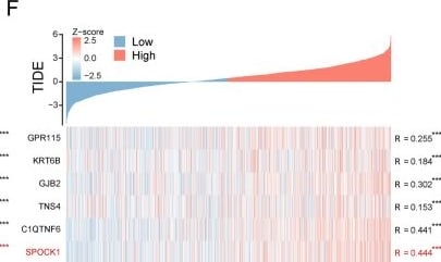
Identification of SPOCK1 as a prognostic key gene within the key modules. A Venn plot of the intersection of differentially expressed genes (DEGs) that are abnormally upregulated in tumors and genes within the red and green modules. B Determination of the optimal lambda when the partial likelihood of deviance reaches a minimum. And LASSO coefficient of module candidate gene profiles. C Screening of the top 10 prognosis-related genes in terms of relative importance using the stochastic survival forest algorithm. D Intersection Venn plot of prognostic key genes. Hub genes’ correlation heatmap with (E) EMT score and (F) TIDE score. (***p < 0.001; **p < 0.01; *p < 0.05; ns. no significance) Image collected and cropped by CiteAb from the following open publication (https://pubmed.ncbi.nlm.nih.gov/38087364), licensed under a CC-BY license. Not internally tested by R&D Systems.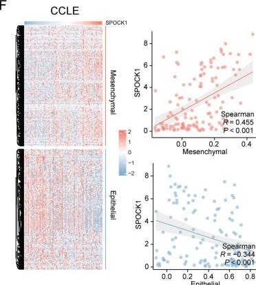
Detection of Testican 1/SPOCK1 by Immunohistochemistry
SPOCK1 is associated with invasive properties. In the GSE166722 dataset, (A) SPOCK1 expression levels in invasive phenotype and inert phenotype LUAD. B ROC curves showing the diagnostic efficacy of SPOCK1 in differentiating between aggressive and inert patients. C Differential expression levels of SPOCK1 in different aggressive pathological histological subtypes; (D) Differential expression levels of SPOCK1 in patients with or without lymph node metastasis. E Heatmap of the distribution of SPOCK1 and EMT-related markers in the combined cohort and (F) NSCLC cell lines, and the mesenchymal score and epithelial score were assessed based on the ssSGEA algorithm, and correlations were calculated (spearman test). G The potential association between SPOCK1 and dysregulated oncogenic pathways in the combined cohort and (H) NSCLC cell lines. (***p < 0.001; **p < 0.01; *p < 0.05; ns. no significance.) Image collected and cropped by CiteAb from the following open publication (https://pubmed.ncbi.nlm.nih.gov/38087364), licensed under a CC-BY license. Not internally tested by R&D Systems.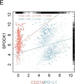
Detection of Testican 1/SPOCK1 by Flow Cytometry
Role of SPOCK1 in the tumor microenvironment. A Heatmap of the distribution of 29 TME associated signatures with SPOCK1 expression values. B Scatter plot of the correlation between 4 TME scores and SPOCK1 expression values. C Histogram of correlation between SPOCK1 and 5 immune features and chord plot of correlation with TGF-beta 1 (TGFB1), TGF-beta 2 (TGFB2), and TGF-beta 3 (TGFB3) (spearman test). D Distribution of SPOCK1 expression in TME subtypes defined by Bagaev et al. E Correlation between SPOCK1 and CD274 (mRNA level) and PD- L1 (protein level, from TCPA database) correlation scatter plots. (***p < 0.001; **p < 0.01; *p < 0.05; ns. no significance.) Image collected and cropped by CiteAb from the following open publication (https://pubmed.ncbi.nlm.nih.gov/38087364), licensed under a CC-BY license. Not internally tested by R&D Systems.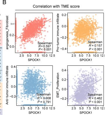
Detection of Testican 1/SPOCK1 by Flow Cytometry
Role of SPOCK1 in the tumor microenvironment. A Heatmap of the distribution of 29 TME associated signatures with SPOCK1 expression values. B Scatter plot of the correlation between 4 TME scores and SPOCK1 expression values. C Histogram of correlation between SPOCK1 and 5 immune features and chord plot of correlation with TGF-beta 1 (TGFB1), TGF-beta 2 (TGFB2), and TGF-beta 3 (TGFB3) (spearman test). D Distribution of SPOCK1 expression in TME subtypes defined by Bagaev et al. E Correlation between SPOCK1 and CD274 (mRNA level) and PD- L1 (protein level, from TCPA database) correlation scatter plots. (***p < 0.001; **p < 0.01; *p < 0.05; ns. no significance.) Image collected and cropped by CiteAb from the following open publication (https://pubmed.ncbi.nlm.nih.gov/38087364), licensed under a CC-BY license. Not internally tested by R&D Systems.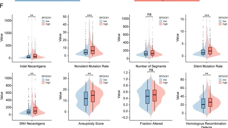
Detection of Testican 1/SPOCK1 by Flow Cytometry
Relationship of SPOCK1 with methylation and mutation. Boxplot indicates the extent of variation in promoter methylation levels in LUAD samples from the UALCAN database, (A) in normal and tumor, (B) TP53 mutant and TP53 wild-type groups. C Exploration of the mutational landscape of SPOCK1 in LUAD using the cBioPortal database. Mutation waterfall plots of the top 20 genes in the (D) high SPCOK1 group and (E) low SPCOK1 group in terms of mutation frequency. F Boxplots show the difference of DNA damage measures between different levels of SPOCK1 expression groups (Wilcon rank-sum test). (***p < 0.001; **p < 0.01; *p < 0.05; ns. no significance.) Image collected and cropped by CiteAb from the following open publication (https://pubmed.ncbi.nlm.nih.gov/38087364), licensed under a CC-BY license. Not internally tested by R&D Systems.Applications for Human Testican 1/SPOCK1 Antibody (316801)
Application
Recommended Usage
Immunoprecipitation
25 µg/mL
Sample: Conditioned cell culture medium spiked with Recombinant Human Testican 1/SPOCK1 (Catalog # 2327-PI), see our available Western blot detection antibodies
Sample: Conditioned cell culture medium spiked with Recombinant Human Testican 1/SPOCK1 (Catalog # 2327-PI), see our available Western blot detection antibodies
Western Blot
1 µg/mL
Sample: Recombinant Human Testican 1/SPOCK1 (Catalog # 2327-PI) under non-reducing conditions
Sample: Recombinant Human Testican 1/SPOCK1 (Catalog # 2327-PI) under non-reducing conditions
Formulation, Preparation, and Storage
Purification
Protein A or G purified from hybridoma culture supernatant
Reconstitution
Reconstitute at 0.5 mg/mL in sterile PBS. For liquid material, refer to CoA for concentration.
Loading...
Formulation
Lyophilized from a 0.2 μm filtered solution in PBS with Trehalose. See Certificate of Analysis for details.<br/>*Small pack size (-SP) is supplied either lyophilized or as a 0.2 µm filtered solution in PBS.
Shipping
Lyophilized product is shipped at ambient temperature. Liquid small pack size (-SP) is shipped with polar packs. Upon receipt, store immediately at the temperature recommended below.
Stability & Storage
Use a manual defrost freezer and avoid repeated freeze-thaw cycles.
- 12 months from date of receipt, -20 to -70 °C as supplied.
- 1 month, 2 to 8 °C under sterile conditions after reconstitution.
- 6 months, -20 to -70 °C under sterile conditions after reconstitution.
Calculators
Background: Testican 1/SPOCK1
Additional Testican 1/SPOCK1 Products
Product Documents for Human Testican 1/SPOCK1 Antibody (316801)
Certificate of Analysis
To download a Certificate of Analysis, please enter a lot or batch number in the search box below.
Note: Certificate of Analysis not available for kit components.
Product Specific Notices for Human Testican 1/SPOCK1 Antibody (316801)
For research use only
Related Research Areas
Citations for Human Testican 1/SPOCK1 Antibody (316801)
 Powered by Bioz
Customer Reviews for Human Testican 1/SPOCK1 Antibody (316801)
There are currently no reviews for this product. Be the first to review Human Testican 1/SPOCK1 Antibody (316801) and earn rewards!
Have you used Human Testican 1/SPOCK1 Antibody (316801)?
Submit a review and receive an Amazon gift card!
$25/€18/£15/$25CAN/¥2500 for a review with an image
$10/€7/£6/$10CAN/¥1110 for a review without an image
Submit a review
Protocols
Find general support by application which include: protocols, troubleshooting, illustrated assays, videos and webinars.
- Cellular Response to Hypoxia Protocols
- Immunoprecipitation Protocol
- R&D Systems Quality Control Western Blot Protocol
- Troubleshooting Guide: Western Blot Figures
- Western Blot Conditions
- Western Blot Protocol
- Western Blot Protocol for Cell Lysates
- Western Blot Troubleshooting
- Western Blot Troubleshooting Guide
- View all Protocols, Troubleshooting, Illustrated assays and Webinars
Loading...