
Vitamin D BP (VitD BP), also called VDB, DBP, Group Specific Component (Gc) or macrophage-activating factor (DBP- or Gc-MAF), is a member of the albumin family. These plasma/body fluid proteins have a conserved pattern of cysteine motifs that form binding domains. VitD BP is predominantly produced by hepatic parenchymal cells. It transports VitD, activates macrophages and osteoclasts, and enhances C5a chemotactic activity. Over 80 variant alleles have been identified, some of which correlate with osteoporosis or prostate cancer. Human and mouse VitD BP share 78% aa identity.
Human Vitamin D BP Antibody (359501)
R&D Systems | Catalog # MAB3778


Scientific images may be resized, cropped, or adjusted for brightness or contrast for presentation purposes. These updates are limited to presentation and do not affect the underlying scientific interpretation of the data. Where available, additional source data may be provided upon request.
Key Product Details
Species Reactivity
Validated:
Human
Cited:
Human, Mouse
Applications
Validated:
Western Blot
Cited:
Western Blot
Label
Unconjugated
Antibody Source
Monoclonal Mouse IgG2B Clone # 359501
Loading...
Product Specifications
Immunogen
Human plasma-derived Vitamin D BP
Accession # P02774
Accession # P02774
Specificity
Detects human plasma-derived Vitamin D BP in direct ELISAs and Western blots.
Clonality
Monoclonal
Host
Mouse
Isotype
IgG2B
Scientific Data Images for Human Vitamin D BP Antibody (359501)
Detection of Vitamin D BP by Western Blot
Legumain is required for VDBP processing and regulation. (A) Purified VDBP from human plasma (1.9 μM) was incubated in legumain assay buffer (pH 5.8) at 37 °C with or without purified active bovine legumain (2 μM) for 5 h before gel electrophoresis and immunoblotting of VDBP (n = 1). (B–H) Wild-type (Lgmn+/+) and legumain-deficient (Lgmn−/−) mice were treated with 50 µg/kg 25(OH)D3 (n = 6–7) or an equal volume vehicle (n = 7, control) subcutaneously every two to three days (four times in total). Tissues were harvested 24 h after the final injection (day 8). (B) One representative immunoblot of VDBP and GAPDH (housekeeping) in kidney and liver (n = 4). (C–F) Quantification of VDBP immunoband (IB) intensity as arbitrary units (ARBU) relative to GAPDH in immunoblots represented in (B) (n = 4). (C) Hepatic VDBP 45 kDa immunoband. (D) Renal VDBP 45 kDa immunoband. (E) Hepatic VDBP 55 kDa immunoband. (F) Renal VDBP 55 kDa immunoband. (G) Plasma VDBP concentration (μg/mL) was measured by ELISA (n = 6–7). (H) Hepatic VDBP mRNA expression relative to the geometric mean of CT values of four housekeeping controls (2− delta delta CT, n = 5). (C–H) Data represent mean ± SEM. Two-way ANOVA. # p < 0.05, ## p < 0.01, ### p < 0.001 vs. different genotype, same treatment. Numbers (n) represent individual biological replicates. Image collected and cropped by CiteAb from the following open publication (https://www.mdpi.com/2073-4409/13/1/36), licensed under a CC-BY license. Not internally tested by R&D Systems.Applications for Human Vitamin D BP Antibody (359501)
Application
Recommended Usage
Western Blot
1 µg/mL
Sample: Human plasma-derived Vitamin D BP
Sample: Human plasma-derived Vitamin D BP
Reviewed Applications
Read 1 review rated 5 using MAB3778 in the following applications:
Formulation, Preparation, and Storage
Purification
Protein A or G purified from hybridoma culture supernatant
Reconstitution
Reconstitute at 0.5 mg/mL in sterile PBS. For liquid material, refer to CoA for concentration.
Loading...
Formulation
Lyophilized from a 0.2 μm filtered solution in PBS and NaCl with Trehalose. *Small pack size (SP) is supplied either lyophilized or as a 0.2 µm filtered solution in PBS.
Shipping
Lyophilized product is shipped at ambient temperature. Liquid small pack size (-SP) is shipped with polar packs. Upon receipt, store immediately at the temperature recommended below.
Stability & Storage
Use a manual defrost freezer and avoid repeated freeze-thaw cycles.
- 12 months from date of receipt, -20 to -70 °C as supplied.
- 1 month, 2 to 8 °C under sterile conditions after reconstitution.
- 6 months, -20 to -70 °C under sterile conditions after reconstitution.
Calculators
Background: Vitamin D BP
Long Name
Vitamin D Binding Protein
Alternate Names
DBP, DBP-MAF, Gc, Gc-MAF, VDBG, VDBP
Gene Symbol
GC
UniProt
Additional Vitamin D BP Products
Product Documents for Human Vitamin D BP Antibody (359501)
Certificate of Analysis
To download a Certificate of Analysis, please enter a lot or batch number in the search box below.
Note: Certificate of Analysis not available for kit components.
Product Specific Notices for Human Vitamin D BP Antibody (359501)
For research use only
Citations for Human Vitamin D BP Antibody (359501)
 Powered by Bioz
Customer Reviews for Human Vitamin D BP Antibody (359501) (1)
5 out of 5
1 Customer Rating
Have you used Human Vitamin D BP Antibody (359501)?
Submit a review and receive an Amazon gift card!
$25/€18/£15/$25CAN/¥2500 for a review with an image
$10/€7/£6/$10CAN/¥1110 for a review without an image
Submit a review
Customer Images
Showing
1
-
1 的
1 review
Showing All
Filter By:
-
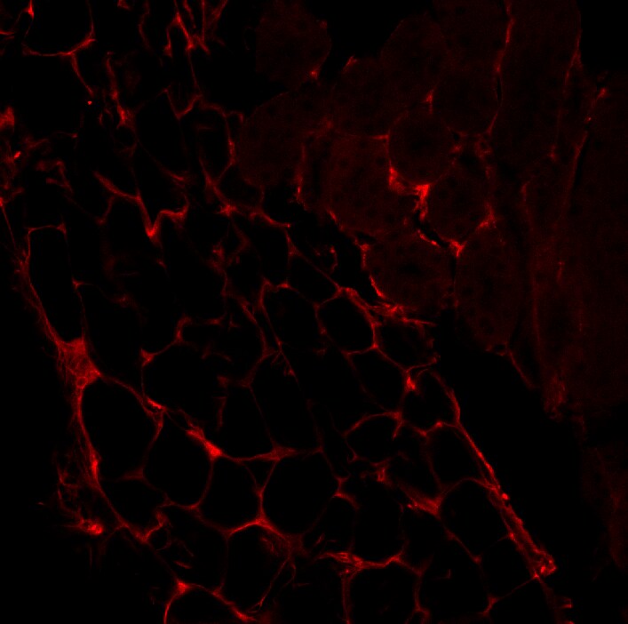
Application: Immunohistochemistry-FrozenSample Tested: Kidney tissueSpecies: MouseVerified Customer | Posted 09/09/2019VDBP (red) in mouse renal tissue.MAB3778 concentration used was 1 µg/ml. Secondary antibody was Alexa Fluor conjugated goat anti-mouse 594 (red; 1:1000).

There are no reviews that match your criteria.
Protocols
Find general support by application which include: protocols, troubleshooting, illustrated assays, videos and webinars.
- Cellular Response to Hypoxia Protocols
- R&D Systems Quality Control Western Blot Protocol
- Troubleshooting Guide: Western Blot Figures
- Western Blot Conditions
- Western Blot Protocol
- Western Blot Protocol for Cell Lysates
- Western Blot Troubleshooting
- Western Blot Troubleshooting Guide
- View all Protocols, Troubleshooting, Illustrated assays and Webinars
Loading...