MitoBrilliant™ 646
Tocris Bioscience | Catalog # 7700

Key Product Details
Description
Wavelength
Product Description
MitoBrilliant probes have been developed to overcome some common limitations encountered with standard mitochondrial trackers, offering clearer answers to scientific questions. They are next-generation fluorescent stains for the localization and tracking of mitochondria in both live and fixed cells. The range harnesses Janelia Fluor® dye technology, conferring some of the properties that make these widely used dyes, into mitochondrial stains.Key information: MitoBrilliant™ 646 is a red-fluorescent mitochondrial stain for both live and fixed cells.
Used for: mitochondrial staining for both live and fixed cells/tissue.
Application: flow cytometry, super-resolution microscopy-STED, high content screening, IHC/ICC.
Properties and Photophysical Data: MitoBrilliant™ 646 is retained in mitochondria with exceptionally clear staining and is retained in the mitochondria of live cells following loss of the mitochondrial membrane potential. Excitation and emission maxima (λ) are 655 nm and 668 nm, respectively; extinction coefficient = 125,000 M-1cm-1.
Two dyes that accumulate in the mitochondria of live cells in a mitochondrial membrane potential (Δψm) dependent manner are also available: MitoBrilliant™ Live 646 (red emission) and MitoBrilliant™ Live 549 (yellow/orange emission).
Please refer to the protocol for guidelines on product use, and download the MitoBrilliant Product Guide to view data for each of the dyes in different applications.
MitoBrilliant™ is a trademark of Bio-Techne Corp.
Scientific Data Images for MitoBrilliant™ 646

Live-cell image of mitochondria stained with MitoBrilliant™ 646
HeLa cells were incubated with MitoBrilliant™ 646 (100 nM) for 40 minutes and counterstained with DAPI (Catalog # 5748). Image was taken using an LSM880 Confocal using a 63x objective. Scale bar = 10 µm.
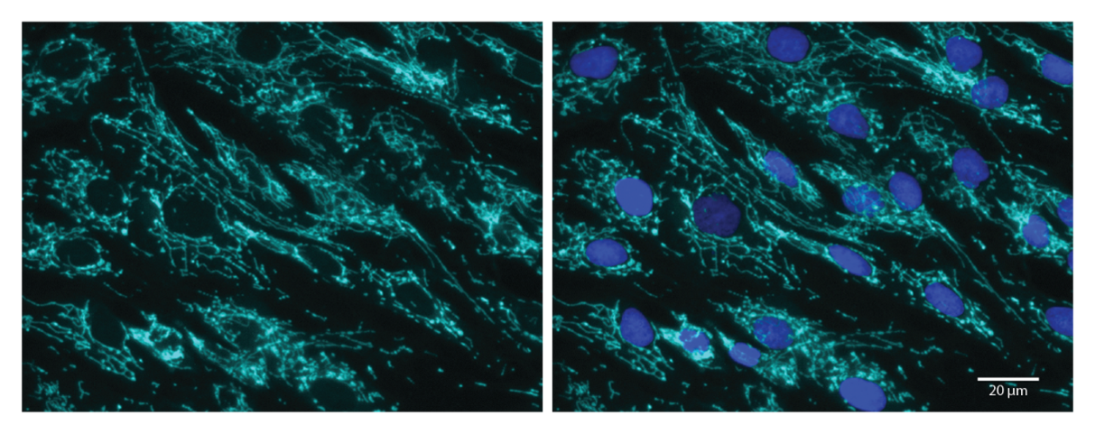
Fixed-cell image of mitochondria stained with MitoBrilliant™ 646.
Dermal fibroblast cells were incubated with MitoBrilliant™ 646 (100 nM) for 1 hour at 37°C, fixed with ice-cold acetone-methanol (1:1) and counterstained with Hoechst 33342 (Catalog # 5117). Image was taken using an Echo Revolve microscope using a 20x objective. Scale bar = 20 µm.| Product Name | Core dye structure | Abs/Em (nm) | Δψm dependent | Live/Fixed cell use | Image without wash step | Demonstrated Applications |
MitoBrilliant™ 646 Cat. No. 7700 |
Janelia Fluor® technology | 655/668 | No* | Suitable for both live and fixed cell work | Yes, but replacing media recommended |
Fixed-cell imaging, Live-cell imaging, Flow Cytometry, IHC/ICC, Super-resolution microscopy – STED, High-content screening |
|---|---|---|---|---|---|---|
|
MitoBrilliant™ Live 646 Cat. No. 7417 |
Janelia Fluor® technology | 648/662 | Yes | Live-cell work only | Yes, but replacing media recommended |
Live-cell imaging, Flow Cytometry, High-content screening |
|
MitoBrilliant™ Live 549 Cat. No. 7693 |
Janelia Fluor® technology | 550/568 | Yes | Live-cell work only | Yes, but replacing media recommended |
Live-cell imaging, Flow Cytometry, High-content screening |
Optical Data

| Emission Color | Red |
| λabs | 655 nm |
| λem | 668 nm |
| Extinction Coefficient (ε) | 125000 M-1cm-1 |
| Closest Laser line | 640 nm |
| Application | Fixed-cell imaging, Live-cell imaging, Flow Cytometry, IHC/ICC, Super-resolution microscopy – STED, |
Spectra Viewer
Plan Your Experiments
Use our spectra viewer to interactively plan your experiments, assessing multiplexing options. View the excitation and emission spectra for our fluorescent dye range and other commonly used dyes.
Spectra Viewer
Product Specifications for MitoBrilliant™ 646
Molecular Weight
Storage
Purity
The technical data provided above is for guidance only. For batch specific data refer to the Certificate of Analysis.
Solubility
| Solvent | Max Conc. mg/mL | Max Conc. mM | |
|---|---|---|---|
| Solubility | |||
| DMSO | 4.94 | 10 |
Preparing Stock Solutions for MitoBrilliant™ 646
The following data is based on the product molecular weight 493.55.
Batch specific molecular weights may vary from batch to batch due to the degree of hydration, which all affect the solvent volumes required to prepare stock solutions.
| Concentration / Solvent Volume / Mass | 1 mg | 5 mg | 10 mg |
|---|---|---|---|
| 0.1 mM | 20.26 mL | 101.31 mL | 202.61 mL |
| 0.5 mM | 4.05 mL | 20.26 mL | 40.52 mL |
| 1 mM | 2.03 mL | 10.13 mL | 20.26 mL |
| 5 mM | 0.41 mL | 2.03 mL | 4.05 mL |
Calculators
Background References
References are publications that support the biological activity of the product. See our Citations tab to view 11 publications citing the usage of this product.
There are currently no references for this product.
Product Documents for MitoBrilliant™ 646
Certificate of Analysis
To download a Certificate of Analysis, please enter a lot or batch number in the search box below.
Product Specific Notices for MitoBrilliant™ 646
For research use only
Citations for MitoBrilliant™ 646
 Powered by Bioz
Customer Reviews for MitoBrilliant™ 646 (1)
Have you used MitoBrilliant™ 646?
Submit a review and receive an Amazon gift card!
$25/€18/£15/$25CAN/¥2500 Yen for a review with an image
$10/€7/£6/$10CAN/¥1110 Yen for a review without an image
Submit a review
Customer Images
-
Species: HumanVerified Customer | Posted 11/20/2023MitoBrilliant™ 646 produced excellent results as both a live and fixed cell stain. As a live cell stain MitoBrilliant™ 646 produced a stable signal within 20 minutes. For use as fixed-cell stain optimal conditions were found to be a 1-hour incubation in live cells followed by fixation with ice-cold acetone-methanol.Fixed-cell image of mitochondria stained with MitoBrilliant™ 646: Dermal fibroblast cells were incubated with MitoBrilliant™ 646 (100 nM) for 1 hour at 37°C, fixed with ice-cold acetone-methanol (1:1) and counterstained with Hoechst 33342. Image was taken using an Echo Revolve microscope using a 20x objective. Scale bar = 20 µm.

There are no reviews that match your criteria.