NKG2D, also known as CD314, is a type II transmembrane protein with an extracellular C-type lectin-like domain. It occurs as a disulfide-linked homodimer that associates with the transmembrane DAP10 (DNAX-activator protein 10) adapter protein to deliver an activating signal. This protein shares approximately 25% amino acid sequence identity with a number of other type II lectin-like proteins that are encoded by genes within the natural killer complex on mouse chromosome 6. NKG2D is expressed on NK cells, where it functions as an activating receptor to trigger cytolytic activity and cytokine secretion, and on some T cell subsets, where it acts as a co‑stimulatory receptor complementing T cell receptor signaling. Several ligands have now been identified for mouse NKG2D including H60 and Rae 1 alpha, beta, gamma, delta, and epsilon. All of these ligands are cell-surface proteins distantly related to MHC class I. However, they do not bind peptide or associate with beta 2-microglobulin. Ligand expression is up-regulated in many transformed cell lines and also during conditions of stress such as heat shock or viral infection. In vivo, tumor models demonstrate that NKG2D functions in anti-tumor surveillance (1-5).
Mouse NKG2D/CD314 Antibody (191004)
R&D Systems | Catalog # MAB1547

Key Product Details
Species Reactivity
Validated:
Mouse
Cited:
Human, Mouse, Transgenic Mouse
Applications
Validated:
Western Blot, Blockade of Receptor-ligand Interaction, Flow Cytometry, CyTOF-ready
Cited:
Immunohistochemistry, Immunohistochemistry-Frozen, Western Blot, Neutralization, Bioassay, Functional Assay
Label
Unconjugated
Antibody Source
Monoclonal Rat IgG2B Clone # 191004
Loading...
Product Specifications
Immunogen
Mouse myeloma cell line NS0-derived recombinant mouse NKG2D
Phe94-Val232
Accession # O54709
Phe94-Val232
Accession # O54709
Specificity
Detects mouse NKG2D in direct ELISAs and Western blots. Does not cross-react with recombinant human NKG2D.
Clonality
Monoclonal
Host
Rat
Isotype
IgG2B
Endotoxin Level
<0.10 EU per 1 μg of the antibody by the LAL method.
Scientific Data Images for Mouse NKG2D/CD314 Antibody (191004)
Detection of NKG2D/CD314 by Flow Cytometry
NK Require NKG2D to Control Early Virus Dissemination & for Optimal Cytotoxicity but Not Activation(A) B6 mice infected with 3,000 pfu ECTV. On day 5 PI, mice pulsed with BrdU for 3 h & their spleens analyzed by flow cytometry. Plots are gated on CD3ɛ− NK1.1+. Data correspond to pools of three mice from three individual experiments.(B) Increased viral titers of infected mice with NKG2D blockade in vivo. Intact B6 mice, B6 mice with NKG2D blockade, B6 mice depleted of NK, B6 mice depleted of T (treated with anti-CD4 + anti-CD8 mAbs), or depleted of T & with NKG2D blockade infected with 3,000 pfu ECTV & the viral titers in spleen determined on day 3 PI. Data are the average ± SD of three individual mice & are representative of two similar experiments.(C) The indicated mice infected with 3,000 pfu ECTV, & the NK responses in the D-LN determined at 2 d PI. Upper panel: Dot plot showing the proportion of NK (DX5+, CD3e−) in the D-LN of infected & control uninfected mice. Lower panel: GzB & IFN-gamma production by NK. correspond to the DX5+, CD3ɛ− gate of the upper panels. Data correspond to pools of three mice & are representative of three similar experiments.(D) NK purified from spleens (5 d PI) of ECTV-infected intact or NKG2D-blocked mice & stained as indicated. Filled histogram: isotype-matched control Ig; gray line: NKG2D-blocked mice; black line: intact mice.(E) NK purified from spleens of intact or NKG2D-blocked mice as indicated & used as effectors in a 4-h 51Cr release assay against the indicated targets. Data correspond to pools of three mice & are representative of three experiments.(F) As in (D), but the NK purified from untreated mice & a neutralizing anti-NKG2D mAb was added to the in vitro cytotoxicity assays, as indicated. Image collected & cropped by CiteAb from the following open publication (https://pubmed.ncbi.nlm.nih.gov/18266471), licensed under a CC-BY license. Not internally tested by R&D Systems.
Detection of NKG2D/CD314 by Flow Cytometry
NK Require NKG2D to Control Early Virus Dissemination & for Optimal Cytotoxicity but Not Activation(A) B6 mice infected with 3,000 pfu ECTV. On day 5 PI, mice pulsed with BrdU for 3 h & their spleens analyzed by flow cytometry. Plots are gated on CD3ɛ− NK1.1+. Data correspond to pools of three mice from three individual experiments.(B) Increased viral titers of infected mice with NKG2D blockade in vivo. Intact B6 mice, B6 mice with NKG2D blockade, B6 mice depleted of NK, B6 mice depleted of T (treated with anti-CD4 + anti-CD8 mAbs), or depleted of T & with NKG2D blockade infected with 3,000 pfu ECTV & the viral titers in spleen determined on day 3 PI. Data are the average ± SD of three individual mice & are representative of two similar experiments.(C) The indicated mice infected with 3,000 pfu ECTV, & the NK responses in the D-LN determined at 2 d PI. Upper panel: Dot plot showing the proportion of NK (DX5+, CD3e−) in the D-LN of infected & control uninfected mice. Lower panel: GzB & IFN-gamma production by NK. correspond to the DX5+, CD3ɛ− gate of the upper panels. Data correspond to pools of three mice & are representative of three similar experiments.(D) NK purified from spleens (5 d PI) of ECTV-infected intact or NKG2D-blocked mice & stained as indicated. Filled histogram: isotype-matched control Ig; gray line: NKG2D-blocked mice; black line: intact mice.(E) NK purified from spleens of intact or NKG2D-blocked mice as indicated & used as effectors in a 4-h 51Cr release assay against the indicated targets. Data correspond to pools of three mice & are representative of three experiments.(F) As in (D), but the NK purified from untreated mice & a neutralizing anti-NKG2D mAb was added to the in vitro cytotoxicity assays, as indicated. Image collected & cropped by CiteAb from the following open publication (https://pubmed.ncbi.nlm.nih.gov/18266471), licensed under a CC-BY license. Not internally tested by R&D Systems.Applications for Mouse NKG2D/CD314 Antibody (191004)
Application
Recommended Usage
Blockade of Receptor-ligand Interaction
In a functional ELISA, 0.05-0.15 µg/mL of this antibody will block 50% of the binding of 100 ng/mL of biotinylated Recombinant Mouse Rae-1 gamma Fc Chimera to immobilized Recombinant Mouse NKG2D Fc Chimera (Catalog # 139‑NK) coated at 4 µg/mL (100 µL/well). At 2 μg/mL, this antibody will block >90% of the binding.
CyTOF-ready
Ready to be labeled using established conjugation methods. No BSA or other carrier proteins that could interfere with conjugation.
Flow Cytometry
2.5 µg/106 cells
Sample: NK1.1+ mouse splenic NK cells
Sample: NK1.1+ mouse splenic NK cells
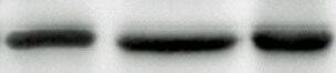
Western Blot
1 µg/mL
Sample: Recombinant Mouse NKG2D/CD314 Fc Chimera (Catalog # 139-NK)
under non-reducing conditions only
Sample: Recombinant Mouse NKG2D/CD314 Fc Chimera (Catalog # 139-NK)
under non-reducing conditions only
Reviewed Applications
Read 1 review rated 5 using MAB1547 in the following applications:
Flow Cytometry Panel Builder
Bio-Techne Knows Flow Cytometry
Save time and reduce costly mistakes by quickly finding compatible reagents using the Panel Builder Tool.
Advanced Features
- Spectra Viewer - Custom analysis of spectra from multiple fluorochromes
- Spillover Popups - Visualize the spectra of individual fluorochromes
- Antigen Density Selector - Match fluorochrome brightness with antigen density

Formulation, Preparation, and Storage
Purification
Protein A or G purified from hybridoma culture supernatant
Reconstitution
Reconstitute at 0.5 mg/mL in sterile PBS. For liquid material, refer to CoA for concentration.
Loading...
Formulation
Lyophilized from a 0.2 μm filtered solution in PBS with Trehalose. *Small pack size (SP) is supplied either lyophilized or as a 0.2 µm filtered solution in PBS.
Shipping
Lyophilized product is shipped at ambient temperature. Liquid small pack size (-SP) is shipped with polar packs. Upon receipt, store immediately at the temperature recommended below.
Stability & Storage
Use a manual defrost freezer and avoid repeated freeze-thaw cycles.
- 12 months from date of receipt, -20 to -70 °C as supplied.
- 1 month, 2 to 8 °C under sterile conditions after reconstitution.
- 6 months, -20 to -70 °C under sterile conditions after reconstitution.
Calculators
Background: NKG2D/CD314
References
- Bauer, S. et al. (1999) Science 285:727.
- Wu, J. et al. (1999) Science 285:730.
- Diefenbach, A. et al. (2001) Nature 413:165.
- Vivier, E. et al. (2002) Curr. Opin. Immunol. 14:306.
- NKG2D and its Ligands; www.RnDSystems.com.
Long Name
Natural Killer G2D
Alternate Names
CD314, D12S2489E, KLRK1
Gene Symbol
KLRK1
UniProt
Additional NKG2D/CD314 Products
Product Documents for Mouse NKG2D/CD314 Antibody (191004)
Certificate of Analysis
To download a Certificate of Analysis, please enter a lot or batch number in the search box below.
Note: Certificate of Analysis not available for kit components.
Product Specific Notices for Mouse NKG2D/CD314 Antibody (191004)
For research use only
Related Research Areas
Citations for Mouse NKG2D/CD314 Antibody (191004)
 Powered by Bioz
Customer Reviews for Mouse NKG2D/CD314 Antibody (191004) (1)
5 out of 5
1 Customer Rating
Have you used Mouse NKG2D/CD314 Antibody (191004)?
Submit a review and receive an Amazon gift card!
$25/€18/£15/$25CAN/¥2500 Yen for a review with an image
$10/€7/£6/$10CAN/¥1110 Yen for a review without an image
Submit a review
Customer Images
Showing
1
-
1 的
1 review
Showing All
Filter By:
-
Application: Western BlotSample Tested: Dendritic epidermal T cellsSpecies: MouseVerified Customer | Posted 10/24/2021

There are no reviews that match your criteria.
Protocols
Find general support by application which include: protocols, troubleshooting, illustrated assays, videos and webinars.
- 7-Amino Actinomycin D (7-AAD) Cell Viability Flow Cytometry Protocol
- Cellular Response to Hypoxia Protocols
- Extracellular Membrane Flow Cytometry Protocol
- Flow Cytometry Protocol for Cell Surface Markers
- Flow Cytometry Protocol for Staining Membrane Associated Proteins
- Flow Cytometry Staining Protocols
- Flow Cytometry Troubleshooting Guide
- Intracellular Flow Cytometry Protocol Using Alcohol (Methanol)
- Intracellular Flow Cytometry Protocol Using Detergents
- Intracellular Nuclear Staining Flow Cytometry Protocol Using Detergents
- Intracellular Staining Flow Cytometry Protocol Using Alcohol Permeabilization
- Intracellular Staining Flow Cytometry Protocol Using Detergents to Permeabilize Cells
- Propidium Iodide Cell Viability Flow Cytometry Protocol
- Protocol for Liperfluo
- Protocol for the Characterization of Human Th22 Cells
- Protocol for the Characterization of Human Th9 Cells
- Protocol: Annexin V and PI Staining by Flow Cytometry
- Protocol: Annexin V and PI Staining for Apoptosis by Flow Cytometry
- R&D Systems Quality Control Western Blot Protocol
- Troubleshooting Guide: Fluorokine Flow Cytometry Kits
- Troubleshooting Guide: Western Blot Figures
- Western Blot Conditions
- Western Blot Protocol
- Western Blot Protocol for Cell Lysates
- Western Blot Troubleshooting
- Western Blot Troubleshooting Guide
- View all Protocols, Troubleshooting, Illustrated assays and Webinars
Loading...
Associated Pathways

