
Mouse PCPE-1 Antibody (261845)
R&D Systems | Catalog # MAB2239

Scientific images may be resized, cropped, or adjusted for brightness or contrast for presentation purposes. These updates are limited to presentation and do not affect the underlying scientific interpretation of the data. Where available, additional source data may be provided upon request.
Key Product Details
Species Reactivity
Validated:
Cited:
Applications
Validated:
Cited:
Label
Antibody Source
Product Specifications
Immunogen
Gln25-Ala468
Accession # Q61398
Specificity
Clonality
Host
Isotype
Scientific Data Images for Mouse PCPE-1 Antibody (261845)
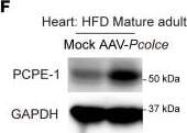
Detection of PCPE-1 by Western Blot
The BATokine PCPE-1 contributes to the pathogenesis of HFpEF.Littermate control (Con) or BAT Pcolce-KO mice were fed with a high-fat diet (HFD) and studied during 41–47 weeks of age (middle-aged). BAT adeno-associated virus (AAV) encoding Pcolce was injected in the BAT of C57BL/6NCrSlc mice fed with an HFD and studied at 15 weeks of age (mature adult). BAT-PCOLCE KI mice were also fed with an HFD and studied at 23–25 weeks of age (mature adult). (A, F, and J) Western blot analysis of mouse (A and F) or human (J) PCPE-1 expression in the heart of the indicated mice. (B, G, and K) Echocardiographic findings in mice: E/e’ (marker for diastolic dysfunction), IVSTd (interventricular septum thickness), LVDs (left ventricular systolic dimension), FS (fractional shortening) (n = 14, 10 for B), (n =7, 7 for G), and (n = 8, 7 for K). (C, H, and L) Masson’s trichrome stain of the heart and its quantification (n =5, 5 for C), (n = 5, 6 for H), and (n = 4, 6 for L) in mice. Scale bar: 50 μm. (D, I, and M) ELISA for collagen type I (shown as Collagen 1) testing hearts in mice (n = 9, 10 for D), (n = 5, 6 for I), and (n = 8, 7 for M). (E) Experimental design of the AAV-Pcolce mouse. (N) Schematic demonstrating that ROS/DNA damage/c-Fos/c-Jun signaling upregulates PCPE-1 expression in BAT with aging. PCPE-1 produced from BAT promotes fibrosis in the heart. All data were analyzed by an independent-samples t test. Data information: Representative Masson’s trichrome–stained images from 1 series of observations (C, H, and L), and other data were obtained from 1 representative analytical experiment out of at least 2 independent experiments showing similar results. *P < 0.05, **P < 0.01. Values are presented as mean ± SEM. NS = not significant. Image collected and cropped by CiteAb from the following open publication (https://pubmed.ncbi.nlm.nih.gov/41955016), licensed under a CC-BY license. Not internally tested by R&D Systems.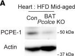
Detection of PCPE-1 by Western Blot
The BATokine PCPE-1 contributes to the pathogenesis of HFpEF.Littermate control (Con) or BAT Pcolce-KO mice were fed with a high-fat diet (HFD) and studied during 41–47 weeks of age (middle-aged). BAT adeno-associated virus (AAV) encoding Pcolce was injected in the BAT of C57BL/6NCrSlc mice fed with an HFD and studied at 15 weeks of age (mature adult). BAT-PCOLCE KI mice were also fed with an HFD and studied at 23–25 weeks of age (mature adult). (A, F, and J) Western blot analysis of mouse (A and F) or human (J) PCPE-1 expression in the heart of the indicated mice. (B, G, and K) Echocardiographic findings in mice: E/e’ (marker for diastolic dysfunction), IVSTd (interventricular septum thickness), LVDs (left ventricular systolic dimension), FS (fractional shortening) (n = 14, 10 for B), (n =7, 7 for G), and (n = 8, 7 for K). (C, H, and L) Masson’s trichrome stain of the heart and its quantification (n =5, 5 for C), (n = 5, 6 for H), and (n = 4, 6 for L) in mice. Scale bar: 50 μm. (D, I, and M) ELISA for collagen type I (shown as Collagen 1) testing hearts in mice (n = 9, 10 for D), (n = 5, 6 for I), and (n = 8, 7 for M). (E) Experimental design of the AAV-Pcolce mouse. (N) Schematic demonstrating that ROS/DNA damage/c-Fos/c-Jun signaling upregulates PCPE-1 expression in BAT with aging. PCPE-1 produced from BAT promotes fibrosis in the heart. All data were analyzed by an independent-samples t test. Data information: Representative Masson’s trichrome–stained images from 1 series of observations (C, H, and L), and other data were obtained from 1 representative analytical experiment out of at least 2 independent experiments showing similar results. *P < 0.05, **P < 0.01. Values are presented as mean ± SEM. NS = not significant. Image collected and cropped by CiteAb from the following open publication (https://pubmed.ncbi.nlm.nih.gov/41955016), licensed under a CC-BY license. Not internally tested by R&D Systems.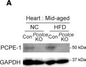
Detection of PCPE-1 by Western Blot
Depletion of PCPE-1 ameliorates HFpEF in obesity and aged mouse models.(A–E) Littermate control (Con) or systemic Pcolce-KO mice fed with a high-fat diet (HFD) from 4 weeks of age or a normal chow diet (NCD) and studied at 41–46 weeks of age (middle-aged). (F–J) Systemic Pcolce-KO mice (74–80 wk) fed with an NCD. (A and F) Western blot analysis of PCPE-1 expression in the heart. (B and G) Echocardiographic findings in the indicated mice: E/e’ (marker for diastolic dysfunction), IVSTd (interventricular septum thickness), LVDs (left ventricular systolic dimension), FS (fractional shortening) (n = 6, 8, 8, 9 for E/e’ and n = 6, 8, 13, 12 for other parameters in B; n = 9, 12 for G). (C and H) Masson’s trichrome stain of the heart. Scale bar: 50 μm. Right panels indicate quantification of relative fibrotic area (C) (n = 7, 5) (H) (n = 4, 5). (D and I) ELISA for cardiac collagen type I (shown as Collagen 1) (n = 10, 9 for D) and (n = 4, 5 for I). (E and J) Treadmill examination test (TMET) showing the running distance (n = 13, 17 for E) and (n = 9, 12 for J). Two-way ANOVA followed by Tukey’s multiple-comparison (B and D) or Dunnett T3 multiple comparison test (C); others: independent-samples t test. Data information: Representative Masson’s trichrome–stained images from 1 series of observations (C and H), and other data were obtained from 1 representative analytical experiment out of at least 2 independent experiments showing similar results. *P < 0.05, **P < 0.01. Values are presented as mean ± SEM. NS = not significant. Image collected and cropped by CiteAb from the following open publication (https://pubmed.ncbi.nlm.nih.gov/41955016), licensed under a CC-BY license. Not internally tested by R&D Systems.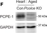
Detection of PCPE-1 by Western Blot
Depletion of PCPE-1 ameliorates HFpEF in obesity and aged mouse models.(A–E) Littermate control (Con) or systemic Pcolce-KO mice fed with a high-fat diet (HFD) from 4 weeks of age or a normal chow diet (NCD) and studied at 41–46 weeks of age (middle-aged). (F–J) Systemic Pcolce-KO mice (74–80 wk) fed with an NCD. (A and F) Western blot analysis of PCPE-1 expression in the heart. (B and G) Echocardiographic findings in the indicated mice: E/e’ (marker for diastolic dysfunction), IVSTd (interventricular septum thickness), LVDs (left ventricular systolic dimension), FS (fractional shortening) (n = 6, 8, 8, 9 for E/e’ and n = 6, 8, 13, 12 for other parameters in B; n = 9, 12 for G). (C and H) Masson’s trichrome stain of the heart. Scale bar: 50 μm. Right panels indicate quantification of relative fibrotic area (C) (n = 7, 5) (H) (n = 4, 5). (D and I) ELISA for cardiac collagen type I (shown as Collagen 1) (n = 10, 9 for D) and (n = 4, 5 for I). (E and J) Treadmill examination test (TMET) showing the running distance (n = 13, 17 for E) and (n = 9, 12 for J). Two-way ANOVA followed by Tukey’s multiple-comparison (B and D) or Dunnett T3 multiple comparison test (C); others: independent-samples t test. Data information: Representative Masson’s trichrome–stained images from 1 series of observations (C and H), and other data were obtained from 1 representative analytical experiment out of at least 2 independent experiments showing similar results. *P < 0.05, **P < 0.01. Values are presented as mean ± SEM. NS = not significant. Image collected and cropped by CiteAb from the following open publication (https://pubmed.ncbi.nlm.nih.gov/41955016), licensed under a CC-BY license. Not internally tested by R&D Systems.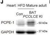
Detection of PCPE-1 by Western Blot
The BATokine PCPE-1 contributes to the pathogenesis of HFpEF.Littermate control (Con) or BAT Pcolce-KO mice were fed with a high-fat diet (HFD) and studied during 41–47 weeks of age (middle-aged). BAT adeno-associated virus (AAV) encoding Pcolce was injected in the BAT of C57BL/6NCrSlc mice fed with an HFD and studied at 15 weeks of age (mature adult). BAT-PCOLCE KI mice were also fed with an HFD and studied at 23–25 weeks of age (mature adult). (A, F, and J) Western blot analysis of mouse (A and F) or human (J) PCPE-1 expression in the heart of the indicated mice. (B, G, and K) Echocardiographic findings in mice: E/e’ (marker for diastolic dysfunction), IVSTd (interventricular septum thickness), LVDs (left ventricular systolic dimension), FS (fractional shortening) (n = 14, 10 for B), (n =7, 7 for G), and (n = 8, 7 for K). (C, H, and L) Masson’s trichrome stain of the heart and its quantification (n =5, 5 for C), (n = 5, 6 for H), and (n = 4, 6 for L) in mice. Scale bar: 50 μm. (D, I, and M) ELISA for collagen type I (shown as Collagen 1) testing hearts in mice (n = 9, 10 for D), (n = 5, 6 for I), and (n = 8, 7 for M). (E) Experimental design of the AAV-Pcolce mouse. (N) Schematic demonstrating that ROS/DNA damage/c-Fos/c-Jun signaling upregulates PCPE-1 expression in BAT with aging. PCPE-1 produced from BAT promotes fibrosis in the heart. All data were analyzed by an independent-samples t test. Data information: Representative Masson’s trichrome–stained images from 1 series of observations (C, H, and L), and other data were obtained from 1 representative analytical experiment out of at least 2 independent experiments showing similar results. *P < 0.05, **P < 0.01. Values are presented as mean ± SEM. NS = not significant. Image collected and cropped by CiteAb from the following open publication (https://pubmed.ncbi.nlm.nih.gov/41955016), licensed under a CC-BY license. Not internally tested by R&D Systems.Applications for Mouse PCPE-1 Antibody (261845)
Immunoprecipitation
Sample: Conditioned cell culture medium spiked with Recombinant Mouse PCPE‑1, see our available Western blot detection antibodies
Western Blot
Sample: Recombinant Mouse PCPE‑1
Formulation, Preparation, and Storage
Purification
Reconstitution
Reconstitute at 0.5 mg/mL in sterile PBS. For liquid material, refer to CoA for concentration.
Formulation
Shipping
Stability & Storage
- 12 months from date of receipt, -20 to -70 °C as supplied.
- 1 month, 2 to 8 °C under sterile conditions after reconstitution.
- 6 months, -20 to -70 °C under sterile conditions after reconstitution.
Calculators
Background: PCPE-1
References
- Steiglitz, B.M. et al. (2002) J. Biol. Chem. 277:49820.
- Kessler, E. et al. (1990) Biochem. Biophys. Res. Commun. 173:81.
- Moali, C. et al. (2005) J. Biol. Chem. 280:24188.
- Ge, G. et al. (2006) J. Biol. Chem. 281:10786.
Long Name
Alternate Names
Gene Symbol
UniProt
Additional PCPE-1 Products
Product Documents for Mouse PCPE-1 Antibody (261845)
Certificate of Analysis
To download a Certificate of Analysis, please enter a lot or batch number in the search box below.
Note: Certificate of Analysis not available for kit components.
Product Specific Notices for Mouse PCPE-1 Antibody (261845)
For research use only
Related Research Areas
Citations for Mouse PCPE-1 Antibody (261845)
 Powered by Bioz
Customer Reviews for Mouse PCPE-1 Antibody (261845)
There are currently no reviews for this product. Be the first to review Mouse PCPE-1 Antibody (261845) and earn rewards!
Have you used Mouse PCPE-1 Antibody (261845)?
Submit a review and receive an Amazon gift card!
$25/€18/£15/$25CAN/¥2500 for a review with an image
$10/€7/£6/$10CAN/¥1110 for a review without an image
Submit a review
Protocols
Find general support by application which include: protocols, troubleshooting, illustrated assays, videos and webinars.
- Cellular Response to Hypoxia Protocols
- Immunoprecipitation Protocol
- R&D Systems Quality Control Western Blot Protocol
- Troubleshooting Guide: Western Blot Figures
- Western Blot Conditions
- Western Blot Protocol
- Western Blot Protocol for Cell Lysates
- Western Blot Troubleshooting
- Western Blot Troubleshooting Guide
- View all Protocols, Troubleshooting, Illustrated assays and Webinars