![Western Blot: Rictor Antibody [NB100-56427]](https://resources.rndsystems.com/images/products/RICTOR-Antibody-Western-Blot-NB100-56427-img0002.jpg "Western Blot: Rictor Antibody [NB100-56427]")
Loading...
Key Product Details
Species Reactivity
Validated:
Human
Predicted:
Mouse (93%). Backed by our 100% Guarantee.
Applications
Western Blot
Label
Unconjugated
Antibody Source
Polyclonal Rabbit IgG
Format
BSA Free
Loading...
Product Specifications
Immunogen
A portion of amino acids 1-50 of human Rictor was used as the immunogen.
Clonality
Polyclonal
Host
Rabbit
Isotype
IgG
Scientific Data Images for Rictor Antibody - BSA Free
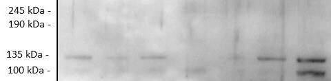
Western Blot: Rictor Antibody [NB100-56427]
Western Blot: Rictor Antibody [NB100-56427] - Analysis of Rictor in Jurkat cell lysate using this antibody.Applications for Rictor Antibody - BSA Free
Application
Recommended Usage
Western Blot
5-7 ug/ml
Reviewed Applications
Read 1 review rated 3 using NB100-56427 in the following applications:
Formulation, Preparation, and Storage
Purification
Protein G purified
Formulation
PBS containing 0.2% gelatin
Format
BSA Free
Preservative
0.05% Sodium Azide
Concentration
0.5 mg/ml
Shipping
The product is shipped with polar packs. Upon receipt, store it immediately at the temperature recommended below.
Stability & Storage
Store at 4C short term. Aliquot and store at -20C long term. Avoid freeze-thaw cycles.
Background: Rictor
Long Name
Rapamycin-insensitive Companion of mTOR
Alternate Names
mAVO3, Pianissimo
Gene Symbol
RICTOR
UniProt
Additional Rictor Products
Product Documents for Rictor Antibody - BSA Free
Certificate of Analysis
To download a Certificate of Analysis, please enter a lot or batch number in the search box below.
Product Specific Notices for Rictor Antibody - BSA Free
This product is for research use only and is not approved for use in humans or in clinical diagnosis. Primary Antibodies are guaranteed for 1 year from date of receipt.
Related Research Areas
Customer Reviews for Rictor Antibody - BSA Free (1)
3 out of 5
1 Customer Rating
Have you used Rictor Antibody - BSA Free?
Submit a review and receive an Amazon gift card!
$25/€18/£15/$25CAN/¥2500 Yen for a review with an image
$10/€7/£6/$10CAN/¥1110 Yen for a review without an image
Submit a review
Customer Images
Showing
1
-
1 的
1 review
Showing All
Filter By:
-
Application: Western BlotSample Tested: Adipose tissueSpecies: MouseVerified Customer | Posted 10/23/2019Mouse white adipose tissue was homogenised and protein content was quantified by a BCA assay. Twenty micrograms of protein were resolved on a 4-12% Bis-Tris gel and transferred to nitrocellulose membranes. Membranes were probed with primary antibody Rictor diluted 1:1000 in 5% BSA 2, before incubation with anti rabbit secondary horseradish peroxidase-conjugated antibody 1:5000. Blots were visualised with Immobilon Western Chemiluminescence HRP Substrate and imaged with Syngene chemiluminescence imaging system.

There are no reviews that match your criteria.
Protocols
Find general support by application which include: protocols, troubleshooting, illustrated assays, videos and webinars.
- Cellular Response to Hypoxia Protocols
- R&D Systems Quality Control Western Blot Protocol
- Troubleshooting Guide: Western Blot Figures
- Western Blot Conditions
- Western Blot Protocol
- Western Blot Protocol for Cell Lysates
- Western Blot Troubleshooting
- Western Blot Troubleshooting Guide
- View all Protocols, Troubleshooting, Illustrated assays and Webinars
Loading...
Associated Pathways

