Secondary Antibodies: General FAQs
Find scientific technical answers for frequently asked questions (FAQs) relating to secondary antibodies:
- What is a secondary antibody?
- Are there any structural differences between primary and secondary antibodies?
- What are antibody classes/sub-classes or isotypes?
- What are the major benefits of using a secondary antibody?
- What is the difference between using a directly conjugated primary antibody vs. a conjugated secondary?
- What species secondary antibody should I use?
- How should I choose a secondary for my primary antibody?
- How do I choose the right format of a secondary antibody?
- When should I use a Fab fragment secondary antibody?
- What label should I choose for a secondary antibody?
- What immunoglobulin class or subclass should I use for my primary antibody?
- What is pre-adsorption?
- What is the advantage of using the pre-adsorbed secondary antibody?
- How are affinity purified antibodies produced?
- Should I use an antigen affinity purified antibody or a whole immunoglobulins fraction?
- Is it necessary to use a specific isotype/subclass of the secondary antibody when using a monoclonal primary antibody?
- Should I use secondary antibodies raised in the same species in my multiple labeling experiment?
- When do I need to use the fragment specific secondary antibodies, such as Fc, F(ab) and F(ab’)2?
- Should I use Fab specific secondary antibodies when using a mouse primary antibody on mouse tissue?
- I am using a polymer based detection kit instead of a standard secondary antibody. I am getting some high background. Do you have a recommendation to help improve my staining?
- Would conjugating a primary antibody with biotin affect its affinity with a secondary antibody?
- In Western blot, can I use BSA-TBST as a blocking and antibody diluent for phospho-antibodies and then 5% milk for diluting and incubating secondary antibodies?
- Why do I get speckling in my IF samples?
- What dilution should I use for my application?
- I have some leftovers of secondary antibody that has been diluted in 5% skim milk. How long can I store before it loses its activity?
- What is a secondary only control?
- Other FAQs about secondary antibodies
A secondary antibody is an antibody directed against another immunoglobulin (antibody) molecule. Under optimal immunoassay conditions, a labeled secondary antibody specifically binds the species and class (isotype) of the primary antibody (indirect detection). In addition to detection of primary antibodies, secondaries are also used for antibody capture in ELISAs and for detection of recombinant protein. Secondary antibody production involves harvesting from an animal immunized with an antibody from another species.
The specifications of the immunizing antibody (e.g. species, subclass, fragment, etc.) determine the specificity of the secondary antibody produced. For example, a goat anti-mouse IgG (H+L) secondary antibody is produced by immunizing a goat with the whole IgG molecule or heavy and light chains of a mouse IgG immunoglobulin molecule (see Figure 1).
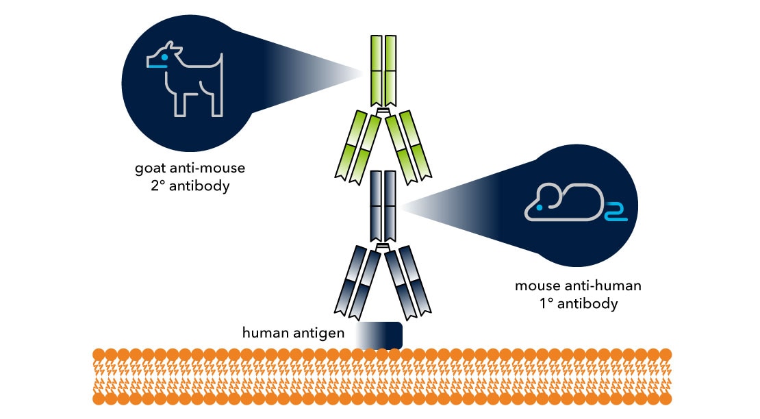
Figure 1. Generation of a Secondary Antibody
Primary and secondary antibodies are similar in structure and they share similar classes/subclasses. Structurally, an antibody is a Y-shaped molecule composed of three equal-sized regions. A flexible hinge joins the antibody stalk (Fc) region to the arms of the antibody [F(ab)]. The two arms functions to bind antigen, while the stalk region determines the antibody's isotype and functional properties. Each arm is composed of one heavy and one light chain. Furthermore, these heavy and light chains are made up of one variable domain (VL or VH) and one constant domain (CLor CH).
The variable regions (V) of the heavy and light chains differ between antibodies and confer antibody specificity for its complementary antigen. The Fc stalk region demonstrates very little variability, but it is important for antibody's interaction with effector molecules and cells (see Figure 2A). In terms of nomenclature, secondary antibodies are named according to their host, format, reactivity, target antigen, specificity, label, and purification grade (see example in Figure 2B).
Goat F(ab) anti-Mouse IgG (H+L) Secondary Antibody [HRP] (Pre-adsorbed)
In this example, here is what each term signifies:
- Goat: Host of the secondary antibody
- F(ab): Format of the secondary
- Mouse: Reactivity, the host of the primary antibody
- IgG: Target, the Ig class of the primary antibody
- H+L: Specificity, target Ig’s region being recognized
- HRP: Detection label conjugated to the antibody
- Pre-adsorbed: Purification grade of the antibody

Figure 2. Structure of an antibody and example of secondary antibody nomenclature
Variation in the Fc portion of the antibody’s heavy chain allows antibodies to be divided into five unique immunoglobulin classes: IgA, IgD, IgE, IgG, and IgM. The heavy chains of the five classes are denoted by their corresponding Greek letter: α (Alpha), δ (Delta), ε (Epsilon), γ (Gamma), and μ (Mu), respectively. Furthermore, the IgG and IgA isotypes can be divided into subclasses due to further heavy chain variation. Unlike an antibody’s heavy chains, the light chains, λ (lambda) and κ (kappa), are shared between all classes. However, it is pertinent to note that each antibody can have only one light chain or the heavy chain, but never both.
Secondary Antibody Classes and Subclasses
| Immunoglobin Classes | IgA | IgD | IgE | IgG | IgM | |
|---|---|---|---|---|---|---|
| Ig Subclasses – Human | IgG1 | IgG2 | IgG3 | IgG4 | IgA1 | |
| Ig Subclasses - Mouse | IgG1 | IgG2a | IgG2b | IgG3 | IgA2 | |
| Light Chains | Kappa (Κ) | Lamba (λ) | ||||
| Heavy Chains | IgG (gamma) | IgM (mu) | IgA (alpha) | IgD (delta) | IgE (epsilon) |
In conjunction with an antigen-specific primary antibody, a secondary antibody allows highly specific amino acid sequences within a target protein to be detected. Hence, it facilitates the detection of protein expression in a cell or tissue sample. The specificity of a secondary for a designated region [e.g. F(ab)] or regions of a primary antibody (e.g. Fc) allows multiple secondaries to bind to a single primary antibody, amplifying signal and increasing assay sensitivity.
Signal amplification arising from the ability of multiple secondaries to bind to a single primary antibody increases assay sensitivity. Their utilization also affords more flexibility in multiple labeling experiment since commercially available secondary antibodies are offered pre-conjugated to a wide range of diverse labels. Both signal amplification and increased assay flexibility are two distinct benefits to consider when designing your experiment. Learn more about: Difference between direct and indirect methods of detection.
Using a directly conjugated primary antibody instead of a standard primary/conjugated secondary set up will allow you to streamline your experiment and stain multiple targets on a single sample with little to no background interference. However, if the protein of interest is expressed at a low level in the sample being examined, it is better to use a conjugated secondary. The conjugated secondary will provide significant signal amplification because multiple secondaries can bind to a single primary. Note that a secondary antibody does not directly bind to the target protein epitope and does not confer target epitope specificity, which is determined by the primary antibody (Figure 3), but rather the labeled secondary determines the method of detection.
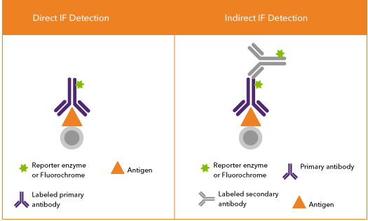
Figure 3. Direct and Indirect Detection
A secondary antibody should be directed against, but not raised in, the same species as the host of the corresponding primary antibody. As an example, a mouse primary antibody requires an anti-mouse secondary raised in any species other than mouse (e.g. rabbit-anti mouse, goat anti-mouse etc.). Whenever possible, secondary antibodies used in a multiple labeling experiment should be derived from the same host species. However, all primary antibodies should be raised in unique or different-different host species. This allows the use of species-specific secondaries directed against one primary antibody which limits cross-reactivity between the secondary and primary antibodies from other species.
To reduce background signal (non-specific staining), it is advised to block samples with serum from the same host species as the secondary antibody. Furthermore, to limit a secondary from binding a primary antibody raised in a closely related species, use a secondary pre-adsorbed against the related species. For example, an anti-mouse secondary may cross-react with a rat primary antibody in a multiple labeling experiment, but if it is pre-adsorbed against rat it may reduce cross-reactivity. (Figure 4).
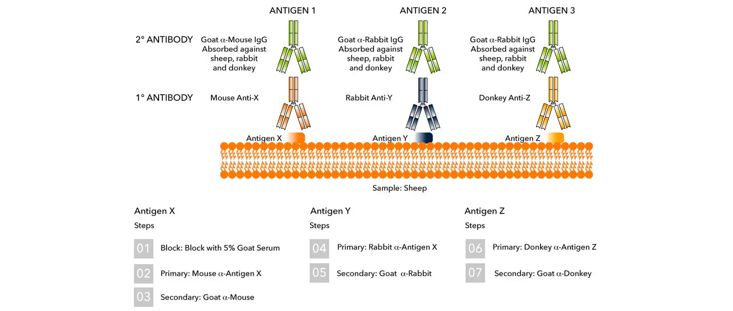
Figure 4. Ideal Combination of Antibodies for Multi-labeling Experiment
Selection criteria will vary depending on the primary antibody that you are using. The general rule is that you will want to select a secondary that matches both the host species and isotype of the primary antibody that you are using.
For example, if your primary is a mouse anti-human VEGF with isotypes IgG2a, the suggested secondary antibody can be a goat anti-mouse IgG2a conjugated to a label of your preference. For assistance on antibody selection, especially when experiments that involve multiple secondaries, you can contact Technical Support via phone, email or Live Chat.
How do I choose the right format of a secondary antibody?
The format refers to the structure of the antibody itself. Whole antibody can be cleaved by pepsin to eliminate the Fc portion of the antibody, producing a F(ab')2 fragment antibody: two arms of the antibody and the hinge region. Further cleavage of the F(ab')2 fragment with β-mercaptoethanol removes one arm of the F(ab')2 fragment, producing a F(ab') fragment: one arm of the antibody and the hinge region.
Note that a F(ab') fragment differs structurally from a F(ab) fragment: a F(ab) fragment is formed by papain cleavage of whole immunoglobulin and lacks the hinge region present in a F(ab') fragment (Figure 5). When considering a secondary, it is important not to confuse the format with the specificity. Consider this example: Goat F(ab')2 Anti-Human IgG F(ab) Secondary Antibody. In this example, the secondary consists of the F(ab')2 portion (format) and is specific for the F(ab) region of the primary antibody it's targeting (specificity).
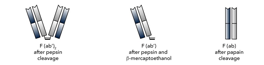
Figure 5: Antibody formats/fragments
Fc receptors expressed on the cell surface of leukocyte populations (e.g. macrophages, B lymphocytes, natural killer cells, etc.) can bind the Fc portion of the antibody which results in an increased non-specific binding (background signal). When the background signal is too high, it may result in false positives or a misinterpretation of data.
To deal with this problem, a F(ab) fragment secondary is recommended for immunostaining assays, especially when staining tissue or cells expressing high amounts of Fc receptor (e.g. lymph node, spleen, peripheral blood, etc.). The lack of the Fc portion of the antibody in F(ab) preparation eliminates secondary reagent's binding to Fc receptors expressed in the sample (see Figure 6).
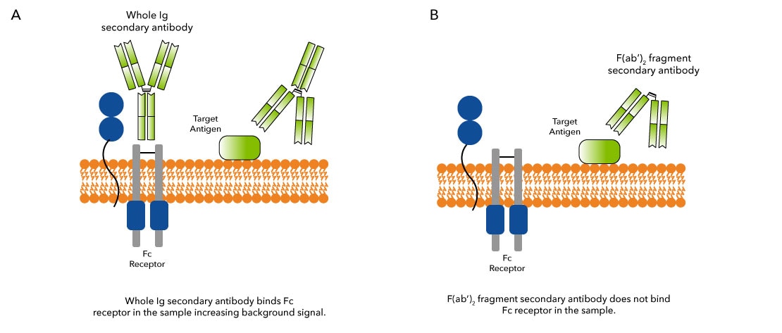
Figure 6: Demonstration of whole molecule vs antibody fragment binding in immunoassay
The choice of label is application dependent. Assays requiring fluorescent detection (e.g. flow cytometry, immunocytochemistry, immunofluorescence, etc.) require an antibody conjugated to a fluorochrome. The excitation and emission spectra of each fluorochrome should be considered when designing each experiment. Common fluorochromes include FITC, PE, APC, DyLight™ AlexaFluor™, or Atto dyes. Enzymatic detection requires a secondary conjugated to HRP, Alkaline Phosphatase (AP), or Biotin. The ability of avidin and streptavidin to bind biotin and form a complex enables signal amplification independent of the host species of the secondary. Because peroxidase is economical and more stable than AP, HRP is more popular for chemiluminescent detection systems. However, the enhanced sensitivity of AP compared to HRP makes AP more common in colorimetric detection assays. Learn more about our wide range of labels at: Conjugated Antibodies.
If you cannot find the product you’re looking for, Bio-Techne offers custom antibody services, such as custom conjugation, to help meet your experimental needs.
When using a primary monoclonal antibody, the secondary antibody should be directed against the class or subclass of the primary antibody. For example, a mouse IgM primary antibody requires an anti-mouse IgM secondary. At times, a specific subclass (e.g. human IgG2, IgG1) may be recommended. In this case, an antibody directed against the more general class (i.e. anti-human IgG) can be used for single labeling experiments, since most class-specific secondaries will recognize individual subclasses.
However, subclass specific secondaries should be used to differentiate between primary antibodies in multiple labeling experiments when more than one subclass specific primary antibody is used. For example, a multiple staining experiment using IgG2 and IgG1 primary antibodies requires anti-IgG2 and anti-IgG1 secondary antibodies.
When a polyclonal primary antibody is used, then an anti-IgG secondary is recommended since most polyclonal antibodies are IgG class immunoglobulins.
Pre-adsorption is a method to increase antibody specificity and minimize non-specific binding by passing an antibody through a column containing immobilized serum proteins or immunoglobulin from potentially cross-reactive species. Reactive antibodies bind to the immobilized proteins, while non-reactive antibodies flow through (See Figure 7).
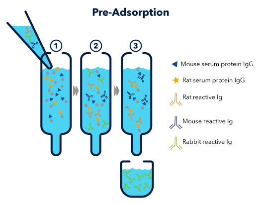
Figure 7: Pre-adsorption process illustrated
To prevent non-specific binding, it is recommended to use a pre-adsorbed secondary when determining protein expression in multiple labeling experiments or when staining immunoglobulin-rich tissues or cells. Using an antibody that is pre-adsorbed against species of the sample being examined (i.e. mouse, rat, porcine, etc.) will minimize non-specific binding of the secondary to the sample.
For example, when staining a mouse tissue, a secondary adsorbed against mouse immunoglobulins or whole serum is recommended as it will not bind with endogenous immunoglobulins in the mouse tissue section.
Due to the high homology of antibody structure between classes, it is recommended class-specific secondaries be affinity purified to minimize cross-reactivity between classes. To produce anti-rabbit IgG affinity purified secondary antibodies, anti-rabbit IgG is passed over a column containing immobilized rabbit IgG. The isolated anti-rabbit IgG antibodies are then passed through an additional column (cross adsorption) containing rabbit IgA, IgD, IgE, and IgM proteins.
This eliminates antibodies cross-reacting with non-IgG isotypes (i.e. IgA, IgD, IgE, and IgM). The resulting antigen affinity purified and cross adsorbed anti-rabbit IgG secondary should demonstrate minimal reactivity with antibody classes other than IgG.
Whole, unpurified immunoglobulin fractions contain the total complement of antibodies. Whole fractions (usually obtained by Protein A purification) can be further antigen affinity purified to remove antibody subclasses or antigen non-specific antibodies from the sample.
Antigen affinity purified secondaries ensure less non-specific binding and lower background signal relative to whole IgG fractions. However, the purification process can eliminate high affinity antibodies due to the strong affinity of some antibodies for the binding matrix. Therefore, an IgG fraction is often recommended for low abundance targets.
Yes, for optimal results, it is crucial that the secondary should match and target the class/subclass of the primary antibody being used.
This is even more important with primary monoclonal antibodies as they only belong to one subclass, thus the secondary should specifically recognize that isotype/subclass.
Yes, if possible, one should always use secondary antibodies raised in the same species in multiplexing experiments. Generating accurate data in double or multiple labeling experiments requires each secondary antibody to specifically recognize one primary antibody.
In addition to cross-reacting with primary antibodies, a secondary can bind to the other secondary antibodies or endogenous immunoglobulin expressed in the sample under investigation. Limiting these reactions is key for to producing accurate data, especially in immunocytochemistry/immunofluorescence (ICC/IF) and immunohistochemistry (IHC) applications involving target identification through tissue and cell imaging.
Fragment specific secondaries can be used when the primary antibody you are trying to detect is comprised of only that specific fragment or when you are trying to only detect that specific fragment from a population of multiple immunoglobulin molecules.
It can also be helpful when an antibody under consideration has an Fc or F(ab) region exposed, due to the specific binding of that primary.
F(ab) specific secondary antibodies may help to reduce background signal by eliminating nonspecific binding of the IgG proteins found in tissues. You will want to make sure that the primary antibody you will be detecting will have an exposed F(ab) region for detection by the secondary.
If not, you may want to consider using a Mouse-on-Mouse blocking reagent or an unconjugated anti-mouse IgG secondary prior to primary incubation as alternative methods for reduction of background signal.
I am using a polymer based detection kit instead of a standard secondary antibody. I am getting some high background. Do you have a recommendation to help improve my staining?
A serum blocking step may not be required when using a polymer based secondary but if you are getting background issues, blocking with 5% goat serum may be employed. We would still recommend performing a peroxidase blocking step as well if the polymer is utilizing a peroxidase detection method.
Would conjugating a primary antibody with biotin affect its affinity with a secondary antibody?
If you are using a low molar access/concentration of biotin for your conjugation reaction, there should not be an impact on the affinity. Also, a majority of the secondaries are polyclonal in nature and can bind more than one epitope on primary antibodies.
In Western blot, can I use BSA-TBST as a blocking and antibody diluent for phospho-antibodies and then 5% milk for diluting and incubating secondary antibodies?
Obtaining a clean western blot for phospho-specific antibodies with milk-based blocking/antibody dilution buffer is a challenge that most researchers experience in their experiments. Phospho-antibody's compatibility with milk-based diluent depend on how strongly the phospho-antibody binds to a protein of interest or if it could be sequestered by phospho-proteins from milk.
Bovine Serum Albumin (BSA) based blocking or dilution buffer is a recommended choice over 5% milk for most of the targets when performing a western blot.
When used at a higher concentration/low dilution, some antibodies exhibit a tendency to form aggregates which results in a speckled signal in immunostaining assays. Aggregates can be removed from the antibody stock by centrifuging the antibody vial at high speed for 5-10 minutes followed by careful pipetting out of the supernatant/antibody solution without touching the bottom of the vial.
Another source of speckled staining is the use of slides and/or coverslips which are not clean. If particulate matter is suspected on the slides or clover slips, double check it by observing them under a microscope. Slides can be cleaned with the use of absolute alcohol and paper wipes or a piece of cotton.
What dilution should I use for my application?
This will vary depending on the product. You can find the recommended dilution range under "Applications/Dilutions" section of the product datasheet (see example below).
Applications/Dilutions
| Dilutions | Western Blot 1:5000 - 1:50000 ELISA 1:1000 - 1:1000000 Immunocytochemistry/Immunofluorescence 1:50 - 1:500 Immunochemistry Immunochemistry-Paraffin 1:40 - 1:400 |
| Readout System | NovaLume-Plus Chemiluminescent Reagent |
| Reviewed Applications | Read 1 rated 5 star using NB7539 in the following applications: Western blot 1 |
| Publications | Read publications using NB7539 in the following applications: WB 1 publication |
The shelf life of prediluted antibodies can vary greatly depending on the number of uses, storage temperature, age of storage buffers, and other handling factors. We recommend keeping the bulk of your secondary in its original vial, only removing enough material for individual use.
If you do decide to store prediluted secondary, you may need to conduct stability tests of the antibody with your primaries to establish a working timeline for your needs. While we offer 100% guarantee on our products when stored properly, storing an antibody in antibody diluent is not what we recommend.
"Secondary Only Control" in immunostaining assays (e.g. ICC/IF, IHC, Flow etc.) refer to a sample which is incubated with PBS or an antibody diluent instead of a primary antibody, everything else in the protocol being the same as for test samples. This control helps to determine the specificity of the staining/signal observed in a given assay. No staining in the secondary only control will reflect that the staining is specific and is being generated from the primary antibody binding to the antigen of interest.
A considerable staining in secondary only control will signify that the staining is non-specific and is originating from secondary binding to endogenous immunoglobulins in the tissue or to some other non-target antigens.
Our sister brand, Novus Biologicals, has several secondary antibodies compatible with the Odyssey Imaging System (from LI-COR). Find them here.
Yes. The antibody product can be stored at -80 ºC instead of -70 ºC. The reason we state -70 ºC as the storage temperature is to accomodate the fact that many -80 ºC freezers do not maintain that temperature uniformly at all times.
The following protocol can be used to thaw antibodies supplied as a Liquid/Frozen formulation: For Volumes < 2 mL, the product should be thawed on benchtop at room temperature.For volumes > 2 mL: Place product into a 37 ºC bead bath, inverting at 10-minute intervals until completely thawed.
For the standard sensitivity substrate, we use 1:1000-1:2000 dilution of the HRP-conjugated secondary antibody. Higher sensitivity substrates might require higher dilutions. 1:2000-1:5000 dilution is a good place to start.
In a direct ELISA, a plate is coated with the analyte of interest and a labeled detection antibody is used to verify the presence of the analyte. The direct ELISA may use a colorimetric, chemiluminescent, or fluorescent reporter.
Environmental stewardship is important to R&D Systems and its employees. R&D Systems is ISO 14001:2015 Certified and as part of this we are continuously reviewing our sustainability practices and "green" options. First and foremost, the energy expended to ship back the styrofoam box is more detrimental to the environment than having the facility re-use or recycle it. Our stance is to encourage our customers to implement a recycling program locally. At R&D Systems we have chosen to reduce the use of styrofoam as much as possible by: doing extensive stability testing in order to determine which products can be shipped with minimal packaging at ambient temperatures, converting the use of non-recyclable packaging materials to recyclable plastics, cardboard, or biodegradable materials, and continuing to investigate alternatives to dry ice shipments, the use of re-usable containers, and gel packs that allow for smaller styrofoam containers. Employees were key to initiating a recycling program at our facilities. Internally, we recycle paper, plastic, cardboard, aluminum, and glass.
R&D Systems has established a policy of not limiting the useful life of a product by providing an expiration date or manufacture date for our protein and antibody products. Under proper storage conditions, proteins and antibodies tend to be stable for many years. These conditions include storing proteins as lyophilized powder, storing the product frozen (-20° C or -80° C) at protein concentrations of greater than 0.1 mg/mL, and limiting the number of freeze/thaw cycles. Please see individual product datasheets for specific instructions. Routine quality control testing by our company ensures that all products have acceptable biological activities at the time of sale. R&D Systems cannot control storage conditions of a product upon receipt by the end user. In lieu of an expiration date, we choose to offer a warranty on our protein and antibody products. All products supplied by R&D Systems are warranted to meet or exceed our published specification when used under normal conditions in your laboratory. Typically, this warranty will extend 6-12 months from time of purchase. Please see individual datasheets for specific stability claims. If the product fails during the stated period, a replacement product or credit will be issued. Click here for details regarding our warranty.
What is trehalose and why is it in the formulation?
- Trehalose is a non-reducing sugar and does not react with amino acids or proteins as part of the Maillard reaction. It is added to stabilize antibodies and other proteins against damage caused by freezing. It can also make proteins more resistant to moisture when lyophilized, resulting in a product that is less likely to precipitate when reconstituted.
Will trehalose affect the performance of the protein or antibody in my specific application?
- We generally have not seen adverse effects in our bioassays or other approved applications. However, customers are advised to run a control in their assay to determine if the concentration of trehalose in the protein or antibody formulation has any adverse effects.
Will trehalose affect my conjugation reaction?
- Trehalose is not usually expected to interfere with conjugation; we often successfully conjugate antibodies which are bottled with Trehalose using primary amine to NHS ester coupling chemistry. However, for optimal conjugation performance, it is best practice to perform buffer change to remove trehalose using desalting column to a compatible conjugation buffer prior to coupling reaction.
Will trehalose included in the formulation affect the animal if it is injected?
- Trehalose is unlikely to have an effect in vivo. It has been approved as an excipient for use in human injectable drugs.
How are your antibodies biotinylated?
- All R&D Systems' biotinylated antibodies are biotinylated via primary amines.
Do you map the locations of biotinylated residues on your Biotin Antibodies?
- No, we do not map the biotinylated residues.
There has been no evidence of ligands leaching off the affinity columns used. The ligand is covalently bound to the resin and remains on the resin when the antibody is eluted.
An isotype control is an antibody that is the same IgG isotype as the primary antibody, but raised against an irrelevant antigen. An isotype control should produce negative staining results, thereby giving you confidence in the staining observed with the target-specific antibody. The Supplemental Products tab on the product page indicates appropriate isotype controls for that product. View an example here.
For IgG antibodies, we estimate that the molecular weight is ~ 150 kDa.