Key Product Details
Applications
Product Summary for DRAQ9 (TM)
Products are shipped at ambient temperature, but on receipt packs should be stored at -20C. Thaw and adjust temperature to above 20C before use.
Product Specifications
Applications
Live Imaging Microscopy (1:500)
Fluorescence Imaging (1:50 - 1:500)
Application Notes
Spectra Viewer
Plan Your Experiments
Use our spectra viewer to interactively plan your experiments, assessing multiplexing options. View the excitation and emission spectra for our fluorescent dye range and other commonly used dyes.
Spectra Viewer
Scientific Data Images for DRAQ9 (TM)
![Live Imaging Microscopy: DRAQ9 (TM) [NBP2-81128]](https://resources.rndsystems.com/images/products/DRAQ9-TM-Live-Imaging-Microscopy-NBP2-81128-img0005.jpg "Live Imaging Microscopy: DRAQ9 (TM) [NBP2-81128]")
Live Imaging Microscopy: DRAQ9 (TM) [NBP2-81128]
Live Imaging Microscopy: DRAQ9 (TM) [NBP2-81128] - DRAQ9 was diluted in EMEM culture media and applied to live HeLa cells at 2 uM (1:500) for 30 minutes at room temperature and protected from light. Imaging was done immediately after staining without washing the cells.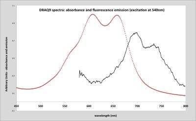
![Live Imaging Microscopy: DRAQ9 (TM) [NBP2-81128]](https://resources.rndsystems.com/images/products/DRAQ9-TM-Immunocytochemistry-Immunofluorescence-NBP2-81128-img0003.jpg "Live Imaging Microscopy: DRAQ9 (TM) [NBP2-81128]")
Live Imaging Microscopy: DRAQ9 (TM) [NBP2-81128]
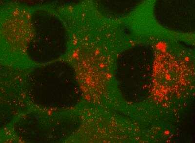
Live Imaging Microscopy: DRAQ9 (TM) [NBP2-81128] - labelling of live U2OS cells for 48 h at 2 uM - showing a number of cells in mitosis.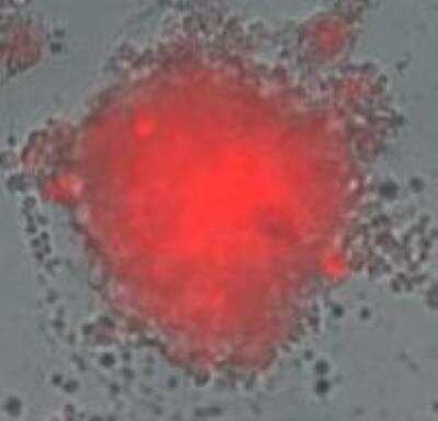
Formulation, Preparation, and Storage
Purification
Concentration
Shipping
Storage
Background: DRAQ9 (TM)
Additional DRAQ9 (TM) Products
Product Documents for DRAQ9 (TM)
Certificate of Analysis
To download a Certificate of Analysis, please enter a lot or batch number in the search box below.
Product Specific Notices for DRAQ9 (TM)
DRAQ9 (TM) is a registered trademark of BioStatus Limited.
This product is for research use only and is not approved for use in humans or in clinical diagnosis. Support products are guaranteed for 6 months from date of receipt.
Citations for DRAQ9 (TM)
 Powered by Bioz
Customer Reviews for DRAQ9 (TM)
There are currently no reviews for this product. Be the first to review DRAQ9 (TM) and earn rewards!
Have you used DRAQ9 (TM)?
Submit a review and receive an Amazon gift card!
$25/€18/£15/$25CAN/¥2500 Yen for a review with an image
$10/€7/£6/$10CAN/¥1110 Yen for a review without an image
Submit a review
Protocols
Find general support by application which include: protocols, troubleshooting, illustrated assays, videos and webinars.
- Appropriate Fixation of IHC/ICC Samples
- Cellular Response to Hypoxia Protocols
- ClariTSA™ Fluorophore Kits
- Detection & Visualization of Antibody Binding
- ICC Cell Smear Protocol for Suspension Cells
- ICC Immunocytochemistry Protocol Videos
- ICC for Adherent Cells
- Immunocytochemistry (ICC) Protocol
- Immunocytochemistry Troubleshooting
- Immunofluorescence of Organoids Embedded in Cultrex Basement Membrane Extract
- Immunohistochemistry (IHC) and Immunocytochemistry (ICC) Protocols
- Preparing Samples for IHC/ICC Experiments
- Preventing Non-Specific Staining (Non-Specific Binding)
- Primary Antibody Selection & Optimization
- Protocol for VisUCyte™ HRP Polymer Detection Reagent
- Protocol for the Fluorescent ICC Staining of Cell Smears - Graphic
- Protocol for the Fluorescent ICC Staining of Cultured Cells on Coverslips - Graphic
- Protocol for the Preparation and Fluorescent ICC Staining of Cells on Coverslips
- Protocol for the Preparation and Fluorescent ICC Staining of Non-adherent Cells
- Protocol for the Preparation and Fluorescent ICC Staining of Stem Cells on Coverslips
- Protocol for the Preparation of a Cell Smear for Non-adherent Cell ICC - Graphic
- TUNEL and Active Caspase-3 Detection by IHC/ICC Protocol
- The Importance of IHC/ICC Controls
- View all Protocols, Troubleshooting, Illustrated assays and Webinars