
FKBP51/FKBP5 Antibody (Hi51B) - BSA Free
Novus Biologicals | Catalog # NB110-96873

![Western Blot: FKBP51/FKBP5 Antibody (Hi51B) [NB110-96873]](https://resources.rndsystems.com/images/products/FKBP51-FKBP5-Antibody-Hi51B-Western-Blot-NB110-96873-img0007.jpg "Western Blot: FKBP51/FKBP5 Antibody (Hi51B) [NB110-96873]")
Scientific images may be resized, cropped, or adjusted for brightness or contrast for presentation purposes. These updates are limited to presentation and do not affect the underlying scientific interpretation of the data. Where available, additional source data may be provided upon request.
Key Product Details
Species Reactivity
Validated:
Cited:
Applications
Validated:
Cited:
Label
Antibody Source
Format
Product Specifications
Immunogen
Localization
Specificity
Clonality
Host
Isotype
Scientific Data Images for FKBP51/FKBP5 Antibody (Hi51B) - BSA Free
Western Blot: FKBP51/FKBP5 Antibody (Hi51B) [NB110-96873]
FKBP51-FKBP5-Antibody-Hi51B-Western-Blot-NB110-96873-img0007.jpg![Immunocytochemistry/ Immunofluorescence: FKBP51/FKBP5 Antibody (Hi51B) [NB110-96873]](https://resources.rndsystems.com/images/products/FKBP51-FKBP5-Antibody-Hi51B-Immunocytochemistry-Immunofluorescence-NB110-96873-img0006.jpg "Immunocytochemistry/ Immunofluorescence: FKBP51/FKBP5 Antibody (Hi51B) [NB110-96873]")
Immunocytochemistry/ Immunofluorescence: FKBP51/FKBP5 Antibody (Hi51B) [NB110-96873]
Immunocytochemistry/Immunofluorescence: FKBP51/FKBP5 Antibody (Hi51B) [NB110-96873] - Immunocytochemistry/Immunofluorescence analysis using Mouse Anti-FKBP51/FKBP5 Monoclonal Antibody, Clone Hi51B (NB110-96873). Tissue: MK cells. Species: Mouse. Primary Antibody: Mouse Anti-FKBP51/FKBP5 Monoclonal Antibody (NB110-96873) at 1:1000. Secondary Antibody: APC Goat Anti-Mouse (red). Counterstain: DAPI (blue) nuclear stain. Cells stained red. Courtesy of: the Hospital Henri Mondor, France.![Western Blot: FKBP51/FKBP5 Antibody (Hi51B) [NB110-96873]](https://resources.rndsystems.com/images/products/FKBP51-FKBP5-Antibody-Hi51B-Western-Blot-NB110-96873-img0005.jpg "Western Blot: FKBP51/FKBP5 Antibody (Hi51B) [NB110-96873]")
Western Blot: FKBP51/FKBP5 Antibody (Hi51B) [NB110-96873]
Western Blot: FKBP51/FKBP5 Antibody (Hi51B) [NB110-96873] - analysis of Human HeLa cell lysates showing detection of FKBP51 protein using Mouse Anti-FKBP51 Monoclonal Antibody, Clone Hi51B. Load: 15 ug protein. Block: 1.5% BSA for 30 minutes at RT. Primary Antibody: Mouse Anti-FKBP51 Monoclonal Antibody at 1:1000 for 2 hours at RT. Secondary Antibody: Sheep Anti-Mouse IgG: HRP for 1 hour at RT.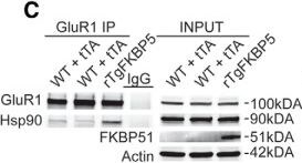
Western Blot: FKBP51/FKBP5 Antibody (Hi51B) [NB110-96873] -
Western Blot: FKBP51/FKBP5 Antibody (Hi51B) [NB110-96873] - FKBP51/Hsp90 bind to GluR1-type AMPA receptors to regulate trafficking. A, Representative Western blottings form biotinylation assays of receptor endocytosis was performed on ex vivo slices, as described in Materials & Methods, rTgFKBP5 (N = 4; n = 8), WT (N = 2; n = 8), & tTA (N = 2; n = 8). Following labeling with Sulfo-NHS-SS biotin & chemical LTD (20 µM NMDA; 5 min) treatment, receptors were permitted to externalize at 30°C for the indicated times. B, The quantification ± SEM of multiple acquisitions is shown for GluR1. C, Representative Western blottings from anti-GluR1 co-immunoprecipitations & corresponding inputs from control & rTgFKBP5 mice immunoblotted as indicated. rTgFKBP5 (N = 2), WT (N = 2), & tTA (N = 2) total from two independent experiments. D, Representative Western blottings of anti-GluR1 co-immunoprecipitations & corresponding inputs from HEK293T cells transfected with GluR1 & FKBP51 or empty vector (EV) for 48 h were immunoblotted with antibodies as indicated. Just before harvest, cells were treated with 100 µM AMPA or PBS for 10 min to induce GluR1 receptor internalization. *p = 0.0286 by t-test of this time point. **p < 0.001 by two-way ANOVA. Image collected & cropped by CiteAb from the following publication (https://pubmed.ncbi.nlm.nih.gov/30963102), licensed under a CC-BY license. Not internally tested by Novus Biologicals.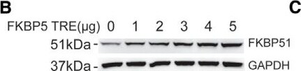
Western Blot: FKBP51/FKBP5 Antibody (Hi51B) [NB110-96873] -
Western Blot: FKBP51/FKBP5 Antibody (Hi51B) [NB110-96873] - Detailed schematic & validation of the FKBP5 TRE transgene. A, To allow for site-directed, single copy insertion into the mouse genome in chromosome 11, the transgenic construct contained flanking attB sites via a PhiC31 integrase. The downstream Mp1 poly A tail will help maintain stable expression. To drive high expression, the transgenic construct included a tetracycline-response element (TRE) promoter made of seven repeats of the tetracycline operators used to drive high expression of the singly inserted FKBP5 gene in the presence of the tTA, & a weak minimal CMV promoter which produces low basal expression. B, Western blotting from HEK293T cells transfected with increasing amounts of FKBP5 TRE plasmid, as indicated, for 48 h. C, HEK293T cells were transfected with the indicated amounts of FKBP5 TRE & tTA plasmid, to ensure the tTA would drive high FKBP51 expression. Image collected & cropped by CiteAb from the following publication (https://pubmed.ncbi.nlm.nih.gov/30963102), licensed under a CC-BY license. Not internally tested by Novus Biologicals.
Western Blot: FKBP51/FKBP5 Antibody (Hi51B) [NB110-96873] -
Western Blot: FKBP51/FKBP5 Antibody (Hi51B) [NB110-96873] - FKBP51 expression & distribution in rTgFKBP5 mice. A, Expression of human & mouse FKBP5 in rTgFKBP5 mice expressed as fold change ± SEM compared to WT mice using qPCR; ***p < 0.001 by t test (N = 10) with three technical replicates. B, Western blotting showing FKBP51 levels in the hippocampus from rTgFKBP5, WT, FKBP5, & tTA mice. C, Western blotting showing levels of FKBP51 levels in the rTgFKBP5 hippocampus from 1 to 10 µg of protein loaded compared to 50 µg of protein from WT or FKBP51 mice. GAPDH levels are shown to confirm protein load. See Extended Data Figure 2-1 for more information on the antibody. D, 20× images of anti-FKBP51 staining from rTgFKBP5 mice. The entorhinal cortex (ECX), anterior cortex (ACX), CA1, CA3, & dentate gyrus (DG) are labeled. E, 20× images of anti-FKBP51 staining from rTgFKBP5 mice in the CA1, CA3, DG, ECX, & ACX. Scale bar = 100 µm; 10 µm (inset). F, Western blotting showing FKBP51 levels in the hippocampus, striatum, ACX, posterior cortex (PCX), interbrain, thalamus & hypothalamus, & cerebellum of a rTgFKBP5 mouse. G, Quantitation of FKBP51 proteins levels throughout the hippocampus (HPC), striatum (STR), ACX, PCX, interbrain, thalamus & hypothalamus (INTER), & cerebellum (CER), of rTgFKBP5 mice from multiple exposures. Image collected & cropped by CiteAb from the following publication (https://pubmed.ncbi.nlm.nih.gov/30963102), licensed under a CC-BY license. Not internally tested by Novus Biologicals.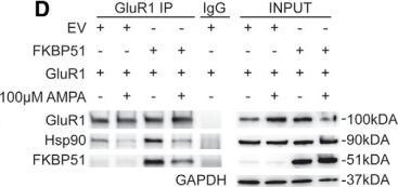
Western Blot: FKBP51/FKBP5 Antibody (Hi51B) [NB110-96873] -
Western Blot: FKBP51/FKBP5 Antibody (Hi51B) [NB110-96873] - FKBP51/Hsp90 bind to GluR1-type AMPA receptors to regulate trafficking. A, Representative Western blottings form biotinylation assays of receptor endocytosis was performed on ex vivo slices, as described in Materials & Methods, rTgFKBP5 (N = 4; n = 8), WT (N = 2; n = 8), & tTA (N = 2; n = 8). Following labeling with Sulfo-NHS-SS biotin & chemical LTD (20 µM NMDA; 5 min) treatment, receptors were permitted to externalize at 30°C for the indicated times. B, The quantification ± SEM of multiple acquisitions is shown for GluR1. C, Representative Western blottings from anti-GluR1 co-immunoprecipitations & corresponding inputs from control & rTgFKBP5 mice immunoblotted as indicated. rTgFKBP5 (N = 2), WT (N = 2), & tTA (N = 2) total from two independent experiments. D, Representative Western blottings of anti-GluR1 co-immunoprecipitations & corresponding inputs from HEK293T cells transfected with GluR1 & FKBP51 or empty vector (EV) for 48 h were immunoblotted with antibodies as indicated. Just before harvest, cells were treated with 100 µM AMPA or PBS for 10 min to induce GluR1 receptor internalization. *p = 0.0286 by t-test of this time point. **p < 0.001 by two-way ANOVA. Image collected & cropped by CiteAb from the following publication (https://pubmed.ncbi.nlm.nih.gov/30963102), licensed under a CC-BY license. Not internally tested by Novus Biologicals.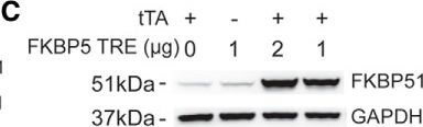
Western Blot: FKBP51/FKBP5 Antibody (Hi51B) [NB110-96873] -
Western Blot: FKBP51/FKBP5 Antibody (Hi51B) [NB110-96873] - Detailed schematic & validation of the FKBP5 TRE transgene. A, To allow for site-directed, single copy insertion into the mouse genome in chromosome 11, the transgenic construct contained flanking attB sites via a PhiC31 integrase. The downstream Mp1 poly A tail will help maintain stable expression. To drive high expression, the transgenic construct included a tetracycline-response element (TRE) promoter made of seven repeats of the tetracycline operators used to drive high expression of the singly inserted FKBP5 gene in the presence of the tTA, & a weak minimal CMV promoter which produces low basal expression. B, Western blotting from HEK293T cells transfected with increasing amounts of FKBP5 TRE plasmid, as indicated, for 48 h. C, HEK293T cells were transfected with the indicated amounts of FKBP5 TRE & tTA plasmid, to ensure the tTA would drive high FKBP51 expression. Image collected & cropped by CiteAb from the following publication (https://pubmed.ncbi.nlm.nih.gov/30963102), licensed under a CC-BY license. Not internally tested by Novus Biologicals.Applications for FKBP51/FKBP5 Antibody (Hi51B) - BSA Free
Immunocytochemistry/ Immunofluorescence
Western Blot
Formulation, Preparation, and Storage
Purification
Formulation
Format
Preservative
Concentration
Shipping
Stability & Storage
Background: FKBP51
Long Name
Alternate Names
Gene Symbol
UniProt
Additional FKBP51 Products
Product Documents for FKBP51/FKBP5 Antibody (Hi51B) - BSA Free
Certificate of Analysis
To download a Certificate of Analysis, please enter a lot or batch number in the search box below.
Product Specific Notices for FKBP51/FKBP5 Antibody (Hi51B) - BSA Free
This product is for research use only and is not approved for use in humans or in clinical diagnosis. Primary Antibodies are guaranteed for 1 year from date of receipt.
Related Research Areas
Citations for FKBP51/FKBP5 Antibody (Hi51B) - BSA Free
 Powered by Bioz
Customer Reviews for FKBP51/FKBP5 Antibody (Hi51B) - BSA Free
There are currently no reviews for this product. Be the first to review FKBP51/FKBP5 Antibody (Hi51B) - BSA Free and earn rewards!
Have you used FKBP51/FKBP5 Antibody (Hi51B) - BSA Free?
Submit a review and receive an Amazon gift card!
$25/€18/£15/$25CAN/¥2500 for a review with an image
$10/€7/£6/$10CAN/¥1110 for a review without an image
Submit a review
Protocols
Find general support by application which include: protocols, troubleshooting, illustrated assays, videos and webinars.
- Appropriate Fixation of IHC/ICC Samples
- Cellular Response to Hypoxia Protocols
- ClariTSA™ Fluorophore Kits
- Detection & Visualization of Antibody Binding
- ICC Cell Smear Protocol for Suspension Cells
- ICC Immunocytochemistry Protocol Videos
- ICC for Adherent Cells
- Immunocytochemistry (ICC) Protocol
- Immunocytochemistry Troubleshooting
- Immunofluorescence of Organoids Embedded in Cultrex Basement Membrane Extract
- Immunohistochemistry (IHC) and Immunocytochemistry (ICC) Protocols
- Preparing Samples for IHC/ICC Experiments
- Preventing Non-Specific Staining (Non-Specific Binding)
- Primary Antibody Selection & Optimization
- Protocol for VisUCyte™ HRP Polymer Detection Reagent
- Protocol for the Fluorescent ICC Staining of Cell Smears - Graphic
- Protocol for the Fluorescent ICC Staining of Cultured Cells on Coverslips - Graphic
- Protocol for the Preparation and Fluorescent ICC Staining of Cells on Coverslips
- Protocol for the Preparation and Fluorescent ICC Staining of Non-adherent Cells
- Protocol for the Preparation and Fluorescent ICC Staining of Stem Cells on Coverslips
- Protocol for the Preparation of a Cell Smear for Non-adherent Cell ICC - Graphic
- R&D Systems Quality Control Western Blot Protocol
- TUNEL and Active Caspase-3 Detection by IHC/ICC Protocol
- The Importance of IHC/ICC Controls
- Troubleshooting Guide: Western Blot Figures
- Western Blot Conditions
- Western Blot Protocol
- Western Blot Protocol for Cell Lysates
- Western Blot Troubleshooting
- Western Blot Troubleshooting Guide
- View all Protocols, Troubleshooting, Illustrated assays and Webinars