
Host Cell Factor 1/HCFC1 Antibody
Novus Biologicals | Catalog # NB100-68209

![Western Blot: Host Cell Factor 1/HCFC1 Antibody [NB100-68209]](https://resources.rndsystems.com/images/products/Host-Cell-Factor-1-HCFC1-Antibody-Western-Blot-NB100-68209-img0010.jpg "Western Blot: Host Cell Factor 1/HCFC1 Antibody [NB100-68209]")
Scientific images may be resized, cropped, or adjusted for brightness or contrast for presentation purposes. These updates are limited to presentation and do not affect the underlying scientific interpretation of the data. Where available, additional source data may be provided upon request.
Key Product Details
Validated by
Species Reactivity
Validated:
Cited:
Applications
Validated:
Cited:
Label
Antibody Source
Product Specifications
Immunogen
Clonality
Host
Isotype
Scientific Data Images for Host Cell Factor 1/HCFC1 Antibody
Western Blot: Host Cell Factor 1/HCFC1 Antibody [NB100-68209]
Western Blot: Host Cell Factor 1/HCFC1 Antibody [NB100-68209] - Samples: Whole cell lysate (5, 15 and 50 ug for WB; 1 mg for IP, 20% of IP loaded) from HeLa cells. Antibodies: Affinity purified rabbit anti-HCF1 antibody NB100-68209 used for WB at 0.04 ug/ml (A) and 1 ug/ml (B) and used for IP at 3 ug/mg lysate. HCF1 was also immunoprecipitated by rabbit anti-HCF1 antibody NB100-68210, which recognizes a downstream epitope. Detection: Chemiluminescence with exposure times of 30 seconds (A) and 10 seconds (B).![Knockdown Validated: Host Cell Factor 1/HCFC1 Antibody [NB100-68209]](https://resources.rndsystems.com/images/products/Host-Cell-Factor-1-HCFC1-Antibody-Knockdown-Validated-NB100-68209-img0011.jpg "Western Blot: Host Cell Factor 1/HCFC1 Antibody [NB100-68209]")
![Immunoprecipitation: Host Cell Factor 1/HCFC1 Antibody [NB100-68209]](https://resources.rndsystems.com/images/products/Host-Cell-Factor-1-HCFC1-Antibody-Immunoprecipitation-NB100-68209-img0012.jpg "Immunoprecipitation: Host Cell Factor 1/HCFC1 Antibody [NB100-68209]")
Immunoprecipitation: Host Cell Factor 1/HCFC1 Antibody [NB100-68209]
Host-Cell-Factor-1-HCFC1-Antibody-Immunoprecipitation-NB100-68209-img0012.jpg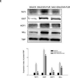
Western Blot: Host Cell Factor 1/HCFC1 Antibody [NB100-68209] -
Western Blot: Host Cell Factor 1/HCFC1 Antibody [NB100-68209] - 5-FU-produced ROS involved in histone methyltransferase-mediated Nrf2 expression. E. Expression patterns of TET1, OGT, HCF1, & MLL in NAC pre-treated SNUC5/5-FUR cells determined by WB analysis. *Significantly different from SNUC5 cells (p<0.05), & #significantly different from SNUC5/5-FUR cells (p<0.05). Also, SNUC5 cells pre-treated with 500 μM of NAC & incubated for an additional 1 h at 37°. Cells then treated with 140 μM 5-FU for 48 h. 10 μM of DHR123 was added to plate & incubated for an additional 30 min at 37°C. Image collected & cropped by CiteAb from the following publication (https://www.oncotarget.com/lookup/doi/10.18632/oncotarget.9745), licensed under a CC-BY license. Not internally tested by Novus Biologicals.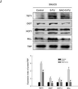
Western Blot: Host Cell Factor 1/HCFC1 Antibody [NB100-68209] -
Western Blot: Host Cell Factor 1/HCFC1 Antibody [NB100-68209] - 5-FU-produced ROS involved in histone methyltransferase-mediated Nrf2 expression. J. Expression of TET1 & interaction proteins detected by WB analysis. *Significantly different from SNUC5 cells (p<0.05), & #significantly different from 5-FU-treated SNUC5 cells (p<0.05). Image collected & cropped by CiteAb from the following publication (https://www.oncotarget.com/lookup/doi/10.18632/oncotarget.9745), licensed under a CC-BY license. Not internally tested by Novus Biologicals.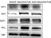
Western Blot: Host Cell Factor 1/HCFC1 Antibody [NB100-68209] -
Western Blot: Host Cell Factor 1/HCFC1 Antibody [NB100-68209] - 5-FU-produced ROS involved in histone methyltransferase-mediated Nrf2 expression. F. The intracellular ROS level was detected by flow cytometry. Image collected & cropped by CiteAb from the following publication (https://www.oncotarget.com/lookup/doi/10.18632/oncotarget.9745), licensed under a CC-BY license. Not internally tested by Novus Biologicals.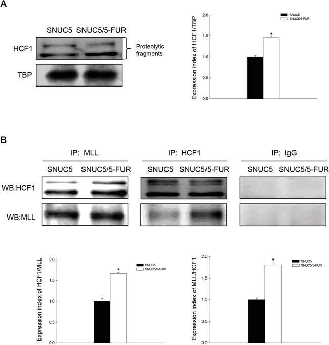
Western Blot: Host Cell Factor 1/HCFC1 Antibody [NB100-68209] -
Western Blot: Host Cell Factor 1/HCFC1 Antibody [NB100-68209] - Effect of MLL-HCF1 interaction on histone methylation & Nrf2 expression in SNUC5/5-FUR cellsA. HCF1 expression was determined by Western blotting. *Significantly different from SNUC5 cells (p<0.05). B. Interaction between MLL & HCF1was examined by immunoprecipitation analysis using anti-MLL & anti-HCF1 antibodies followed by Western blotting with anti-HCF1 & anti-MLL antibodies. *Significantly different from SNUC5 cells (p<0.05). C. Interaction between MLL & HCF1was assessed by PLA. Each green spot represents for a single interaction (MLL & HCF1) & DNA was stained with DAPI. *Significantly different from SNUC5 cells (p<0.05). The cells were transfected with non-targeting siRNA (siControl) or siHCF1 RNA for 24 h. D. Expression pattern of MLL mRNA in SNUC5/5-FUR cells was determined by RT-PCR analysis. *Significantly different from siControl-transfected cells (p<0.05). E. Expression pattern of HCF1, MLL, & Nrf2 in SNUC5/5-FUR cells was determined by Western blot analysis. *Significantly different from siControl-transfected cells (p<0.05). Image collected & cropped by CiteAb from the following publication (https://www.oncotarget.com/lookup/doi/10.18632/oncotarget.9745), licensed under a CC-BY license. Not internally tested by Novus Biologicals.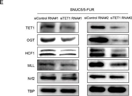
Western Blot: Host Cell Factor 1/HCFC1 Antibody [NB100-68209] -
Western Blot: Host Cell Factor 1/HCFC1 Antibody [NB100-68209] - Relation of OGT-TET1 interaction on histone methylation & Nrf2 expression in SNUC5/5-FUR cellsA. Interaction between OGT & TET1 was examined by immunoprecipitation analyses using anti-OGT & anti-TET1 antibodies followed by Western blotting with anti-TET1 & anti-OGT antibodies. B. Interaction between OGT & TET1 was assessed by using PLA. Each green spot represents for a single interaction (OGT & TET1) & DNA was stained with DAPI. *Significantly different from SNUC5 cells (p<0.05). C. The O-GlcNAcylated TET1 or O-GlcNAcylated OGT was examined by immunoprecipitation analyses using anti-O-GlcNAc antibody followed by Western blotting with anti-TET1 & anti-OGT antibodies. The cells were transfected with non-targeting siRNA (siControl) or siOGT RNA or siTET1 RNA for 24 h. D. Expression pattern of OGT & TET1 in SNUC5/5-FUR cells was determined by Western blot analysis. *Significantly different from siControl-transfected cells (p<0.05). E. Expression pattern of TET1, OGT, HCF1, MLL, & Nrf2 in SNUC5/5-FUR cells was determined by Western blot analysis. *Significantly different from siControl-transfected cells (p<0.05). Image collected & cropped by CiteAb from the following publication (https://www.oncotarget.com/lookup/doi/10.18632/oncotarget.9745), licensed under a CC-BY license. Not internally tested by Novus Biologicals.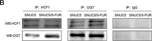
Western Blot: Host Cell Factor 1/HCFC1 Antibody [NB100-68209] -
Western Blot: Host Cell Factor 1/HCFC1 Antibody [NB100-68209] - Involvement of OGT in histone methylase MLL/COMPASS-like complex-mediated Nrf2 expression in SNUC5/5-FUR cellsA. OGT & O-GlcNAc expression was determined by Western blotting. *Significantly different from SNUC5 cells (p<0.05). B. Interaction between HCF1 & OGT was examined by immunoprecipitation analysis using anti-HCF1 & anti-OGT antibodies followed by Western blotting with anti-HCF1 & anti-OGT antibodies. C. Interaction between HCF1 & OGT was assessed by using PLA. Each green spot represents for a single interaction (HCF1 & OGT) & DNA was stained with DAPI. *Significantly different from SNUC5 cells (p<0.05). D. Interaction between MLL & OGT was examined by immunoprecipitation analysis using anti-MLL & anti-OGT antibodies followed by Western blotting with anti-OGT & anti-MLL antibodies. E. Interaction between MLL & OGT was assessed by using PLA. Each green spot represents for a single interaction (MLL & OGT) & DNA was stained with DAPI. *Significantly different from SNUC5 cells (p<0.05). The cells were transfected with non-targeting siRNA (siControl) or siOGT RNA for 24 h. F. Expression pattern of MLL mRNA in SNUC5/5-FUR cells was determined by RT-PCR analysis. *Significantly different from siControl-transfected cells (p<0.05). G. Expression pattern of OGT, MLL, HFC1, & Nrf2 in SNUC5/5-FUR cells was determined by Western blot analysis. *Significantly different from siControl-transfected cells (p<0.05). Image collected & cropped by CiteAb from the following publication (https://www.oncotarget.com/lookup/doi/10.18632/oncotarget.9745), licensed under a CC-BY license. Not internally tested by Novus Biologicals.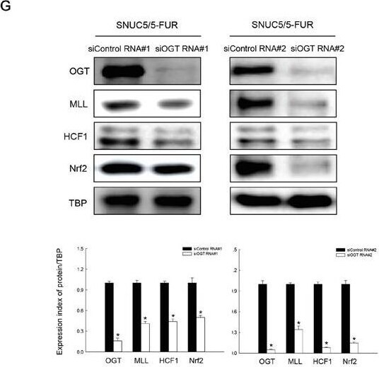
Western Blot: Host Cell Factor 1/HCFC1 Antibody [NB100-68209] -
Western Blot: Host Cell Factor 1/HCFC1 Antibody [NB100-68209] - Involvement of OGT in histone methylase MLL/COMPASS-like complex-mediated Nrf2 expression in SNUC5/5-FUR cellsA. OGT & O-GlcNAc expression was determined by Western blotting. *Significantly different from SNUC5 cells (p<0.05). B. Interaction between HCF1 & OGT was examined by immunoprecipitation analysis using anti-HCF1 & anti-OGT antibodies followed by Western blotting with anti-HCF1 & anti-OGT antibodies. C. Interaction between HCF1 & OGT was assessed by using PLA. Each green spot represents for a single interaction (HCF1 & OGT) & DNA was stained with DAPI. *Significantly different from SNUC5 cells (p<0.05). D. Interaction between MLL & OGT was examined by immunoprecipitation analysis using anti-MLL & anti-OGT antibodies followed by Western blotting with anti-OGT & anti-MLL antibodies. E. Interaction between MLL & OGT was assessed by using PLA. Each green spot represents for a single interaction (MLL & OGT) & DNA was stained with DAPI. *Significantly different from SNUC5 cells (p<0.05). The cells were transfected with non-targeting siRNA (siControl) or siOGT RNA for 24 h. F. Expression pattern of MLL mRNA in SNUC5/5-FUR cells was determined by RT-PCR analysis. *Significantly different from siControl-transfected cells (p<0.05). G. Expression pattern of OGT, MLL, HFC1, & Nrf2 in SNUC5/5-FUR cells was determined by Western blot analysis. *Significantly different from siControl-transfected cells (p<0.05). Image collected & cropped by CiteAb from the following publication (https://www.oncotarget.com/lookup/doi/10.18632/oncotarget.9745), licensed under a CC-BY license. Not internally tested by Novus Biologicals.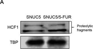
Western Blot: Host Cell Factor 1/HCFC1 Antibody [NB100-68209] -
Western Blot: Host Cell Factor 1/HCFC1 Antibody [NB100-68209] - Effect of MLL-HCF1 interaction on histone methylation & Nrf2 expression in SNUC5/5-FUR cellsA. HCF1 expression was determined by Western blotting. *Significantly different from SNUC5 cells (p<0.05). B. Interaction between MLL & HCF1was examined by immunoprecipitation analysis using anti-MLL & anti-HCF1 antibodies followed by Western blotting with anti-HCF1 & anti-MLL antibodies. *Significantly different from SNUC5 cells (p<0.05). C. Interaction between MLL & HCF1was assessed by PLA. Each green spot represents for a single interaction (MLL & HCF1) & DNA was stained with DAPI. *Significantly different from SNUC5 cells (p<0.05). The cells were transfected with non-targeting siRNA (siControl) or siHCF1 RNA for 24 h. D. Expression pattern of MLL mRNA in SNUC5/5-FUR cells was determined by RT-PCR analysis. *Significantly different from siControl-transfected cells (p<0.05). E. Expression pattern of HCF1, MLL, & Nrf2 in SNUC5/5-FUR cells was determined by Western blot analysis. *Significantly different from siControl-transfected cells (p<0.05). Image collected & cropped by CiteAb from the following publication (https://www.oncotarget.com/lookup/doi/10.18632/oncotarget.9745), licensed under a CC-BY license. Not internally tested by Novus Biologicals.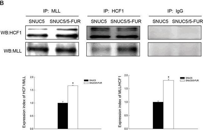
Western Blot: Host Cell Factor 1/HCFC1 Antibody [NB100-68209] -
Western Blot: Host Cell Factor 1/HCFC1 Antibody [NB100-68209] - Effect of MLL-HCF1 interaction on histone methylation & Nrf2 expression in SNUC5/5-FUR cellsA. HCF1 expression was determined by Western blotting. *Significantly different from SNUC5 cells (p<0.05). B. Interaction between MLL & HCF1was examined by immunoprecipitation analysis using anti-MLL & anti-HCF1 antibodies followed by Western blotting with anti-HCF1 & anti-MLL antibodies. *Significantly different from SNUC5 cells (p<0.05). C. Interaction between MLL & HCF1was assessed by PLA. Each green spot represents for a single interaction (MLL & HCF1) & DNA was stained with DAPI. *Significantly different from SNUC5 cells (p<0.05). The cells were transfected with non-targeting siRNA (siControl) or siHCF1 RNA for 24 h. D. Expression pattern of MLL mRNA in SNUC5/5-FUR cells was determined by RT-PCR analysis. *Significantly different from siControl-transfected cells (p<0.05). E. Expression pattern of HCF1, MLL, & Nrf2 in SNUC5/5-FUR cells was determined by Western blot analysis. *Significantly different from siControl-transfected cells (p<0.05). Image collected & cropped by CiteAb from the following publication (https://www.oncotarget.com/lookup/doi/10.18632/oncotarget.9745), licensed under a CC-BY license. Not internally tested by Novus Biologicals.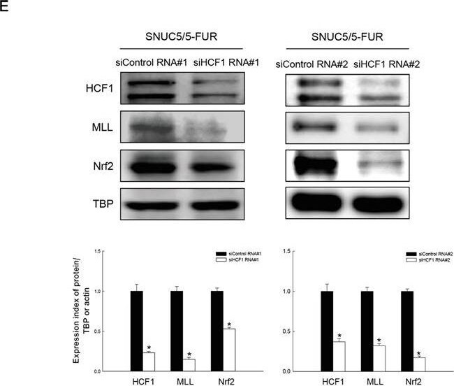
Knockdown Validated: Host Cell Factor 1/HCFC1 Antibody [NB100-68209] -
Effect of MLL-HCF1 interaction on histone methylation and Nrf2 expression in SNUC5/5-FUR cellsA. HCF1 expression was determined by Western blotting. *Significantly different from SNUC5 cells (p<0.05). B. Interaction between MLL and HCF1was examined by immunoprecipitation analysis using anti-MLL and anti-HCF1 antibodies followed by Western blotting with anti-HCF1 and anti-MLL antibodies. *Significantly different from SNUC5 cells (p<0.05). C. Interaction between MLL and HCF1was assessed by PLA. Each green spot represents for a single interaction (MLL and HCF1) and DNA was stained with DAPI. *Significantly different from SNUC5 cells (p<0.05). The cells were transfected with non-targeting siRNA (siControl) or siHCF1 RNA for 24 h. D. Expression pattern of MLL mRNA in SNUC5/5-FUR cells was determined by RT-PCR analysis. *Significantly different from siControl-transfected cells (p<0.05). E. Expression pattern of HCF1, MLL, and Nrf2 in SNUC5/5-FUR cells was determined by Western blot analysis. *Significantly different from siControl-transfected cells (p<0.05). Image collected and cropped by CiteAb from the following open publication (https://pubmed.ncbi.nlm.nih.gov/27259240), licensed under a CC-BY license. Not internally tested by Novus Biologicals.Applications for Host Cell Factor 1/HCFC1 Antibody
Immunoprecipitation
Western Blot
Formulation, Preparation, and Storage
Purification
Formulation
Preservative
Concentration
Shipping
Stability & Storage
Background: Host Cell Factor 1/HCFC1
Additional Host Cell Factor 1/HCFC1 Products
Product Documents for Host Cell Factor 1/HCFC1 Antibody
Certificate of Analysis
To download a Certificate of Analysis, please enter a lot or batch number in the search box below.
Product Specific Notices for Host Cell Factor 1/HCFC1 Antibody
This product is for research use only and is not approved for use in humans or in clinical diagnosis. Primary Antibodies are guaranteed for 1 year from date of receipt.
Related Research Areas
Citations for Host Cell Factor 1/HCFC1 Antibody
 Powered by Bioz
Customer Reviews for Host Cell Factor 1/HCFC1 Antibody
There are currently no reviews for this product. Be the first to review Host Cell Factor 1/HCFC1 Antibody and earn rewards!
Have you used Host Cell Factor 1/HCFC1 Antibody?
Submit a review and receive an Amazon gift card!
$25/€18/£15/$25CAN/¥2500 for a review with an image
$10/€7/£6/$10CAN/¥1110 for a review without an image
Submit a review
Protocols
Find general support by application which include: protocols, troubleshooting, illustrated assays, videos and webinars.
- Cellular Response to Hypoxia Protocols
- Immunoprecipitation Protocol
- R&D Systems Quality Control Western Blot Protocol
- Troubleshooting Guide: Western Blot Figures
- Western Blot Conditions
- Western Blot Protocol
- Western Blot Protocol for Cell Lysates
- Western Blot Troubleshooting
- Western Blot Troubleshooting Guide
- View all Protocols, Troubleshooting, Illustrated assays and Webinars
FAQs for Host Cell Factor 1/HCFC1 Antibody
-
Q: Hi, I'm trying to use your HCFC1 antibody for a western blot. I calculated the predicted size of the bands based on the cleavage sites in the protein and I'm getting a band that's similar to the one listed on your website, but 30 kd larger than what's calculated. I would appreciate any help clarifying this.
A: HCFC1 is subject to a number of post-translational modifications that would cause the band to migrate differently than the expected molecular weight. Our image for this antibody shows a number of bands to reflect the different fragments produced by proteolytic cleavage of the protein.