Human IL-38/IL-1F10 Antibody
R&D Systems | Catalog # AF2427


Scientific images may be resized, cropped, or adjusted for brightness or contrast for presentation purposes. These updates are limited to presentation and do not affect the underlying scientific interpretation of the data. Where available, additional source data may be provided upon request.
Key Product Details
Species Reactivity
Validated:
Cited:
Applications
Validated:
Cited:
Label
Antibody Source
Product Specifications
Immunogen
Cys2-Trp152
Accession # Q8WWZ1
Specificity
Clonality
Host
Isotype
Scientific Data Images for Human IL-38/IL-1F10 Antibody
Human IL‑38/IL‑1F10 ELISA Standard Curve.
Recombinant Human IL-38/IL-1F10 protein was serially diluted 2-fold and captured by Mouse Anti-Human IL-38/IL-1F10 Monoclonal Antibody(Catalog # MAB24271) coated on a Clear Polystyrene Microplate (Catalog # DY990). Goat Anti-Human IL-38/IL-1F10 Antigen Affinity-purified Polyclonal Antibody (Catalog # AF2427) was biotinylated and incubated with the protein captured on the plate. Detection of the standard curve was achieved by incubating Streptavidin-HRP (Catalog # DY998) followed by Substrate Solution (Catalog # DY999) and stopping the enzymatic reaction with Stop Solution (Catalog # DY994).
Human IL-38 / IL-1F10 ELISA Standard Curve
Human IL-38/IL-1F10 was serially diluted and captured by Mouse Anti-Human IL‑38/IL‑1F10 Monoclonal Antibody (Catalog # MAB24271) coated on a Clear Polystyrene Microplate (Catalog # DY990). Goat Anti-Human IL‑38/IL‑1F10 Antigen Affinity-purified Polyclonal Antibody (Catalog # AF2427) was biotinylated and incubated with the protein captured on the plate. Detection of the standard curve was achieved by incubating Streptavidin-HRP (Catalog # DY998)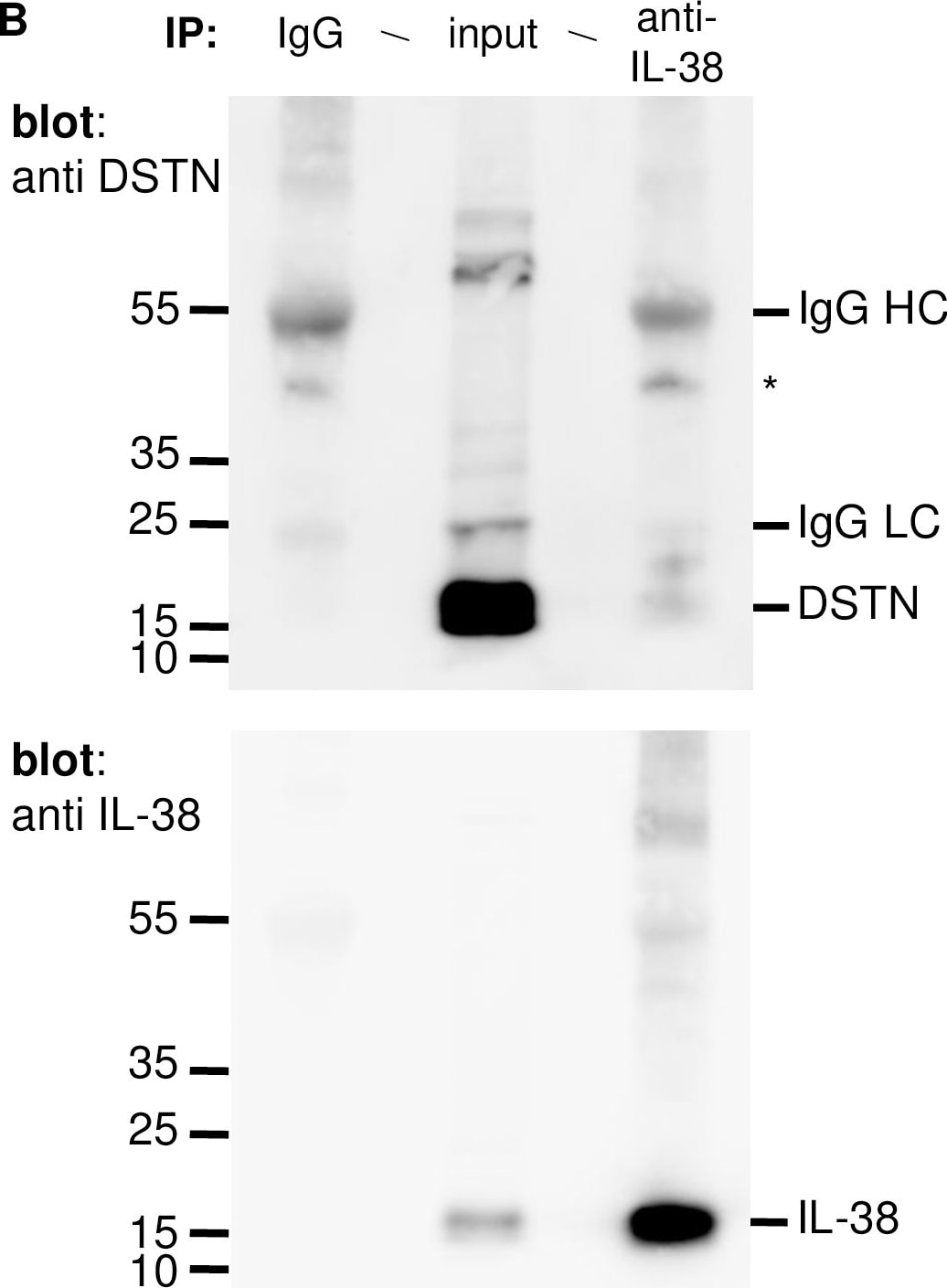
Detection of IL-38/IL-1F10 by Western Blot
Interaction of IL-38 and DSTN in HEK293T cells.Association of IL-38 and DSTN was examined by co-IP in HEK293T cells co-transfected with pcDNA3.1/hIL-38 and pcDNA3.1/hDSTN. A. Cells were treated with DSP, lyzed and immunoprecipitated with a polyclonal rabbit anti-DSTN antibody (right lane), or with normal rabbit IgG (left lane) as a negative control. Whole-cell lysates (input; middle lane) and immunoprecipitated proteins were fractionated by SDS-PAGE and IL-38 was detected by Western blotting (upper panel). The membrane was then stripped and reprobed with the anti-DSTN antibody (lower panel). B. Cells were treated with DSP, lyzed and immunoprecipitated with a polyclonal goat anti-IL-38 antibody (right lane), or with normal goat IgG (left lane) as a negative control. Whole-cell lysates (input; middle lane) and immunoprecipitated proteins were fractionated by SDS-PAGE and DSTN was detected by Western blotting (upper panel). The membrane was then stripped and reprobed with the anti-IL-38 antibody (lower panel). The positions of IL-38 and DSTN, as well as of the heavy (HC) and light (LC) chains of the IgGs used for IP are indicated on the right of the blots. *indicates a non-specific band present also in the negative control IP. Molecular mass markers are indicated on the left of each blot. Image collected and cropped by CiteAb from the following open publication (https://pubmed.ncbi.nlm.nih.gov/31770407), licensed under a CC-BY license. Not internally tested by R&D Systems.Applications for Human IL-38/IL-1F10 Antibody
ELISA
This antibody functions as an ELISA detection antibody when paired with Mouse Anti-Human IL‑38/IL‑1F10 Monoclonal Antibody(Catalog # MAB24271).
This product is intended for assay development on various assay platforms requiring antibody pairs. We recommend the Human IL-38/IL-1F10 DuoSet ELISA Kit (Catalog # DY9110-05) for convenient development of a sandwich ELISA.
Western Blot
Sample: Recombinant Human IL‑38/IL‑1F10
Formulation, Preparation, and Storage
Purification
Reconstitution
Reconstitute at 0.2 mg/mL in sterile PBS. For liquid material, refer to CoA for concentration.
Formulation
Shipping
Stability & Storage
- 12 months from date of receipt, -20 to -70 °C as supplied.
- 1 month, 2 to 8 °C under sterile conditions after reconstitution.
- 6 months, -20 to -70 °C under sterile conditions after reconstitution.
Calculators
Background: IL-38/IL-1F10
Long Name
Alternate Names
Gene Symbol
UniProt
Additional IL-38/IL-1F10 Products
Product Documents for Human IL-38/IL-1F10 Antibody
Certificate of Analysis
To download a Certificate of Analysis, please enter a lot or batch number in the search box below.
Note: Certificate of Analysis not available for kit components.
Product Specific Notices for Human IL-38/IL-1F10 Antibody
For research use only
Related Research Areas
Citations for Human IL-38/IL-1F10 Antibody
 Powered by Bioz
Customer Reviews for Human IL-38/IL-1F10 Antibody
There are currently no reviews for this product. Be the first to review Human IL-38/IL-1F10 Antibody and earn rewards!
Have you used Human IL-38/IL-1F10 Antibody?
Submit a review and receive an Amazon gift card!
$25/€18/£15/$25CAN/¥2500 for a review with an image
$10/€7/£6/$10CAN/¥1110 for a review without an image
Submit a review
Protocols
Find general support by application which include: protocols, troubleshooting, illustrated assays, videos and webinars.
- Cellular Response to Hypoxia Protocols
- ELISA Sample Preparation & Collection Guide
- ELISA Troubleshooting Guide
- How to Run an R&D Systems DuoSet ELISA
- How to Run an R&D Systems Quantikine ELISA
- How to Run an R&D Systems Quantikine™ QuicKit™ ELISA
- Quantikine HS ELISA Kit Assay Principle, Alkaline Phosphatase
- Quantikine HS ELISA Kit Principle, Streptavidin-HRP Polymer
- R&D Systems Quality Control Western Blot Protocol
- Sandwich ELISA (Colorimetric) – Biotin/Streptavidin Detection Protocol
- Sandwich ELISA (Colorimetric) – Direct Detection Protocol
- Troubleshooting Guide: ELISA
- Troubleshooting Guide: Western Blot Figures
- Western Blot Conditions
- Western Blot Protocol
- Western Blot Protocol for Cell Lysates
- Western Blot Troubleshooting
- Western Blot Troubleshooting Guide
- View all Protocols, Troubleshooting, Illustrated assays and Webinars
Associated Pathways