BID is a 195 amino acid member of the Bcl-2 family of proteins that regulates outer mitochondrial membrane permeability (1). BID is a pro-apoptotic member that causes cytochrome c to be released from the mitochondria intermembrane space into the cytosol. In healthy cells BID is cytosolic. In response to Fas ligand or TNF, BID is cleaved by caspase-8 and it then relocates to the mitochondria outer membrane (2, 3). Cleavage of BID by caspase-8 generates a new N-terminal that contains a terminal glycine. It appears that the glycine is myristoylated and myristoylation serves to target BID to the mitochondria (4). BID may then interact with another pro-apoptotic Bcl-2 family member Bak (5). Interaction of BID with Bak causes altered mitochondrial membrane permeability. A 9‑13 amino acid stretch called the BH3 region (Bcl-2 homology region) appears to mediate the BID interaction with other Bcl-2 family members. BID is neutralized by binding to the anti-apoptotic member Bcl-xL.

Key Product Details
Validated by
Biological Validation
Species Reactivity
Validated:
Human, Mouse
Cited:
Human, Mouse, Plant - Arabidopsis thaliana
Applications
Validated:
Western Blot, Simple Western, Immunoprecipitation
Cited:
Western Blot, Immunocytochemistry
Label
Unconjugated
Antibody Source
Polyclonal Goat IgG
Loading...
Product Specifications
Immunogen
E. coli-derived recombinant mouse BID
Met1-Asp195
Accession # AAC71064
Met1-Asp195
Accession # AAC71064
Specificity
Detects human and mouse BID, a fragment generated in cells treated with anti-Fas and the carboxyl‑terminal portion of recombinant BID generated by cleavage with Caspase 8 in Western blots.
Clonality
Polyclonal
Host
Goat
Isotype
IgG
Scientific Data Images for BID Antibody
Detection of Human BID by Western Blot.
Western blot shows Jurkat human acute T cell leukemia cell line treated with apoptosis inducer anti-Fas for the indicated times. PVDF membrane was probed with 1 µg/mL Goat Anti-Human/Mouse BID Antigen Affinity-purified Polyclonal Antibody (Catalog # AF860) followed by HRP-conjugated Anti-Goat IgG Secondary Antibody (Catalog # HAF109). A specific band was detected for BID at approximately 20 kDa (as indicated). For additional reference short (5 seconds, left panel) and long (60 seconds, right panel) exposures are shown. This experiment was conducted under reducing conditions and using Immunoblot Buffer Group 2.
Detection of Human and Mouse BID by Western Blot.
Western blot shows lysates of SVEC4-10 mouse vascular endothelial cell line and Jurkat human acute T cell leukemia cell line. PVDF membrane was probed with 1 µg/mL of Goat Anti-Human/Mouse BID Antigen Affinity-purified Polyclonal Antibody (Catalog # AF860) followed by HRP-conjugated Anti-Goat IgG Secondary Antibody (Catalog # HAF017). A specific band was detected for BID at approximately 20 kDa (as indicated). This experiment was conducted under reducing conditions and using Immunoblot Buffer Group 1.
Detection of Human BID by Simple WesternTM.
Simple Western lane view shows lysates of Jurkat human acute T cell leukemia cell line, loaded at 0.2 mg/mL. A specific band was detected for BID at approximately 29 kDa (as indicated) using 10 µg/mL of Goat Anti-Human/Mouse BID Antigen Affinity-purified Polyclonal Antibody (Catalog # AF860) followed by 1:50 dilution of HRP-conjugated Anti-Goat IgG Secondary Antibody (Catalog # HAF109). This experiment was conducted under reducing conditions and using the 12-230 kDa separation system.
Detection of Human BID by Western Blot
Survivin is predominantly expressed in obese hASCs and is influenced by the inflammatory environment. (a) Survivin protein expression and quantification in total adipose tissue (AT), adipocyte fraction (AD) and stromal-vascular fraction (SVF) from SAT of lean and obese patients. Fumarylacetoacetase (FAA) was used as a loading control. Ponceau S staining is also provided. *P<0.001 versus total AT and AD. (b) Survivin and p53 protein levels and quantification in hASCs from lean and obese patients. GAPDH was used as a loading control. *P<0.01 versus lean. (c) Survivin secreted levels and quantification in medium from lean- and obese-derived hASCs. Ponceau S staining was used to check loading. *P<0.05 versus lean conditioned medium. (d) Total cell extracts of lean- and obese-derived hASCs were subjected to immunoblotting with antibodies against apoptotic and antiapoptotic markers and GAPDH was used as a loading control. No significant differences were found. (e) Lean hASCs were cultured in RPMI medium (C: control) or for 24 h in conditioned medium (CM) from macrophages (M0, M1 and M2) and cells were subjected to immunobloting with survivin and GAPDH antibodies. *P<0.05 versus CM from control, and M0; #P<0.05 versus M2. (f) Left panel, lean hASCs were cultured in RPMI medium (C: control) or RPMI supplemented with IL-1 beta (IL1B) or IL-1 beta and IL-1 beta receptor antagonist (a); right panel, with CM of M1 macrophages or CM of M1 and IL-1 beta receptor antagonist (a). Survivin protein levels were measured and GAPDH was used as a loading control. Quantification of three experiments is shown. *P<0.05 versus control (c); #P<0.05 as indicated in figure. Data information: Values are expressed as mean±S.E.M. Statistical analysis: unpaired Student’s t-test. n=3–4 patients for each group. Image collected and cropped by CiteAb from the following open publication (https://pubmed.ncbi.nlm.nih.gov/28518147), licensed under a CC-BY license. Not internally tested by R&D Systems.
Detection of Human BID by Western Blot
Survivin is predominantly expressed in obese hASCs and is influenced by the inflammatory environment. (a) Survivin protein expression and quantification in total adipose tissue (AT), adipocyte fraction (AD) and stromal-vascular fraction (SVF) from SAT of lean and obese patients. Fumarylacetoacetase (FAA) was used as a loading control. Ponceau S staining is also provided. *P<0.001 versus total AT and AD. (b) Survivin and p53 protein levels and quantification in hASCs from lean and obese patients. GAPDH was used as a loading control. *P<0.01 versus lean. (c) Survivin secreted levels and quantification in medium from lean- and obese-derived hASCs. Ponceau S staining was used to check loading. *P<0.05 versus lean conditioned medium. (d) Total cell extracts of lean- and obese-derived hASCs were subjected to immunoblotting with antibodies against apoptotic and antiapoptotic markers and GAPDH was used as a loading control. No significant differences were found. (e) Lean hASCs were cultured in RPMI medium (C: control) or for 24 h in conditioned medium (CM) from macrophages (M0, M1 and M2) and cells were subjected to immunobloting with survivin and GAPDH antibodies. *P<0.05 versus CM from control, and M0; #P<0.05 versus M2. (f) Left panel, lean hASCs were cultured in RPMI medium (C: control) or RPMI supplemented with IL-1 beta (IL1B) or IL-1 beta and IL-1 beta receptor antagonist (a); right panel, with CM of M1 macrophages or CM of M1 and IL-1 beta receptor antagonist (a). Survivin protein levels were measured and GAPDH was used as a loading control. Quantification of three experiments is shown. *P<0.05 versus control (c); #P<0.05 as indicated in figure. Data information: Values are expressed as mean±S.E.M. Statistical analysis: unpaired Student’s t-test. n=3–4 patients for each group Image collected and cropped by CiteAb from the following open publication (https://pubmed.ncbi.nlm.nih.gov/28518147), licensed under a CC-BY license. Not internally tested by R&D Systems.Applications for BID Antibody
Application
Recommended Usage
Immunoprecipitation
4 µg/106 cells
Sample: Jurkat human acute T cell leukemia cell line treated with anti-Fas, see our available Western blot detection antibodies
Sample: Jurkat human acute T cell leukemia cell line treated with anti-Fas, see our available Western blot detection antibodies
Simple Western
10 µg/mL
Sample: Jurkat human acute T cell leukemia cell line
Sample: Jurkat human acute T cell leukemia cell line
Western Blot
1 µg/mL
Sample: Jurkat human acute T cell leukemia cell line was treated or untreated with anti-Fas and SVEC4‑10 mouse vascular endothelial cell line
Sample: Jurkat human acute T cell leukemia cell line was treated or untreated with anti-Fas and SVEC4‑10 mouse vascular endothelial cell line
Reviewed Applications
Read 4 reviews rated 4 using AF860 in the following applications:
Formulation, Preparation, and Storage
Purification
Antigen Affinity-purified
Reconstitution
Reconstitute at 0.2 mg/mL in sterile PBS. For liquid material, refer to CoA for concentration.
Loading...
Formulation
Lyophilized from a 0.2 μm filtered solution in PBS with Trehalose. *Small pack size (SP) is supplied either lyophilized or as a 0.2 µm filtered solution in PBS.
Shipping
Lyophilized product is shipped at ambient temperature. Liquid small pack size (-SP) is shipped with polar packs. Upon receipt, store immediately at the temperature recommended below.
Stability & Storage
Use a manual defrost freezer and avoid repeated freeze-thaw cycles.
- 12 months from date of receipt, -20 to -70 °C as supplied.
- 1 month, 2 to 8 °C under sterile conditions after reconstitution.
- 6 months, -20 to -70 °C under sterile conditions after reconstitution.
Calculators
Background: BID
References
- Gross, A. et al. (1999) Genes and Develop. 13:1899.
- Luo, X., et al. (1998) Cell 94:481.
- Li, H. et al. (1998) Cell 94:491.
- Zha, J. et al. (2000) Science 290:1761.
- Wei, M.C. et al. (2000) Genes Dev. 14:2060.
Long Name
BH3 Interacting Domain Death Agonist
Alternate Names
apoptic death agonist, BH3 interacting domain death agonist, BH3-interacting domain death agonist, BID isoform ES(1b), BID isoform L(2), BID isoform Si6, desmocollin type 4, FP497, Human BID coding sequence, MGC15319, MGC42355, p22 BID, tbid
Gene Symbol
BID
UniProt
Additional BID Products
Product Documents for BID Antibody
Certificate of Analysis
To download a Certificate of Analysis, please enter a lot or batch number in the search box below.
Note: Certificate of Analysis not available for kit components.
Product Specific Notices for BID Antibody
For research use only
Related Research Areas
Citations for BID Antibody
 Powered by Bioz
Customer Reviews for BID Antibody (4)
4 out of 5
4 Customer Ratings
Have you used BID Antibody?
Submit a review and receive an Amazon gift card!
$25/€18/£15/$25CAN/¥2500 Yen for a review with an image
$10/€7/£6/$10CAN/¥1110 Yen for a review without an image
Submit a review
Customer Images
Showing
1
-
4 的
4 reviews
Showing All
Filter By:
-
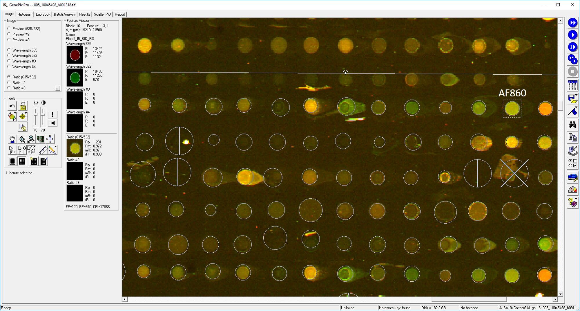
Application: MicroarraysSample Tested: EDTA PlasmaSpecies: HumanVerified Customer | Posted 03/11/2019

-
Application: ELISASample Tested: PlasmaSpecies: HumanVerified Customer | Posted 11/10/2018
-
Application: MicroarraySample Tested: EDTA PlasmaSpecies: HumanVerified Customer | Posted 11/09/2018
-
Application: Western BlotSample Tested: See PMID 21048194Species: HumanVerified Customer | Posted 01/09/2015
There are no reviews that match your criteria.
Protocols
Find general support by application which include: protocols, troubleshooting, illustrated assays, videos and webinars.
- Cellular Response to Hypoxia Protocols
- Immunoprecipitation Protocol
- R&D Systems Quality Control Western Blot Protocol
- Troubleshooting Guide: Western Blot Figures
- Western Blot Conditions
- Western Blot Protocol
- Western Blot Protocol for Cell Lysates
- Western Blot Troubleshooting
- Western Blot Troubleshooting Guide
- View all Protocols, Troubleshooting, Illustrated assays and Webinars
Loading...
Associated Pathways
