
Myocardin (MYOCD) is a transcriptional co-activator necessary for differentiation of smooth muscle cells. MYOCD functions by binding the transcription factor Serum Response Factor (SRF) and stimulating smooth muscle cell-specific gene expression.

Scientific images may be resized, cropped, or adjusted for brightness or contrast for presentation purposes. These updates are limited to presentation and do not affect the underlying scientific interpretation of the data. Where available, additional source data may be provided upon request.
Key Product Details
Validated by
Biological Validation
Species Reactivity
Validated:
Human, Mouse
Cited:
Human, Mouse, Porcine, Canine, Transgenic Mouse
Applications
Validated:
Western Blot
Cited:
Immunohistochemistry, Immunohistochemistry-Paraffin, Western Blot
Label
Unconjugated
Antibody Source
Monoclonal Mouse IgG2B Clone # 355521
Loading...
Product Specifications
Immunogen
E. coli-derived recombinant human Myocardin
Met97-Leu290
Accession # Q8IZQ8
Met97-Leu290
Accession # Q8IZQ8
Specificity
Detects human and mouse Myocardin in Western blots.
Clonality
Monoclonal
Host
Mouse
Isotype
IgG2B
Scientific Data Images for Myocardin Antibody (355521)
Detection of Human Myocardin by Western Blot.
Western blot shows lysates of MCF-7 human breast cancer cell line, Raji human Burkitt's lymphoma cell line, HeLa human cervical epithelial carcinoma cell line, and C2C12 mouse myoblast cell line. PVDF membrane was probed with 1 µg/mL of Human Myocardin Monoclonal Antibody (Catalog # MAB4028) followed by HRP-conjugated Anti-Mouse IgG Secondary Antibody (Catalog # HAF007). A specific band was detected for Myocardin at approximately 105 kDa (as indicated). This experiment was conducted under reducing conditions and using Immunoblot Buffer Group 1.
Detection of Mouse Myocardin by Western Blot
Laminin-111 but not laminin-alpha 4 blocking antibody affects pericyte differentiation. (a) Immunoblots show that laminin-111 blockage (Ln Ab) significantly enhances the expression of PDGFR beta, SMA, and SM22-alpha, but not myocardin in pericytes. Full blots of these proteins are shown in Supplementary Figure 14b. Rabbit IgG treated cells were used as a Ctr. All bands were normalized to actin (n=5–6). (b) Immunoblots show that laminin-alpha 4 blockage (Anti-Ln alpha 4) does not change the expression of PDGFR beta, SMA, SM22-alpha, or myocardinin in pericytes. Full blots of these proteins are shown in Supplementary Figure 14c. Rabbit IgG treated cells were used as a Ctr. All bands were normalized to actin (n=3). Data are shown as mean ± sd. *p<0.05 versus the Ctrs by student’s t-test. Image collected and cropped by CiteAb from the following publication (https://pubmed.ncbi.nlm.nih.gov/24583950), licensed under a CC-BY license. Not internally tested by R&D Systems.
Detection of Mouse Myocardin by Western Blot
Astrocytic laminin mediates pericyte differentiation via integrin alpha 2. (a) Immunoblots show that integrin alpha 2 blockage (ITGA2) but not integrin beta 1 blockage significantly increases the expression of PDGFR beta, SMA, and SM22-alpha, but not myocardin in pericytes. Full blots of these proteins are shown in Supplementary Figure 14d. Rabbit IgG treated cells were used as a Ctr. All bands were normalized to actin (n=6). (b) Schematic diagram of shRNA designed to target ITGA2 mRNA. (c) Immunoblot analysis shows that all three ITGA2-specific shRNAs (#1–3) dramatically reduce ITGA2 at protein level and ITGA2-specific shRNA-3 (#1) is the most efficient one. Full blots of ITGA2 and actin are shown in Supplementary Figure 14e. Scramble shRNA was used as a Ctr. (d) Immunoblot analysis shows that transduction of pericytes with lenti-shRNA-1 (#1) significantly enhances the expression of PDGFR beta, SMA, and SM22-alpha, but does not affect myocardin level. Full blots of these proteins are shown in Supplementary Figure 14f. Scramble shRNA was used as a Ctr. All bands were normalized to actin (n=4–5). Data are shown as mean ± sd. *p<0.05 versus the Ctrs by student’s t-test. Image collected and cropped by CiteAb from the following publication (https://pubmed.ncbi.nlm.nih.gov/24583950), licensed under a CC-BY license. Not internally tested by R&D Systems.
Detection of Mouse Myocardin by Western Blot
Laminin-111 but not laminin-alpha 4 blocking antibody affects pericyte differentiation. (a) Immunoblots show that laminin-111 blockage (Ln Ab) significantly enhances the expression of PDGFR beta, SMA, and SM22-alpha, but not myocardin in pericytes. Full blots of these proteins are shown in Supplementary Figure 14b. Rabbit IgG treated cells were used as a Ctr. All bands were normalized to actin (n=5–6). (b) Immunoblots show that laminin-alpha 4 blockage (Anti-Ln alpha 4) does not change the expression of PDGFR beta, SMA, SM22-alpha, or myocardinin in pericytes. Full blots of these proteins are shown in Supplementary Figure 14c. Rabbit IgG treated cells were used as a Ctr. All bands were normalized to actin (n=3). Data are shown as mean ± sd. *p<0.05 versus the Ctrs by student’s t-test. Image collected and cropped by CiteAb from the following publication (https://pubmed.ncbi.nlm.nih.gov/24583950), licensed under a CC-BY license. Not internally tested by R&D Systems.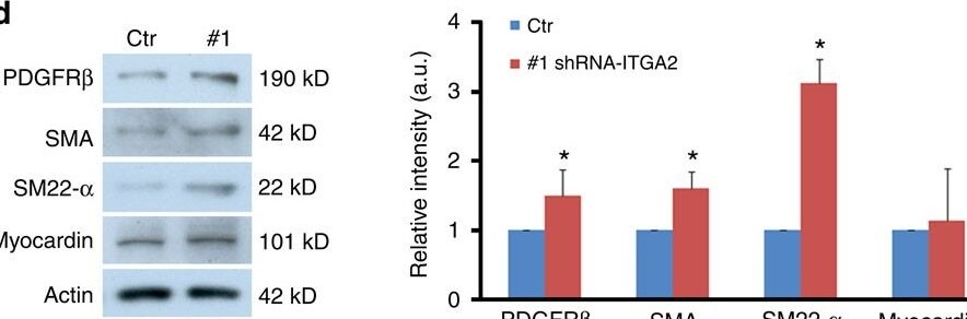
Detection of Mouse Myocardin by Western Blot
Astrocytic laminin mediates pericyte differentiation via integrin alpha 2. (a) Immunoblots show that integrin alpha 2 blockage (ITGA2) but not integrin beta 1 blockage significantly increases the expression of PDGFR beta, SMA, and SM22-alpha, but not myocardin in pericytes. Full blots of these proteins are shown in Supplementary Figure 14d. Rabbit IgG treated cells were used as a Ctr. All bands were normalized to actin (n=6). (b) Schematic diagram of shRNA designed to target ITGA2 mRNA. (c) Immunoblot analysis shows that all three ITGA2-specific shRNAs (#1–3) dramatically reduce ITGA2 at protein level and ITGA2-specific shRNA-3 (#1) is the most efficient one. Full blots of ITGA2 and actin are shown in Supplementary Figure 14e. Scramble shRNA was used as a Ctr. (d) Immunoblot analysis shows that transduction of pericytes with lenti-shRNA-1 (#1) significantly enhances the expression of PDGFR beta, SMA, and SM22-alpha, but does not affect myocardin level. Full blots of these proteins are shown in Supplementary Figure 14f. Scramble shRNA was used as a Ctr. All bands were normalized to actin (n=4–5). Data are shown as mean ± sd. *p<0.05 versus the Ctrs by student’s t-test. Image collected and cropped by CiteAb from the following publication (https://pubmed.ncbi.nlm.nih.gov/24583950), licensed under a CC-BY license. Not internally tested by R&D Systems.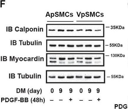
Detection of Myocardin by Western Blot
Differences in cell behavior between pig arterial smooth muscle cells (ApSMCs) and venous smooth muscle cells (VpSMCs). (A) The purity of VSMCs was demonstrated with anti-smooth muscle actin immunofluorescent staining, following the protocol described in the Materials and Methods. Scale bars = 200 microns. (B,C) Cell proliferation under the growth medium (GM) or in response to PDGF-BB stimulation was measured following the protocol described in the Materials and Methods. Different letters (a, b) indicated significant difference between two groups. (D) Cell migration assays were performed via scratch assays in serum-free (SF) or 10% FBS containing GM. (E) Migrated cells in scratched areas were counted. Scale bars = 200 microns. (F) Cells were induced to differentiate with a differentiation medium (DM), and 48 h before collecting cell lysates, PDGF-BB was applied in the indicated wells. (G,H) The densitometry of each band was obtained using Image J (NIH version 1.53K), and ratio values were obtained by interested protein bands/loading control bands. (n = 6 for calponin; n = 5 for myocardin.) ****: p < 0.000; ***: p < 0.001; **: p < 0.01; *: p < 0.05; ns: no significant differences were found using a Student’s t-test for two-group comparisons and an ANOVA test for multiple group comparisons. Image collected and cropped by CiteAb from the following open publication (https://pubmed.ncbi.nlm.nih.gov/40243809), licensed under a CC-BY license. Not internally tested by R&D Systems.Applications for Myocardin Antibody (355521)
Application
Recommended Usage
Western Blot
1 µg/mL
Sample: MCF-7 human breast cancer cell line, Raji human Burkitt's lymphoma cell line, HeLa human cervical epithelial carcinoma cell line, and C2C12 mouse myoblast cell line
Sample: MCF-7 human breast cancer cell line, Raji human Burkitt's lymphoma cell line, HeLa human cervical epithelial carcinoma cell line, and C2C12 mouse myoblast cell line
Reviewed Applications
Read 1 review rated 5 using MAB4028 in the following applications:
Formulation, Preparation, and Storage
Purification
Protein A or G purified from hybridoma culture supernatant
Reconstitution
Reconstitute at 0.5 mg/mL in sterile PBS. For liquid material, refer to CoA for concentration.
Loading...
Formulation
Lyophilized from a 0.2 μm filtered solution in PBS with Trehalose. *Small pack size (SP) is supplied either lyophilized or as a 0.2 µm filtered solution in PBS.
Shipping
Lyophilized product is shipped at ambient temperature. Liquid small pack size (-SP) is shipped with polar packs. Upon receipt, store immediately at the temperature recommended below.
Stability & Storage
Use a manual defrost freezer and avoid repeated freeze-thaw cycles.
- 12 months from date of receipt, -20 to -70 °C as supplied.
- 1 month, 2 to 8 °C under sterile conditions after reconstitution.
- 6 months, -20 to -70 °C under sterile conditions after reconstitution.
Calculators
Background: Myocardin
Alternate Names
BSAC2A, MYCD, MYOCD, Srfcp
Gene Symbol
MYOCD
UniProt
Additional Myocardin Products
Product Documents for Myocardin Antibody (355521)
Certificate of Analysis
To download a Certificate of Analysis, please enter a lot or batch number in the search box below.
Note: Certificate of Analysis not available for kit components.
Product Specific Notices for Myocardin Antibody (355521)
For research use only
Related Research Areas
Citations for Myocardin Antibody (355521)
 Powered by Bioz
Customer Reviews for Myocardin Antibody (355521) (1)
5 out of 5
1 Customer Rating
Have you used Myocardin Antibody (355521)?
Submit a review and receive an Amazon gift card!
$25/€18/£15/$25CAN/¥2500 for a review with an image
$10/€7/£6/$10CAN/¥1110 for a review without an image
Submit a review
Customer Images
Showing
1
-
1 的
1 review
Showing All
Filter By:
-
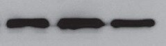
Application: Western BlotSample Tested: C2C12 mouse myoblast cell lineSpecies: MouseVerified Customer | Posted 09/01/2021

There are no reviews that match your criteria.
Protocols
Find general support by application which include: protocols, troubleshooting, illustrated assays, videos and webinars.
- Cellular Response to Hypoxia Protocols
- R&D Systems Quality Control Western Blot Protocol
- Troubleshooting Guide: Western Blot Figures
- Western Blot Conditions
- Western Blot Protocol
- Western Blot Protocol for Cell Lysates
- Western Blot Troubleshooting
- Western Blot Troubleshooting Guide
- View all Protocols, Troubleshooting, Illustrated assays and Webinars
Loading...