TGF-beta RI/ALK-5 Antibody (141231)
R&D Systems | Catalog # MAB5871

Key Product Details
Validated by
Biological Validation
Species Reactivity
Validated:
Human, Mouse
Cited:
Human, Mouse
Applications
Validated:
Western Blot
Cited:
Immunohistochemistry-Frozen, Western Blot, Flow Cytometry, Immunocytochemistry
Label
Unconjugated
Antibody Source
Monoclonal Rat IgG2A Clone # 141231
Loading...
Product Specifications
Immunogen
S. frugiperda insect ovarian cell line Sf 21-derived recombinant mouse TGF‑ beta RI/ALK-5
Ala21-Glu121
Accession # BAA05023
Ala21-Glu121
Accession # BAA05023
Specificity
Detects human and mouse TGF-beta RI/ALK-5 in Western blots. In direct ELISAs and Western blots, this antibody shows no cross-reactivity with rrMIS RII, rhTGF‑ beta RII, rhTGF‑ beta RIIb, or rhTGF‑ beta RIII.
Clonality
Monoclonal
Host
Rat
Isotype
IgG2A
Scientific Data Images for TGF-beta RI/ALK-5 Antibody (141231)
Detection of Mouse TGF-beta RI/ALK-5 by Western Blot
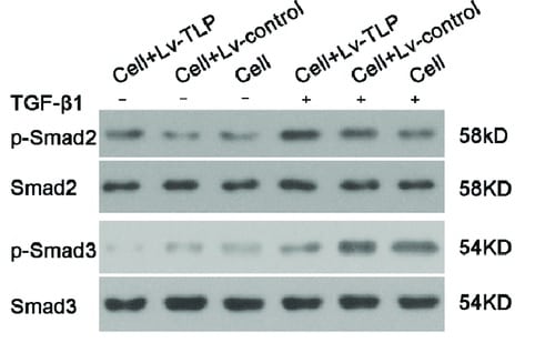
TGFRI and TGFRII association in fibulin-6 KD cells upon TGF-beta stimulus.scr siRNA or fibulin-6 siRNA transfected nCF, after TGF-beta stimulation are subjected to (a) FACS analysis. No difference in the amount of TGF beta RI and TGF beta RII on the surface of control and fibulin-6 KD cells was observed. (b) Immuno-precipitation of TGFRII from cell lysates of scr transfected and TGF-beta stimulated cells, shows more accumulation of TGFRI after western blotting compared to non TGF-beta treated nCF. (c) Immuno-precipitation of TGFRII followed by WB for TGFRI from cell lysates of scr transfected or fibulin-6 KD cells after TGF-beta stimulation display reduced association of receptors in fibulin-6 KD condition. The control input lanes shows no difference and IgG control is also clean (n = 4, p < 0.05). (d) Phosphorylation status of TGFRI was analyzed by western blot using phospho-TGFRI specific antibody. Densitometric analysis of western blot display decreased phosphorylation of TGFRI in fibulin-6 KD cells after TGF-beta stimulation compared to control cells (n = 5, p < 0.01, nonparametric Mann-Whitney U test). Image collected and cropped by CiteAb from the following publication (https://pubmed.ncbi.nlm.nih.gov/28209981), licensed under a CC-BY license. Not internally tested by R&D Systems.
Detection of Mouse TGF-beta RI/ALK-5 by Western Blot
TGFRI and TGFRII association in fibulin-6 KD cells upon TGF-beta stimulus.scr siRNA or fibulin-6 siRNA transfected nCF, after TGF-beta stimulation are subjected to (a) FACS analysis. No difference in the amount of TGF beta RI and TGF beta RII on the surface of control and fibulin-6 KD cells was observed. (b) Immuno-precipitation of TGFRII from cell lysates of scr transfected and TGF-beta stimulated cells, shows more accumulation of TGFRI after western blotting compared to non TGF-beta treated nCF. (c) Immuno-precipitation of TGFRII followed by WB for TGFRI from cell lysates of scr transfected or fibulin-6 KD cells after TGF-beta stimulation display reduced association of receptors in fibulin-6 KD condition. The control input lanes shows no difference and IgG control is also clean (n = 4, p < 0.05). (d) Phosphorylation status of TGFRI was analyzed by western blot using phospho-TGFRI specific antibody. Densitometric analysis of western blot display decreased phosphorylation of TGFRI in fibulin-6 KD cells after TGF-beta stimulation compared to control cells (n = 5, p < 0.01, nonparametric Mann-Whitney U test). Image collected and cropped by CiteAb from the following publication (https://pubmed.ncbi.nlm.nih.gov/28209981), licensed under a CC-BY license. Not internally tested by R&D Systems.
Detection of Mouse TGF-beta RI/ALK-5 by Flow Cytometry
TGFRI and TGFRII association in fibulin-6 KD cells upon TGF-beta stimulus.scr siRNA or fibulin-6 siRNA transfected nCF, after TGF-beta stimulation are subjected to (a) FACS analysis. No difference in the amount of TGF beta RI and TGF beta RII on the surface of control and fibulin-6 KD cells was observed. (b) Immuno-precipitation of TGFRII from cell lysates of scr transfected and TGF-beta stimulated cells, shows more accumulation of TGFRI after western blotting compared to non TGF-beta treated nCF. (c) Immuno-precipitation of TGFRII followed by WB for TGFRI from cell lysates of scr transfected or fibulin-6 KD cells after TGF-beta stimulation display reduced association of receptors in fibulin-6 KD condition. The control input lanes shows no difference and IgG control is also clean (n = 4, p < 0.05). (d) Phosphorylation status of TGFRI was analyzed by western blot using phospho-TGFRI specific antibody. Densitometric analysis of western blot display decreased phosphorylation of TGFRI in fibulin-6 KD cells after TGF-beta stimulation compared to control cells (n = 5, p < 0.01, nonparametric Mann-Whitney U test). Image collected and cropped by CiteAb from the following publication (https://pubmed.ncbi.nlm.nih.gov/28209981), licensed under a CC-BY license. Not internally tested by R&D Systems.Applications for TGF-beta RI/ALK-5 Antibody (141231)
Application
Recommended Usage
Western Blot
1 µg/mL
Sample: Recombinant Mouse TGF-beta RI/ALK‑5 Fc Chimera (Catalog # 587-RI) and Recombinant Human TGF-beta RI/ALK‑5 Fc Chimera (Catalog # 3025-BR)
Sample: Recombinant Mouse TGF-beta RI/ALK‑5 Fc Chimera (Catalog # 587-RI) and Recombinant Human TGF-beta RI/ALK‑5 Fc Chimera (Catalog # 3025-BR)
Reviewed Applications
Read 3 reviews rated 3.7 using MAB5871 in the following applications:
Formulation, Preparation, and Storage
Purification
Protein A or G purified from hybridoma culture supernatant
Reconstitution
Reconstitute at 0.5 mg/mL in sterile PBS. For liquid material, refer to CoA for concentration.
Loading...
Formulation
Lyophilized from a 0.2 μm filtered solution in PBS with Trehalose. *Small pack size (SP) is supplied either lyophilized or as a 0.2 µm filtered solution in PBS.
Shipping
Lyophilized product is shipped at ambient temperature. Liquid small pack size (-SP) is shipped with polar packs. Upon receipt, store immediately at the temperature recommended below.
Stability & Storage
Use a manual defrost freezer and avoid repeated freeze-thaw cycles.
- 12 months from date of receipt, -20 to -70 °C as supplied.
- 1 month, 2 to 8 °C under sterile conditions after reconstitution.
- 6 months, -20 to -70 °C under sterile conditions after reconstitution.
Calculators
Background: TGF-beta RI/ALK-5
References
- Miyazono, K. et al. (1994) Adv. In Immunol. 55:181.
- Massagùe, J. (1998) Ann. Rev. Biochem. 67:753.
Long Name
Transforming Growth Factor beta Receptor I
Alternate Names
ALK-5, SKR4, TGF-bRI, TGFbetaRI, TGFBR1
Gene Symbol
TGFBR1
UniProt
Additional TGF-beta RI/ALK-5 Products
Product Documents for TGF-beta RI/ALK-5 Antibody (141231)
Certificate of Analysis
To download a Certificate of Analysis, please enter a lot or batch number in the search box below.
Note: Certificate of Analysis not available for kit components.
Product Specific Notices for TGF-beta RI/ALK-5 Antibody (141231)
For research use only
Citations for TGF-beta RI/ALK-5 Antibody (141231)
 Powered by Bioz
Customer Reviews for TGF-beta RI/ALK-5 Antibody (141231) (3)
3.7 out of 5
3 Customer Ratings
Have you used TGF-beta RI/ALK-5 Antibody (141231)?
Submit a review and receive an Amazon gift card!
$25/€18/£15/$25CAN/¥2500 Yen for a review with an image
$10/€7/£6/$10CAN/¥1110 Yen for a review without an image
Submit a review
Customer Images
Showing
1
-
3 的
3 reviews
Showing All
Filter By:
-
Application: Western BlotSample Tested: Dermal fibroblastsSpecies: HumanVerified Customer | Posted 07/17/2021

-
Application: Western BlotSample Tested: Cell LysatesSpecies: MouseVerified Customer | Posted 06/29/2021
-
Application: Western BlotSample Tested: colo357Species: HumanVerified Customer | Posted 06/28/2016Bio-Techne ResponseR&D Systems Technical Service is investigating.
There are no reviews that match your criteria.
Protocols
Find general support by application which include: protocols, troubleshooting, illustrated assays, videos and webinars.
- Cellular Response to Hypoxia Protocols
- R&D Systems Quality Control Western Blot Protocol
- Troubleshooting Guide: Western Blot Figures
- Western Blot Conditions
- Western Blot Protocol
- Western Blot Protocol for Cell Lysates
- Western Blot Troubleshooting
- Western Blot Troubleshooting Guide
- View all Protocols, Troubleshooting, Illustrated assays and Webinars


