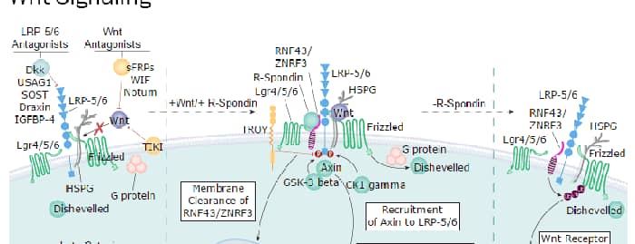
Wnt-3a is one of about 19 vertebrate members of the Wingless-type MMTV integration site (Wnt) family of highly conserved cysteine-rich secreted glycoproteins important for normal developmental processes (1‑3). Wnts bind to receptors of the Frizzled family in conjunction with a coreceptor of the low-density lipoprotein receptor-related protein family (LRP-5 or -6), or the Ryk atypical receptor tyrosine kinase (1, 4). Mouse Wnt-3a is a 44 kDa secreted hydrophobic glycoprotein containing a conserved pattern of 24 cysteine residues (5). Like other Wnts, Wnt-3a is modified by palmitate addition (at Cys 77) following glycosylation, which increases its hydrophobicity, secretion and activity (6, 7). A second site at Ser 209 is modified by palmitoleic acid and also contributes to activity and secretion (8). Mouse Wnt-3a shares 96% amino acid (aa) identity with human Wnt-3a, and 97% with bovine and canine Wnt-3a. The rat Wnt-3a precursor as it is apparently translated shares 100% aa identity with mouse Wnt-3a aa 63-352 (9). Wnt-3a also shares 87% aa identity with Wnt-3. During development, Wnt-3a is morphogen that is thought to coordinate somitogenesis and mesoderm boundary determination, and is expressed at the same locations and times as Wnt-2b and Wnt-5a (10). When Wnt-3a is deleted, mice fail to develop a hippocampus, and show defects in anterior-posterior patterning, somite development and tailbud formation (10-13). Recombinant Wnt-3a promotes proliferation of committed stem cell lineages in vitro, and may help maintain the cells in an undifferentiated state (6, 14) For example, Wnt-3a can induce self-renewal of hematopoietic stem cells, allowing expansion without further differentiation (6).
Wnt-3a Antibody (930769)
R&D Systems | Catalog # MAB9025

Scientific images may be resized, cropped, or adjusted for brightness or contrast for presentation purposes. These updates are limited to presentation and do not affect the underlying scientific interpretation of the data. Where available, additional source data may be provided upon request.
Key Product Details
Species Reactivity
Validated:
Cited:
Applications
Validated:
Cited:
Label
Antibody Source
Product Specifications
Immunogen
Ser19-Lys352
Accession # P27467
Specificity
Clonality
Host
Isotype
Endotoxin Level
Scientific Data Images for Wnt-3a Antibody (930769)
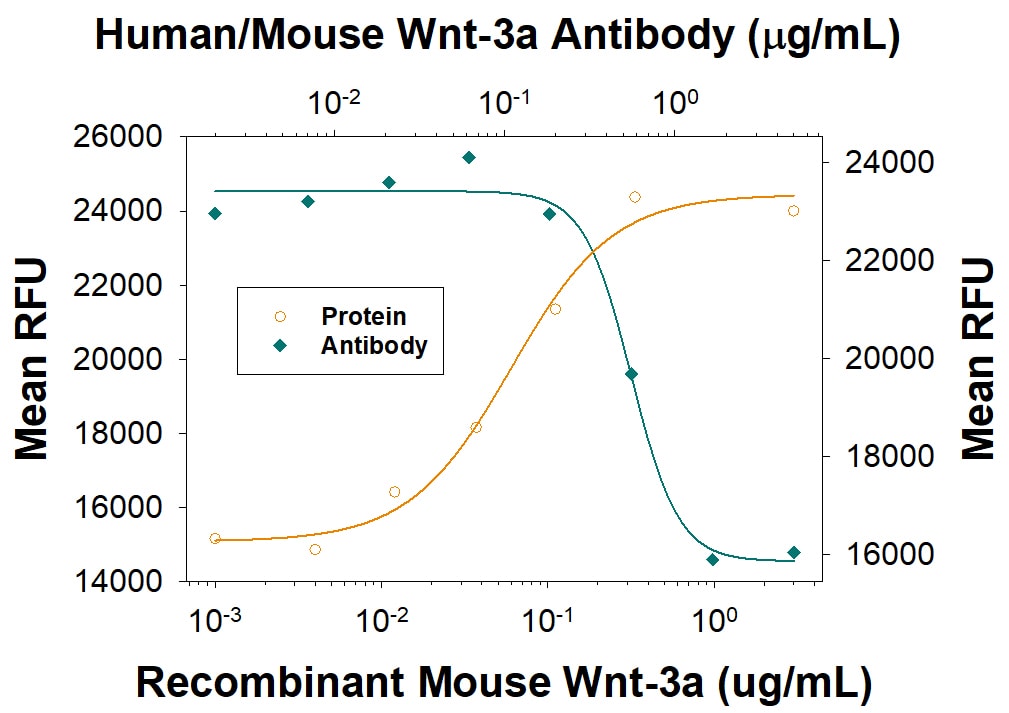
Topflash Reporter Activity Induced by Wnt-3a and Neutralization by Human/Mouse Wnt-3a Antibody.
Recombinant Mouse Wnt-3a (Catalog # 1324-WN) induces Topflash reporter activity in the HEK293T human embryonic kidney cell line in a dose-dependent manner (orange line), as measured by the Topflash Reporter Assay. Under these conditions, Topflash reporter activity elicited by Recombinant Mouse Wnt-3a (0.5 µg/mL) is neutralized (green line) by increasing concentrations of Rat Anti-Human/Mouse Wnt-3a Monoclonal Antibody (Catalog # MAB9025). The ND50 is typically 0.15-0.9 µg/mL.
Detection of Wnt-3a by Western Blot
STAT3 involvement in the Wnt3a-mediated enhancement of IL-4 response.(A) Proportion of differentially expressed genes (using RNA-seq) of bone marrow-derived macrophages (BMDMs) treated with IL-4 (green), Wnt3a (orange), or their combination (purple) compared to untreated cells. (B) Top eight GO terms of differentially expressed genes in Wnt3a and Wnt3a+IL-4-treated BMDMs. (C) Representative western blot image for phosphorylates (pY) and total STAT3 following treatment of BMDMs with Wnt3a for the indicated times (one out of two independent experiments). (D) BMDMs used in Figure 7F were examined for STAT3 expression using qPCR. (E) Representative western blot image for phosphorylates and total STAT3, as well as GAPDH, following treatment of BMDMs with Wnt3a or its conditioned media (CM) for the indicated times (one out of three independent experiments). Wnt CM was produced by treatment of BMDMs with Wnt3a for 16 hr. (F) BMDMs were treated with Wnt3a for 16 hr and added IL-4 for 2.5 hr thereafter, or with CM+IL-4 for 2.5 hr. Some CM was treated for 1 hr with a blocking antibody to Wnt3a prior to their exposure (in combination with IL4) to naïve BMDMs for 2.5 hr. Arg1 expression was determined with qPCR. p-Values were determined via one-way ANOVA, compared with IL-4 treatment, and Dunnett’s multiple comparison test (with repeated measurements). (G) BMDMs were treated with IL-4, with or without Wnt3a, and with blocking antibodies for transforming growth factor beta, C-X-C motif chemokine receptor 3, glycoprotein 103, IL-10 receptor, and hepatocyte growth factor receptor for 16 hr, and Arg1 expression was determined using real-time qPCR (n = 2–3 mice per treatment). Image collected and cropped by CiteAb from the following open publication (https://pubmed.ncbi.nlm.nih.gov/33720008), licensed under a CC-BY license. Not internally tested by R&D Systems.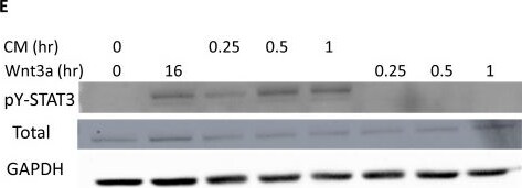
Detection of Wnt-3a by Western Blot
STAT3 involvement in the Wnt3a-mediated enhancement of IL-4 response.(A) Proportion of differentially expressed genes (using RNA-seq) of bone marrow-derived macrophages (BMDMs) treated with IL-4 (green), Wnt3a (orange), or their combination (purple) compared to untreated cells. (B) Top eight GO terms of differentially expressed genes in Wnt3a and Wnt3a+IL-4-treated BMDMs. (C) Representative western blot image for phosphorylates (pY) and total STAT3 following treatment of BMDMs with Wnt3a for the indicated times (one out of two independent experiments). (D) BMDMs used in Figure 7F were examined for STAT3 expression using qPCR. (E) Representative western blot image for phosphorylates and total STAT3, as well as GAPDH, following treatment of BMDMs with Wnt3a or its conditioned media (CM) for the indicated times (one out of three independent experiments). Wnt CM was produced by treatment of BMDMs with Wnt3a for 16 hr. (F) BMDMs were treated with Wnt3a for 16 hr and added IL-4 for 2.5 hr thereafter, or with CM+IL-4 for 2.5 hr. Some CM was treated for 1 hr with a blocking antibody to Wnt3a prior to their exposure (in combination with IL4) to naïve BMDMs for 2.5 hr. Arg1 expression was determined with qPCR. p-Values were determined via one-way ANOVA, compared with IL-4 treatment, and Dunnett’s multiple comparison test (with repeated measurements). (G) BMDMs were treated with IL-4, with or without Wnt3a, and with blocking antibodies for transforming growth factor beta, C-X-C motif chemokine receptor 3, glycoprotein 103, IL-10 receptor, and hepatocyte growth factor receptor for 16 hr, and Arg1 expression was determined using real-time qPCR (n = 2–3 mice per treatment). Image collected and cropped by CiteAb from the following open publication (https://pubmed.ncbi.nlm.nih.gov/33720008), licensed under a CC-BY license. Not internally tested by R&D Systems.Applications for Wnt-3a Antibody (930769)
Neutralization
Formulation, Preparation, and Storage
Purification
Reconstitution
Reconstitute at 0.5 mg/mL in sterile PBS. For liquid material, refer to CoA for concentration.
Formulation
Shipping
Stability & Storage
- 12 months from date of receipt, -20 to -70 °C as supplied.
- 1 month, 2 to 8 °C under sterile conditions after reconstitution.
- 6 months, -20 to -70 °C under sterile conditions after reconstitution.
Calculators
Background: Wnt-3a
References
- Mikels, A.J. and R. Nusse (2006) Oncogene 25:7461.
- Miller, J.R. (2001) Genome Biol. 3:3001.
- Roelink, H. and R. Nusse (1991) Genes Dev. 5:381.
- Lu, W et al. (2004) Cell 119:97.
- Smolich, B.D. et al. (1993) Mol. Biol Cell 4:1267.
- Willert, K. et al. (2003) Nature 423:448.
- Komekado, H. et al. (2007) Genes Cells 12:521.
- Takada, R. et al. (2006) Dev. Cell 11:791.
- Entrez Accession # NP_001100475.
- Dunty, W.C. et al. (2008) Development 135:85.
- Lee, S.M.K. et al. (2000) Development 127:457.
- Takada, S. et al. (1994) Genes Dev. 8:174.
- Ikeya, M. and S. Takada (2001) Mech. Dev. 103:27.
- Nusse, R. (2008) Cell Res. 18:523.
Long Name
Alternate Names
Gene Symbol
UniProt
Additional Wnt-3a Products
Product Documents for Wnt-3a Antibody (930769)
Certificate of Analysis
To download a Certificate of Analysis, please enter a lot or batch number in the search box below.
Note: Certificate of Analysis not available for kit components.
Product Specific Notices for Wnt-3a Antibody (930769)
For research use only
Related Research Areas
Citations for Wnt-3a Antibody (930769)
 Powered by Bioz
Customer Reviews for Wnt-3a Antibody (930769)
There are currently no reviews for this product. Be the first to review Wnt-3a Antibody (930769) and earn rewards!
Have you used Wnt-3a Antibody (930769)?
Submit a review and receive an Amazon gift card!
$25/€18/£15/$25CAN/¥2500 for a review with an image
$10/€7/£6/$10CAN/¥1110 for a review without an image
Submit a review