Mouse Pure-Blot anti-Rabbit IgG (H+L) Secondary Antibody (eB182) [FITC]
Novus Biologicals | Catalog # NBP3-11665
Key Product Details
Species Reactivity
Rabbit
Applications
Western Blot, Immunocytochemistry/ Immunofluorescence, Immunoprecipitation
Label
FITC (Excitation = 495 nm, Emission = 519 nm)
Antibody Source
Monoclonal Mouse IgG Clone # eB182
Loading...
Product Specifications
Immunogen
Rabbit IgG
Clonality
Monoclonal
Host
Mouse
Isotype
IgG
Description
This secondary antibody was prepared from tissue culture supernatant by Protein G affinity chromatography. Assay by Immunoelectrophoresis resulted in a single precipitin arc against anti-fluorescein and Anti-Rabbit Serum.
Store vial at 4C prior to restoration. For extended storage aliquot contents and freeze at -20C or below. Avoid cycles of freezing and thawing. Centrifuge product if not completely clear after standing at room temperature. This product is stable for several weeks at 4C as an undiluted liquid. Dilute only prior to immediate use.
Store vial at 4C prior to restoration. For extended storage aliquot contents and freeze at -20C or below. Avoid cycles of freezing and thawing. Centrifuge product if not completely clear after standing at room temperature. This product is stable for several weeks at 4C as an undiluted liquid. Dilute only prior to immediate use.
Scientific Data Images
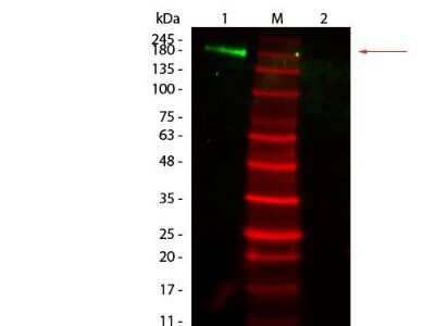
Western Blot: Mouse Pure-Blot anti-Rabbit IgG (H+L) Secondary Antibody (eB182) [FITC] [NBP3-11665] - Western Blot of Mouse Pure-Blot anti-Rabbit IgG Secondary antibody (eB182) [FITC]. Lane 1: Rabbit IgG, Non-reduced. Lane 2: Rabbit IgG, Reduced. Load: 50 ng per lane. Primary antibody: none. Secondary antibody: Mouse Pure-Blot anti-Rabbit IgG Secondary antibody (eB182) [FITC] at 1:1,000 for 60 min at RT. Block for 30 min at RT. Predicted/Observed size: 160 kDa for Rabbit IgG, Non-reduced. Other band(s): none.
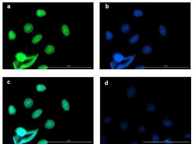
Immunocytochemistry/Immunofluorescence: Mouse Pure-Blot anti-Rabbit IgG (H+L) Secondary Antibody (eB182) [FITC] [NBP3-11665] - Immunofluorescence microscopy of BCL3 in Caco-2 cells using FITC-conjugated Mouse Pure-Blot anti-Rabbit IgG Secondary antibody (eB182) [FITC] for detection. Caco-2 cells were fixed with 4% PFA, blocked (5% mouse serum/0.3% Triton X-100 in 1X PBS ) for 1 hr, then incubated with 15 ug/mL of anti-BCL3 primary antibody at 4C overnight. Following 3 washes in 1X PBS for 5 min each, 5 ug/mL of FITC-conjugated Mouse Pure-Blot anti-Rabbit IgG Secondary antibody (eB182) [FITC] was added and allowed to incubate for 1 hr at room temperature. Nuclei were counterstained with DAPI present in mounting medium. The predicted main localization is nucleoplasm. Additional localization in some cell types includes vesicles and midbody. (a) BCL3 (b) DAPI (c) merged DAPI/BCL3 (d) secondary antibody only. Image taken at 40X magnification.
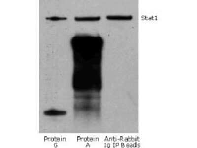
Western Blot: Mouse Pure-Blot anti-Rabbit IgG (H+L) Secondary Antibody (eB182) [FITC] [NBP3-11665] - Jurkat cell lysate (0.5 ml of 1x10e7 cells/ml) was incubated with rabbit anti-human Stat1 and immunoprecipitated using Protein G, Protein A and Anti-Rabbit Ig IP Beads. Precipitate from 5x10e5 cells was subjected to electrophoresis, transferred to a PVDF membrane, and Western blotted with anti-Stat1 using Mouse Pure-Blot anti-Rabbit IgG Secondary antibody (eB182) [FITC].
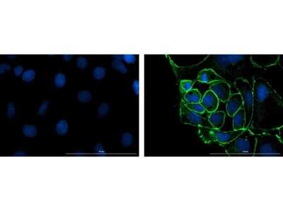
Immunocytochemistry/Immunofluorescence: Mouse Pure-Blot anti-Rabbit IgG (H+L) Secondary Antibody (eB182) [FITC] [NBP3-11665] - Immunofluorescence microscopy of ZO-1 in Caco-2 cells using FITC-conjugated Mouse Pure-Blot anti-Rabbit IgG Secondary antibody (eB182) [FITC] for detection. Caco-2 cells were fixed with 4% PFA, blocked (5% mouse serum/0.3% Triton X-100 in 1X PBS) for 1 hr, then incubated with 15 ug/mL of anti-ZO-1 primary antibody at 4C overnight. Following 3 washes in 1X PBS for 5 min each, 5 ug/mL of FITC-conjugated Mouse Pure-Blot anti-Rabbit IgG Secondary antibody (eB182) [FITC] was added and allowed to incubate for 1 hr at room temperature. Nuclei were counterstained with DAPI present in mounting medium. Predicted cell localization is cell membrane and cell junctions. Image taken at 40X magnification. (right) Merged DAPI (blue)/ZO-1 (green), image shown (left) secondary antibody only.
Applications
Application
Recommended Usage
Immunocytochemistry/ Immunofluorescence
1:500 - 1:2500
Western Blot
1:1000
Application Notes
This secondary antibody has been tested in immunofluorescence microscopy, fluorescent western blotting, and immunoprecipitation and are suitable for fluorescence based plate assays (FLISA, multiplex analysis, including multicolor imaging, utilizing various commercial platforms. Fluorescent Rabbit Pure-Blot Antibody Fluorescein may also be used for detection in immunoassays that do not employ immunoprecipitation. Fluorescent Rabbit Pure-Blot Antibody Fluorescein is provided as a lyophilized powder. To conserve reagent, we recommend incubating the blots from minigels in sealed bags (removing as much air as possible) with minimal volume (2-3 mLs). If used conservatively at 2.5mLs/blot will yield enough reagent for 40 blots. Note that there are three key procedural considerations: 1. Protein A or G should not be used for the immunoprecipitation. Use of protein A or G beads with the rabbit Pure-Blot will result in contaminating bands. For immunoprecipitation, Anti-rat IgG beads or Anti-rabbit IgG beads should be used for rat or rabbit immunoprecipitating antibodies, respectively. 2. Immunoprecipitate should be completely reduced. 3. blocking buffer for Fluorescent Western Blotting should be used as the blocking protein for the immunoblot. All recommended dilutions for listed applications are intended as an initial recommendation, specific conditions for each protein and antibody combination should be specifically optimized by the end user.
Formulation, Preparation, and Storage
Purification
Protein G purified
Reconstitution
Reconstitute with 100 ul deionized water (or equivalent).
Formulation
Lyophilized from 0.02 M Potassium Phosphate, 0.15 M Sodium Chloride, pH 7.2, 10 mg/ml Polyethylene Glycol (PEG-8000)
Preservative
0.01% Sodium Azide
Concentration
LYOPH mg/ml
Shipping
The product is shipped with polar packs. Upon receipt, store it immediately at the temperature recommended below.
Stability & Storage
Store at 4C short term. Aliquot and store at -20C long term. Avoid freeze-thaw cycles.
Calculators
Background: IgG (H+L)
The 4 IgG subclasses, sharing 95% amino acid identity, include IgG1, IgG2, IgG3, and IgG4 for humans and IgG1, IgG2a, IgG2b, and IgG3 for mice. The relative abundance of each human subclass is 60% for IgG1, 32% for IgG2, 4% for IgG3, and 4% for IgG4. In an IgG deficiency, there may be a shortage of one or more subclasses (4).
References
1. Painter RH. (1998) Encyclopedia of Immunology (Second Edition). Elsevier. 1208-1211
2. Chapter 9 - Antibodies. (2012) Immunology for Pharmacy. Mosby 70-78
3. Schroeder H, Cavacini, L. (2010) Structure and Function of Immunoglobulins. J Allergy Clin Immunol. 125(2 0 2): S41-S52. PMID: 20176268
4. Vidarsson G, Dekkers G, Rispens T. (2014) IgG subclasses and allotypes: from structure to effector functions. Front Immunol. 5:520. PMID: 25368619
Alternate Names
IP Detection Reagent
Additional IgG (H+L) Products
Product Documents
Certificate of Analysis
To download a Certificate of Analysis, please enter a lot or batch number in the search box below.
Product Specific Notices
This product is for research use only and is not approved for use in humans or in clinical diagnosis. Secondary Antibodies are guaranteed for 1 year from date of receipt.
Customer Reviews
There are currently no reviews for this product. Be the first to review Mouse Pure-Blot anti-Rabbit IgG (H+L) Secondary Antibody (eB182) [FITC] and earn rewards!
Have you used Mouse Pure-Blot anti-Rabbit IgG (H+L) Secondary Antibody (eB182) [FITC]?
Submit a review and receive an Amazon gift card!
$25/€18/£15/$25CAN/¥2500 Yen for a review with an image
$10/€7/£6/$10CAN/¥1110 Yen for a review without an image
Submit a review
Protocols
Find general support by application which include: protocols, troubleshooting, illustrated assays, videos and webinars.
- Appropriate Fixation of IHC/ICC Samples
- Cellular Response to Hypoxia Protocols
- ClariTSA™ Fluorophore Kits
- Detection & Visualization of Antibody Binding
- ICC Cell Smear Protocol for Suspension Cells
- ICC Immunocytochemistry Protocol Videos
- ICC for Adherent Cells
- Immunocytochemistry (ICC) Protocol
- Immunocytochemistry Troubleshooting
- Immunofluorescence of Organoids Embedded in Cultrex Basement Membrane Extract
- Immunohistochemistry (IHC) and Immunocytochemistry (ICC) Protocols
- Immunoprecipitation Protocol
- Preparing Samples for IHC/ICC Experiments
- Preventing Non-Specific Staining (Non-Specific Binding)
- Primary Antibody Selection & Optimization
- Protocol for VisUCyte™ HRP Polymer Detection Reagent
- Protocol for the Fluorescent ICC Staining of Cell Smears - Graphic
- Protocol for the Fluorescent ICC Staining of Cultured Cells on Coverslips - Graphic
- Protocol for the Preparation and Fluorescent ICC Staining of Cells on Coverslips
- Protocol for the Preparation and Fluorescent ICC Staining of Non-adherent Cells
- Protocol for the Preparation and Fluorescent ICC Staining of Stem Cells on Coverslips
- Protocol for the Preparation of a Cell Smear for Non-adherent Cell ICC - Graphic
- R&D Systems Quality Control Western Blot Protocol
- TUNEL and Active Caspase-3 Detection by IHC/ICC Protocol
- The Importance of IHC/ICC Controls
- Troubleshooting Guide: Western Blot Figures
- Western Blot Conditions
- Western Blot Protocol
- Western Blot Protocol for Cell Lysates
- Western Blot Troubleshooting
- Western Blot Troubleshooting Guide
- View all Protocols, Troubleshooting, Illustrated assays and Webinars
Loading...