

![Western Blot: Luciferase Antibody [NB110-17348]](https://resources.rndsystems.com/images/products/Luciferase-Antibody-Western-Blot-NB110-17348-img0003.jpg "Western Blot: Luciferase Antibody [NB110-17348]")
Scientific images may be resized, cropped, or adjusted for brightness or contrast for presentation purposes. These updates are limited to presentation and do not affect the underlying scientific interpretation of the data. Where available, additional source data may be provided upon request.
Loading...
Key Product Details
Species Reactivity
Firefly
Applications
Western Blot, Immunocytochemistry/ Immunofluorescence
Label
Unconjugated
Antibody Source
Polyclonal Rabbit IgG
Format
BSA Free
Loading...
Product Specifications
Immunogen
This Luciferase Antibody was developed against full length native protein (purified) (Firefly (Photinus pyralis)).
Localization
Peroxisomal
Specificity
This Luciferase Antibody identifies recombinant luciferase in eukaryotic cells transfected with a plasmid bearing the luciferase gene.
Clonality
Polyclonal
Host
Rabbit
Isotype
IgG
Scientific Data Images for Luciferase Antibody - BSA Free
Western Blot: Luciferase Antibody [NB110-17348]
Western Blot: Luciferase Antibody [NB110-17348] - Lysates of HEK-293T cells overexpressing luciferase was separated on SDS-PAGE and probed with Anti-Luciferase The antibody was developed using Goat Anti-Rabbit IgG-Alkaline Phosphatase and a colorimetric substrate. Lanes 1. Antibody dilution 1:1,000 2. Antibody dilution 1:1,000 + luciferase immunizing peptide![Immunocytochemistry/ Immunofluorescence: Luciferase Antibody [NB110-17348]](https://resources.rndsystems.com/images/products/Luciferase-Antibody-Immunocytochemistry-Immunofluorescence-NB110-17348-img0004.jpg "Immunocytochemistry/ Immunofluorescence: Luciferase Antibody [NB110-17348]")
Immunocytochemistry/ Immunofluorescence: Luciferase Antibody [NB110-17348]
Immunocytochemistry/Immunofluorescence: Luciferase Antibody [NB110-17348] - HEK-293T cells overexpressing luciferase were fixed and stained 10 ug/mL Anti-Luciferase. The antibody was developed using Goat Anti-Rabbit IgG, FITC conjugate.Applications for Luciferase Antibody - BSA Free
Application
Recommended Usage
Immunocytochemistry/ Immunofluorescence
1:500-1:1000
Western Blot
1:10000
Application Notes
ICC/IF: A working dilution of 1:5001:1,000 is obtained on methanol-acetone fixed transfected cells using an FITC conjugated secondary antibody. Cells were transfected with a reporter plasmid containing the gene for luciferase.
Reviewed Applications
Read 1 review rated 1 using NB110-17348 in the following applications:
Formulation, Preparation, and Storage
Purification
IgG purified
Formulation
10mM PBS (pH 7.4)
Format
BSA Free
Preservative
0.09% Sodium Azide
Concentration
Please see the vial label for concentration. If unlisted please contact technical services.
Shipping
The product is shipped with polar packs. Upon receipt, store it immediately at the temperature recommended below.
Stability & Storage
Store at 4C short term. Aliquot and store at -20C long term. Avoid freeze-thaw cycles.
Background: Luciferase
The luciferase assay is fast and sensitive, differentiating itself from the CAT (chloramphenicol acetyltransferase) assay because it does not require a radioactive substrate.
References
1. Eun, H. (1996). Marker/Reporter enzymes. Enzymology Primer for Recombinant DNA Technology, 567-645. doi:10.1016/b978-012243740-3/50011-9
2. McNabb, D. S., Reed, R., & Marciniak, R. A. (2005). Dual luciferase assay system for rapid assessment of gene expression in Saccharomyces cerevisiae. Eukaryotic Cell, 4(9), 1539-1549. doi:10.1128/ec.4.9.1539-1549.2005
3. Fraga, H. (2008). Firefly luminescence: A historical perspective and recent developments. Photochemical & Photobiological Sciences, 7(2), 146-158. doi:10.1039/b719181b
4. Younes, A., Lukyanenko, Y. O., Lyashkov, A. E., Lakatta, E. G., & Sollott, S. J. (2011). A bioluminescence method for direct measurement of phosphodiesterase activity. Analytical Biochemistry, 417(1), 36-40. doi:10.1016/j.ab.2011.05.036
Alternate Names
LuC, luciferin 4 monooxygenase, Luciferin 4-monooxygenase
Additional Luciferase Products
Product Documents for Luciferase Antibody - BSA Free
Certificate of Analysis
To download a Certificate of Analysis, please enter a lot or batch number in the search box below.
Product Specific Notices for Luciferase Antibody - BSA Free
This product is for research use only and is not approved for use in humans or in clinical diagnosis. Primary Antibodies are guaranteed for 1 year from date of receipt.
Customer Reviews for Luciferase Antibody - BSA Free (1)
1 out of 5
1 Customer Rating
Have you used Luciferase Antibody - BSA Free?
Submit a review and receive an Amazon gift card!
$25/€18/£15/$25CAN/¥2500 for a review with an image
$10/€7/£6/$10CAN/¥1110 for a review without an image
Submit a review
Customer Images
Showing
1
-
1 的
1 review
Showing All
Filter By:
-
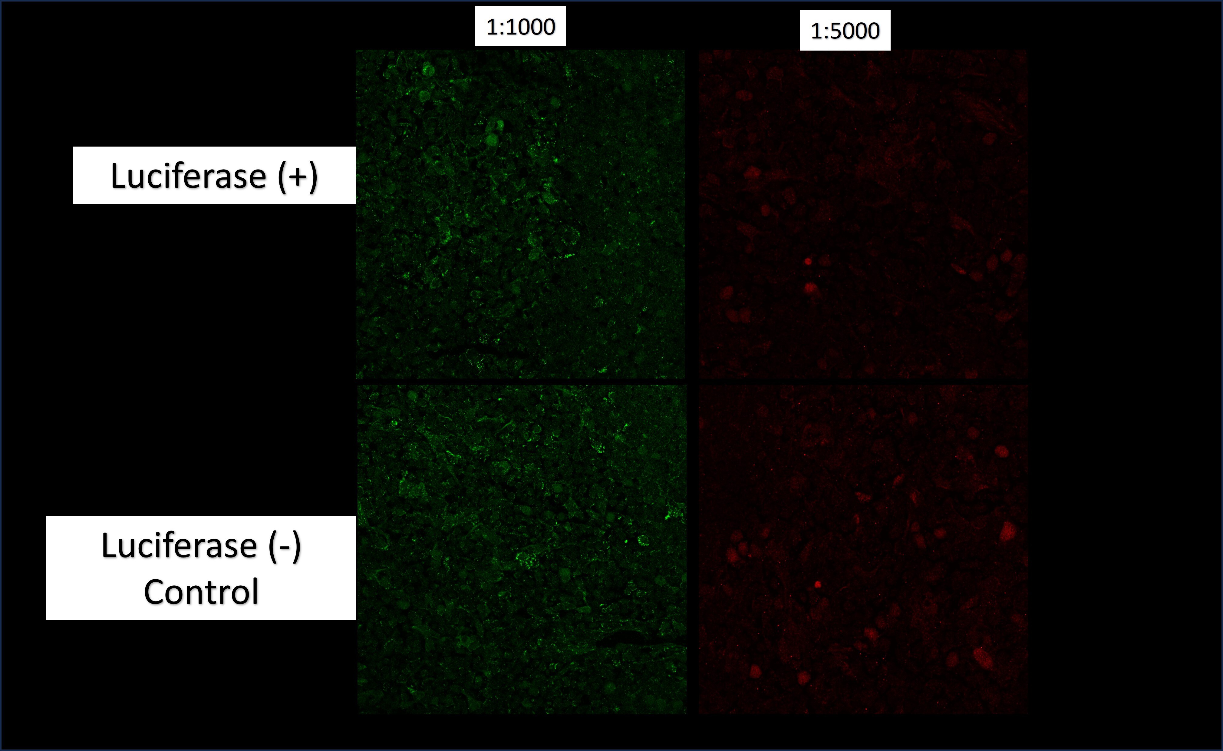
Application: Immunofluorescence-FrozenSample Tested: Mouse spleen, mouse thymus and Thymus tissueSpecies: MouseVerified Customer | Posted 01/31/2024Top: Luc+ mouse thymus stained with anti-luc 1:1K or 1:5K followed by incubation with anti-rabbit 488 L or 555 R. Bottom: WT mouse thymus stained with anti-luc 1:1K or 1:5K followed by incubation with anti-rabbit 488 L or 555 R.Frozen Sections were fixed for 10 min in 4% PFA at room temp followed by incubation in 100% Methanol at -20 C for 10 min. Sections were washed and blocked using 10% Donkey Serum or 2% Fish Gelatin/10% Donkey Serum. Tested Primary Dilutions: 1:500, 1:1000, and 1:5000 overnight at 4 C. Secondary Tested: Preabsorbed Anti-Rabbit 488 or Rabbit 555. Tissue collected from transgenic mouse expressing luciferase from CAG promoter. Luciferase activity was confirmed by measuring luminescence from collected tissue before embedding in OCT.
 Bio-Techne ResponseThis review was submitted through the legacy Novus Innovators Program, reflecting a new species or application tested on a primary antibody.
There are no reviews that match your criteria.
Protocols
Find general support by application which include: protocols, troubleshooting, illustrated assays, videos and webinars.
- Appropriate Fixation of IHC/ICC Samples
- Cellular Response to Hypoxia Protocols
- ClariTSA™ Fluorophore Kits
- Detection & Visualization of Antibody Binding
- ICC Cell Smear Protocol for Suspension Cells
- ICC Immunocytochemistry Protocol Videos
- ICC for Adherent Cells
- Immunocytochemistry (ICC) Protocol
- Immunocytochemistry Troubleshooting
- Immunofluorescence of Organoids Embedded in Cultrex Basement Membrane Extract
- Immunohistochemistry (IHC) and Immunocytochemistry (ICC) Protocols
- Preparing Samples for IHC/ICC Experiments
- Preventing Non-Specific Staining (Non-Specific Binding)
- Primary Antibody Selection & Optimization
- Protocol for VisUCyte™ HRP Polymer Detection Reagent
- Protocol for the Fluorescent ICC Staining of Cell Smears - Graphic
- Protocol for the Fluorescent ICC Staining of Cultured Cells on Coverslips - Graphic
- Protocol for the Preparation and Fluorescent ICC Staining of Cells on Coverslips
- Protocol for the Preparation and Fluorescent ICC Staining of Non-adherent Cells
- Protocol for the Preparation and Fluorescent ICC Staining of Stem Cells on Coverslips
- Protocol for the Preparation of a Cell Smear for Non-adherent Cell ICC - Graphic
- R&D Systems Quality Control Western Blot Protocol
- TUNEL and Active Caspase-3 Detection by IHC/ICC Protocol
- The Importance of IHC/ICC Controls
- Troubleshooting Guide: Western Blot Figures
- Western Blot Conditions
- Western Blot Protocol
- Western Blot Protocol for Cell Lysates
- Western Blot Troubleshooting
- Western Blot Troubleshooting Guide
- View all Protocols, Troubleshooting, Illustrated assays and Webinars
Loading...