
Mouse CD90/Thy1 Antibody
R&D Systems | Catalog # AF7335

Scientific images may be resized, cropped, or adjusted for brightness or contrast for presentation purposes. These updates are limited to presentation and do not affect the underlying scientific interpretation of the data. Where available, additional source data may be provided upon request.
Key Product Details
Species Reactivity
Validated:
Cited:
Applications
Validated:
Cited:
Label
Antibody Source
Product Specifications
Immunogen
Gln20-Lys130
Accession # P01831
Specificity
Clonality
Host
Isotype
Scientific Data Images for Mouse CD90/Thy1 Antibody
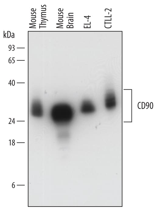
Detection of Mouse CD90/Thy1 by Western Blot.
Western blot shows lysates of mouse thymus tissue, mouse brain tissue, EL-4 mouse lymphoblast cell line, and CTLL-2 mouse cytotoxic T cell line. PVDF membrane was probed with 0.2 µg/mL of Sheep Anti-Mouse CD90/Thy1 Antigen Affinity-purified Polyclonal Antibody (Catalog # AF7335) followed by HRP-conjugated Anti-Sheep IgG Secondary Antibody (Catalog # HAF016). A specific band was detected for CD90/Thy1 at approximately 25-35 kDa (as indicated). This experiment was conducted under reducing conditions and using Immunoblot Buffer Group 1.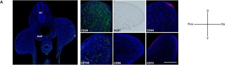
Detection of CD90/Thy1 by Immunohistochemistry
In vivo detection of two MSP limb bud subpopulations based on MSC-am localization. (A) Transversal section of an E10.5 mouse embryo to show the histological organization at this stage; the immunodetections of CD29, SCA-1, CD44, CD105, CD90, and CD73 markers in E10.5 limb buds are shown. The limb bud for each MSC-am marker corresponds to the region inside the square in panel (A). (B) Dot plots of the multiparametric flow cytometry analysis showing the hierarchy to identify sSca+ and sSca– subpopulations in E10.5, E11.5, and E12.5 mouse limbs. The number of positive cells for each subpopulation is expressed in percentage; ±mean standard deviation. (C) Comparison of the expression percentage of the sSca+ subpopulation between E10.5, E11.5, and E12.5 limbs. Statistical significance was set as follows: E10.5 vs. E11.5 p > 0.0061; 10.5 vs. 12.5 p > 0.0023; E11.5 vs. E12.5 p > 0.4079, **p < 0.005. NT, neural tube; AGM, aorta-gonad-mesonephros; Do, dorsal; V, ventral; Pr, proximal; D, distal. In (A) AER is marked with an asterisk. The scale bar is set at 200 μm and is representative for all images. Image collected and cropped by CiteAb from the following open publication (https://pubmed.ncbi.nlm.nih.gov/34336823), licensed under a CC-BY license. Not internally tested by R&D Systems.Applications for Mouse CD90/Thy1 Antibody
Western Blot
Sample: Mouse thymus tissue, mouse brain tissue, EL‑4 mouse lymphoblast cell line, and CTLL‑2 mouse cytotoxic T cell line
Formulation, Preparation, and Storage
Purification
Reconstitution
Sterile PBS to a final concentration of 0.2 mg/mL. For liquid material, refer to CoA for concentration.
Formulation
Shipping
Stability & Storage
- 12 months from date of receipt, -20 to -70 °C as supplied.
- 1 month, 2 to 8 °C under sterile conditions after reconstitution.
- 6 months, -20 to -70 °C under sterile conditions after reconstitution.
Calculators
Background: CD90/Thy1
Alternate Names
Gene Symbol
UniProt
Additional CD90/Thy1 Products
Product Documents for Mouse CD90/Thy1 Antibody
Certificate of Analysis
To download a Certificate of Analysis, please enter a lot or batch number in the search box below.
Note: Certificate of Analysis not available for kit components.
Product Specific Notices for Mouse CD90/Thy1 Antibody
For research use only
Citations for Mouse CD90/Thy1 Antibody
 Powered by Bioz
Customer Reviews for Mouse CD90/Thy1 Antibody
There are currently no reviews for this product. Be the first to review Mouse CD90/Thy1 Antibody and earn rewards!
Have you used Mouse CD90/Thy1 Antibody?
Submit a review and receive an Amazon gift card!
$25/€18/£15/$25CAN/¥2500 for a review with an image
$10/€7/£6/$10CAN/¥1110 for a review without an image
Submit a review
Protocols
Find general support by application which include: protocols, troubleshooting, illustrated assays, videos and webinars.
- Cellular Response to Hypoxia Protocols
- R&D Systems Quality Control Western Blot Protocol
- Troubleshooting Guide: Western Blot Figures
- Western Blot Conditions
- Western Blot Protocol
- Western Blot Protocol for Cell Lysates
- Western Blot Troubleshooting
- Western Blot Troubleshooting Guide
- View all Protocols, Troubleshooting, Illustrated assays and Webinars



