
Reg1, also known as pancreatic thread protein (PTP), pancreatic stone protein (PSP) and lithostathine, is the founding member of the Reg family of proteins, which are secreted proteins with a C-type lectin domain. Reg1 is expressed during islet regeneration. Reg1 functions in an autocrine/paracrine fashion via a cell surface Reg receptor that mediates a growth signal for beta -cell regeneration (1).

Scientific images may be resized, cropped, or adjusted for brightness or contrast for presentation purposes. These updates are limited to presentation and do not affect the underlying scientific interpretation of the data. Where available, additional source data may be provided upon request.
Key Product Details
Species Reactivity
Validated:
Mouse
Cited:
Mouse
Applications
Validated:
Western Blot
Cited:
Immunohistochemistry, Western Blot
Label
Unconjugated
Antibody Source
Polyclonal Sheep IgG
Loading...
Product Specifications
Immunogen
E. coli-derived recombinant mouse Reg1
Gln22-Gly165
Accession # P43137
Gln22-Gly165
Accession # P43137
Specificity
Detects mouse Reg1 in direct ELISAs and Western blots. In Western blots, approximately 10% cross-reactivity with recombinant mouse (rm) Reg2 is observed and less than 5% cross-reactivity with rmReg3A, recombinant human (rh) Reg1B, rhReg4, recombinant rat (rr) Reg3 and rrReg2 is observed.
Clonality
Polyclonal
Host
Sheep
Isotype
IgG
Scientific Data Images for Mouse Reg1 Antibody
Detection of Mouse Reg1 by Western Blot
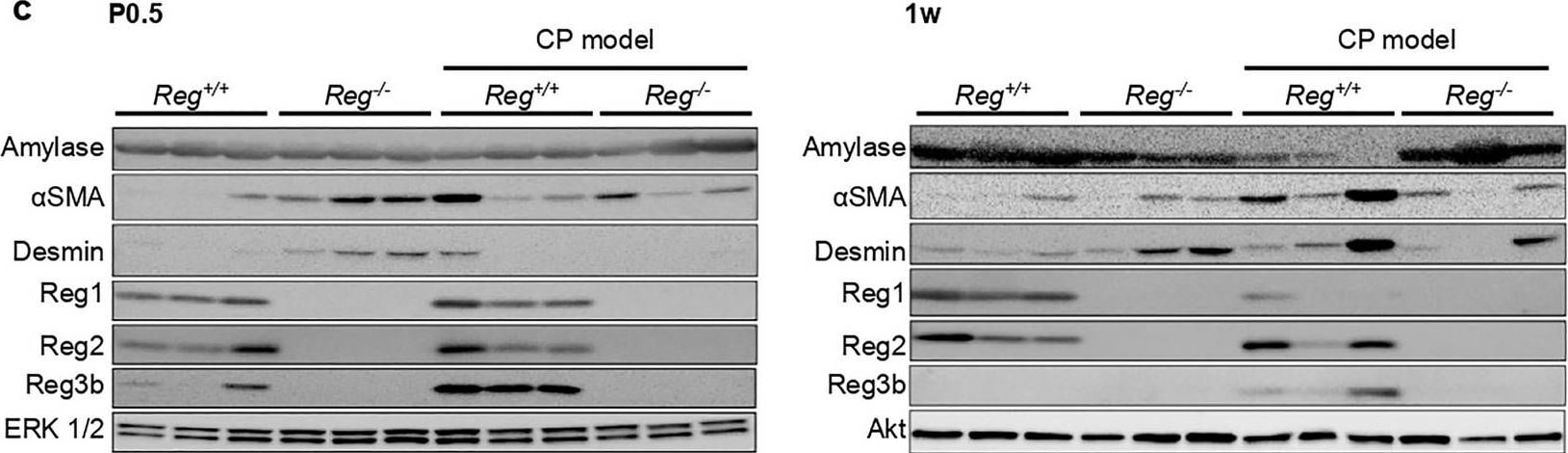
Reg family proteins stimulated PSCs activation contributing to fibrosis in chronic pancreatitis. (a) Immunofluorescence analysis of alpha-smooth muscle actin ( alpha SMA, an activated PSCs marker, red) in the pancreas sections of indicated genotypes at 2 weeks, and 4 weeks (n = 3–4 mice). Nuclei were counterstained by DAPI (blue). (b) The percentage of epithelial cells with alpha SMA positive signals. Results represent mean ± SEM (n = 3–4 mice). Statistical analysis was performed by one-way ANOVA with Tukey’s multiple comparison tests among four groups. **P < 0.01 (c) Cropped images of western blot gels of Amylase, alpha SMA, Desmin, Reg1, Reg2, Reg3b of the pancreas of indicated genotypes at 0.5 day after birth(P0.5), 1w and 4w, p44/42 MAPK (ERK 1/2) and Akt were used as the loading control (n = 3 mice). The samples derived from the same experiment and gels were processed in parallel. Images of the entire gels are presented in Supplementary Fig. 5–7. The right panel showed the respective densitometric quantification analysis of the relative intensity of alpha SMA. Densitometric quantification analysis of other proteins is presented in Supplementary Fig. 4.Results represent mean ± SEM (n = 3 mice). Statistical analysis was performed by one-way ANOVA with Tukey’s multiple comparison tests among four groups. ns: not significant; #P < 0.0001. Image collected and cropped by CiteAb from the following open publication (https://pubmed.ncbi.nlm.nih.gov/37500741), licensed under a CC-BY license. Not internally tested by R&D Systems.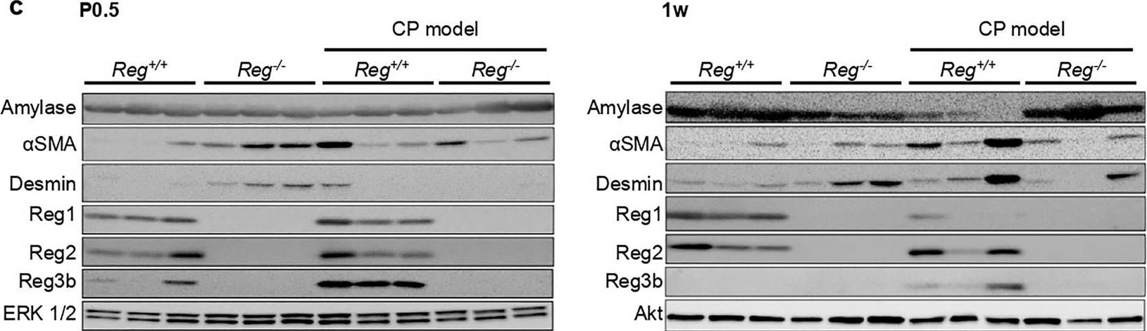
Detection of Mouse Reg1 by Western Blot
Reg family proteins stimulated PSCs activation contributing to fibrosis in chronic pancreatitis. (a) Immunofluorescence analysis of alpha-smooth muscle actin ( alpha SMA, an activated PSCs marker, red) in the pancreas sections of indicated genotypes at 2 weeks, and 4 weeks (n = 3–4 mice). Nuclei were counterstained by DAPI (blue). (b) The percentage of epithelial cells with alpha SMA positive signals. Results represent mean ± SEM (n = 3–4 mice). Statistical analysis was performed by one-way ANOVA with Tukey’s multiple comparison tests among four groups. **P < 0.01 (c) Cropped images of western blot gels of Amylase, alpha SMA, Desmin, Reg1, Reg2, Reg3b of the pancreas of indicated genotypes at 0.5 day after birth(P0.5), 1w and 4w, p44/42 MAPK (ERK 1/2) and Akt were used as the loading control (n = 3 mice). The samples derived from the same experiment and gels were processed in parallel. Images of the entire gels are presented in Supplementary Fig. 5–7. The right panel showed the respective densitometric quantification analysis of the relative intensity of alpha SMA. Densitometric quantification analysis of other proteins is presented in Supplementary Fig. 4.Results represent mean ± SEM (n = 3 mice). Statistical analysis was performed by one-way ANOVA with Tukey’s multiple comparison tests among four groups. ns: not significant; #P < 0.0001. Image collected and cropped by CiteAb from the following open publication (https://pubmed.ncbi.nlm.nih.gov/37500741), licensed under a CC-BY license. Not internally tested by R&D Systems.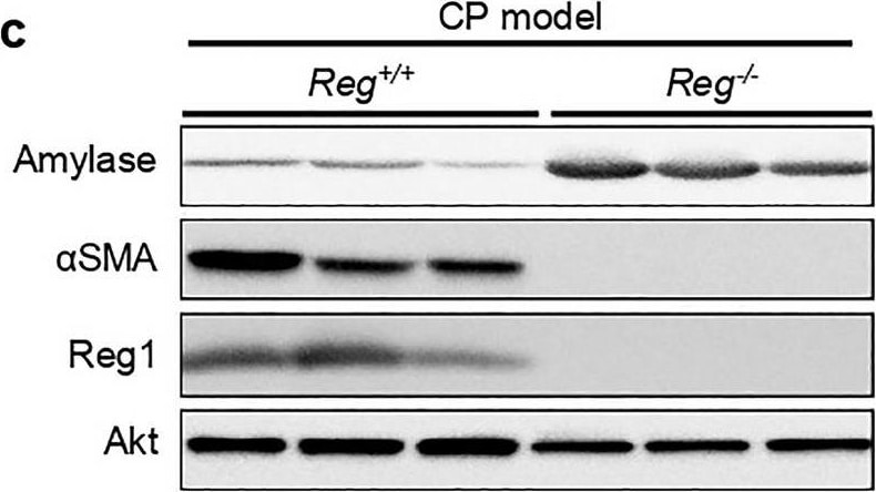
Detection of Mouse Reg1 by Western Blot
Reg deficiency led to the remission of chronic pancreatitis. (a) Representative pancreatic tissue section from mice of the indicated genotype at 8 weeks was stained with H&E or Azan (for collagen, dark blue) and Immunofluorescence staining of alpha-smooth muscle actin ( alpha SMA, an activated PSCs marker, red) (n = 5 mice). Scale bars: H&E, 50 μm; Azan, 100 μm (b) Pancreatic mRNA expression of proinflammatory cytokines interleukin 6(Il6), tumor necrosis factor a (Tnfa) and Il1b of indicated genotypes were assessed by qRT-PCR. The expression amounts of each gene were calculated relative to those of ribosomal protein S3 (Rps3) with the fold change to CP model_Reg+/+ mice. Mean ± SEM (n = 5 mice). Statistical analysis was performed by two-tailed unpaired Student t-test between two groups. *P < 0.05 (c) Cropped images of western blot gels of Amylase, alpha SMA and Reg1 of the pancreas of indicated genotypes at 8 weeks and Akt was used as the loading control (n = 5 mice). The samples derived from the same experiment and gels were processed in parallel. Images of the entire gels and respective densitometric quantification analysis of the relative intensity of protein are presented in Supplementary Fig. 4,8. Image collected and cropped by CiteAb from the following open publication (https://pubmed.ncbi.nlm.nih.gov/37500741), licensed under a CC-BY license. Not internally tested by R&D Systems.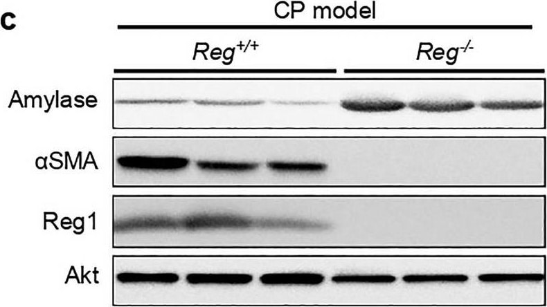
Detection of Mouse Reg1 by Western Blot
Reg deficiency led to the remission of chronic pancreatitis. (a) Representative pancreatic tissue section from mice of the indicated genotype at 8 weeks was stained with H&E or Azan (for collagen, dark blue) and Immunofluorescence staining of alpha-smooth muscle actin ( alpha SMA, an activated PSCs marker, red) (n = 5 mice). Scale bars: H&E, 50 μm; Azan, 100 μm (b) Pancreatic mRNA expression of proinflammatory cytokines interleukin 6(Il6), tumor necrosis factor a (Tnfa) and Il1b of indicated genotypes were assessed by qRT-PCR. The expression amounts of each gene were calculated relative to those of ribosomal protein S3 (Rps3) with the fold change to CP model_Reg+/+ mice. Mean ± SEM (n = 5 mice). Statistical analysis was performed by two-tailed unpaired Student t-test between two groups. *P < 0.05 (c) Cropped images of western blot gels of Amylase, alpha SMA and Reg1 of the pancreas of indicated genotypes at 8 weeks and Akt was used as the loading control (n = 5 mice). The samples derived from the same experiment and gels were processed in parallel. Images of the entire gels and respective densitometric quantification analysis of the relative intensity of protein are presented in Supplementary Fig. 4,8. Image collected and cropped by CiteAb from the following open publication (https://pubmed.ncbi.nlm.nih.gov/37500741), licensed under a CC-BY license. Not internally tested by R&D Systems.Applications for Mouse Reg1 Antibody
Application
Recommended Usage
Western Blot
0.1 µg/mL
Sample: Recombinant Mouse Reg1
Sample: Recombinant Mouse Reg1
Formulation, Preparation, and Storage
Purification
Antigen Affinity-purified
Reconstitution
Reconstitute at 0.2 mg/mL in sterile PBS. For liquid material, refer to CoA for concentration.
Loading...
Formulation
Lyophilized from a 0.2 μm filtered solution in PBS with Trehalose. *Small pack size (SP) is supplied either lyophilized or as a 0.2 µm filtered solution in PBS.
Shipping
Lyophilized product is shipped at ambient temperature. Liquid small pack size (-SP) is shipped with polar packs. Upon receipt, store immediately at the temperature recommended below.
Stability & Storage
Use a manual defrost freezer and avoid repeated freeze-thaw cycles.
- 12 months from date of receipt, -20 to -70 °C as supplied.
- 1 month, 2 to 8 °C under sterile conditions after reconstitution.
- 6 months, -20 to -70 °C under sterile conditions after reconstitution.
Calculators
Background: Reg1
References
- Okamoto, H. and S. Takasawa (2002) Diabetes 51:5462.
Long Name
Regenerating Islet-derived 1
Alternate Names
ICRF, Islet cells regeneration factor, Islet of Langerhans regenerating protein, lithostathine-1-alpha, Pancreatic stone protein, pancreatic stone protein, secretory, Pancreatic thread protein, protein-X, PSPregenerating islet-derived 1 alpha (pancreatic stone protein, pancreatic threadprotein), PSPS1REG-1-alpha, PSPSMGC12447, PTPP19, regenerating islet-derived 1 alpha, Regenerating islet-derived protein 1-alpha, Regenerating protein I alpha, REGlithostathine 1 alpha
Entrez Gene IDs
19692 (Mouse)
Gene Symbol
REG1
UniProt
Additional Reg1 Products
Product Documents for Mouse Reg1 Antibody
Certificate of Analysis
To download a Certificate of Analysis, please enter a lot or batch number in the search box below.
Note: Certificate of Analysis not available for kit components.
Product Specific Notices for Mouse Reg1 Antibody
For research use only
Related Research Areas
Citations for Mouse Reg1 Antibody
 Powered by Bioz
Customer Reviews for Mouse Reg1 Antibody
There are currently no reviews for this product. Be the first to review Mouse Reg1 Antibody and earn rewards!
Have you used Mouse Reg1 Antibody?
Submit a review and receive an Amazon gift card!
$25/€18/£15/$25CAN/¥2500 for a review with an image
$10/€7/£6/$10CAN/¥1110 for a review without an image
Submit a review
Protocols
Find general support by application which include: protocols, troubleshooting, illustrated assays, videos and webinars.
- Cellular Response to Hypoxia Protocols
- R&D Systems Quality Control Western Blot Protocol
- Troubleshooting Guide: Western Blot Figures
- Western Blot Conditions
- Western Blot Protocol
- Western Blot Protocol for Cell Lysates
- Western Blot Troubleshooting
- Western Blot Troubleshooting Guide
- View all Protocols, Troubleshooting, Illustrated assays and Webinars
Loading...