
Neuron-specific beta-III Tubulin Antibody (TuJ-1) [NL637]
R&D Systems | Catalog # NL1195V

Scientific images may be resized, cropped, or adjusted for brightness or contrast for presentation purposes. These updates are limited to presentation and do not affect the underlying scientific interpretation of the data. Where available, additional source data may be provided upon request.
Key Product Details
Species Reactivity
Validated:
Cited:
Applications
Validated:
Cited:
Label
Antibody Source
Product Specifications
Immunogen
Specificity
Clonality
Host
Isotype
Scientific Data Images for Neuron-specific beta-III Tubulin Antibody (TuJ-1) [NL637]
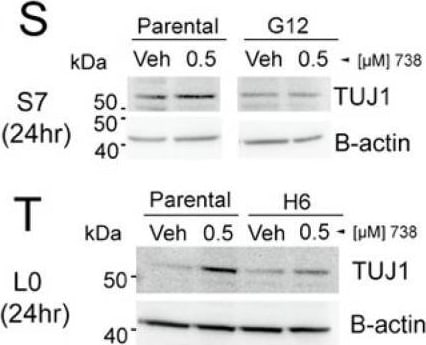
Detection of Neuron-specific beta-III Tubulin NL637 by Western Blot
ACY-738 induced changes in Ki67 and TUJ1 are not observed in ARL13B-KO, cilia-depleted human glioma cells. (A–P) S7 parental cells (left two columns) or ARL13B KO (clone G12) (right two columns) were grown to ~75% confluency and treated with vehicle or 500-nM 738. Cells were fixed after 24 h and immunostained for Ki67 (A–D), TUJ1 (E–H), and ARL13B (I–L). Merged images are displayed M-P. Scale bar in A = 10 µm. (Q) Mean nuclear intensity of Ki67 signal for indicated cell lines and treatments. n = 60–80 cells/group. * p < 0.05, **** p < 0.0001, ANOVA. (R) Mean soma intensity of TUJ1 signal for indicated cell lines and treatments. (S,T) Western blot of S7 (S) and L0 (T) cell lysates 24 h after exposure to vehicle or 500-nM 738. Blots were probed for TUJ1 and beta -actin as loading controls. Image collected and cropped by CiteAb from the following open publication (https://pubmed.ncbi.nlm.nih.gov/33915983), licensed under a CC-BY license. Not internally tested by R&D Systems.Applications for Neuron-specific beta-III Tubulin Antibody (TuJ-1) [NL637]
Immunocytochemistry
Reviewed Applications
Read 2 reviews rated 4.5 using NL1195V in the following applications:
Formulation, Preparation, and Storage
Formulation
Shipping
Stability & Storage
Background: beta-III Tubulin
Alternate Names
Entrez Gene IDs
Gene Symbol
Additional beta-III Tubulin Products
Product Documents for Neuron-specific beta-III Tubulin Antibody (TuJ-1) [NL637]
Certificate of Analysis
To download a Certificate of Analysis, please enter a lot or batch number in the search box below.
Note: Certificate of Analysis not available for kit components.
Product Specific Notices for Neuron-specific beta-III Tubulin Antibody (TuJ-1) [NL637]
For research use only
Related Research Areas
Citations for Neuron-specific beta-III Tubulin Antibody (TuJ-1) [NL637]
 Powered by Bioz
Customer Reviews for Neuron-specific beta-III Tubulin Antibody (TuJ-1) [NL637] (2)
Have you used Neuron-specific beta-III Tubulin Antibody (TuJ-1) [NL637]?
Submit a review and receive an Amazon gift card!
$25/€18/£15/$25CAN/¥2500 for a review with an image
$10/€7/£6/$10CAN/¥1110 for a review without an image
Submit a review
Customer Images
![Neuron-specific beta-III Tubulin Antibody (TuJ-1) [NL637] NL1195V](https://resources.rndsystems.com/images/reviews/review_nl1195v_68036_0.jpg)
-
Application: Immunocytochemistry/ImmunofluorescenceSample Tested: Prostate cancer cells and Human prostatic cell line PNT1ASpecies: HumanVerified Customer | Posted 11/01/2021
![Neuron-specific beta-III Tubulin Antibody (TuJ-1) [NL637] NL1195V](data:image/png;base64,R0lGODlhAQABAAD/ACwAAAAAAQABAAACADs=)
-
Application: Flow CytometrySample Tested: cellsSpecies: MultipleVerified Customer | Posted 05/02/2017
There are no reviews that match your criteria.
Protocols
Find general support by application which include: protocols, troubleshooting, illustrated assays, videos and webinars.
- Appropriate Fixation of IHC/ICC Samples
- Cellular Response to Hypoxia Protocols
- ClariTSA™ Fluorophore Kits
- Detection & Visualization of Antibody Binding
- ICC Cell Smear Protocol for Suspension Cells
- ICC Immunocytochemistry Protocol Videos
- ICC for Adherent Cells
- Immunocytochemistry (ICC) Protocol
- Immunocytochemistry Troubleshooting
- Immunofluorescence of Organoids Embedded in Cultrex Basement Membrane Extract
- Immunohistochemistry (IHC) and Immunocytochemistry (ICC) Protocols
- Preparing Samples for IHC/ICC Experiments
- Preventing Non-Specific Staining (Non-Specific Binding)
- Primary Antibody Selection & Optimization
- Protocol for VisUCyte™ HRP Polymer Detection Reagent
- Protocol for the Fluorescent ICC Staining of Cell Smears - Graphic
- Protocol for the Fluorescent ICC Staining of Cultured Cells on Coverslips - Graphic
- Protocol for the Preparation and Fluorescent ICC Staining of Cells on Coverslips
- Protocol for the Preparation and Fluorescent ICC Staining of Non-adherent Cells
- Protocol for the Preparation and Fluorescent ICC Staining of Stem Cells on Coverslips
- Protocol for the Preparation of a Cell Smear for Non-adherent Cell ICC - Graphic
- TUNEL and Active Caspase-3 Detection by IHC/ICC Protocol
- The Importance of IHC/ICC Controls
- View all Protocols, Troubleshooting, Illustrated assays and Webinars