![Western Blot: RAB35 Antibody [NBP1-79484]](https://resources.rndsystems.com/images/products/RAB35-Antibody-Western-Blot-NBP1-79484-img0001.jpg "Western Blot: RAB35 Antibody [NBP1-79484]")
Loading...
Key Product Details
Species Reactivity
Human
Applications
Western Blot, Immunocytochemistry/ Immunofluorescence
Label
Unconjugated
Antibody Source
Polyclonal Rabbit IgG
Format
BSA Free
Loading...
Product Specifications
Immunogen
Synthetic peptide directed towards the middle region of human RAB35. Peptide sequence: GIQLFETSAKENVNVEEMFNCITELVLRAKKDNLAKQQQQQQNDVVKLTK The peptide sequence for this immunogen was taken from within the described region.
Clonality
Polyclonal
Host
Rabbit
Isotype
IgG
Theoretical MW
23 kDa.
Disclaimer note: The observed molecular weight of the protein may vary from the listed predicted molecular weight due to post translational modifications, post translation cleavages, relative charges, and other experimental factors.
Disclaimer note: The observed molecular weight of the protein may vary from the listed predicted molecular weight due to post translational modifications, post translation cleavages, relative charges, and other experimental factors.
Description
The addition of 50% glycerol is optional for those storing this antibody at -20C and not aliquoting smaller units. However, please note that glycerol may interrupt some downstream antibody applications and should be added with caution.
Scientific Data Images for RAB35 Antibody - BSA Free
Western Blot: RAB35 Antibody [NBP1-79484]
Western Blot: RAB35 Antibody [NBP1-79484] - Human heart cell lysate.![Immunocytochemistry/ Immunofluorescence: RAB35 Antibody [NBP1-79484]](https://resources.rndsystems.com/images/products/RAB35-Antibody-Immunocytochemistry-Immunofluorescence-NBP1-79484-img0004.jpg "Immunocytochemistry/ Immunofluorescence: RAB35 Antibody [NBP1-79484]")
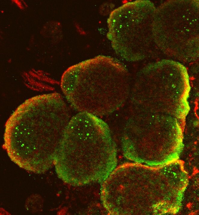
Immunocytochemistry/ Immunofluorescence: RAB35 Antibody [NBP1-79484]
Immunocytochemistry/Immunofluorescence: RAB35 Antibody [NBP1-79484] - Human dorsal root ganglion neurons. Dual ICC stain RAB35 in green, NF200 in red. Note axons are out of view, this is a Z-stack section. RAB35 antibody at 1:500. Strong plasma membrane binding which is consistent with tagged construct expression. Some non-specific binding in the nucleus/nucleolus which may improve with lowered antibody concentrations. Primary incubation overnight at 4C, secondary antibodies (Alexa 488 and 546) at room temperature. ICC/IF image submitted by a verified customer review.![Western Blot: RAB35 Antibody [NBP1-79484]](https://resources.rndsystems.com/images/products/RAB35-Antibody-Western-Blot-NBP1-79484-img0002.jpg "Western Blot: RAB35 Antibody [NBP1-79484]")
Western Blot: RAB35 Antibody [NBP1-79484]
Western Blot: RAB35 Antibody [NBP1-79484] - HeLa cells, HEK293T cells. Antibody at 1:500.Applications for RAB35 Antibody - BSA Free
Application
Recommended Usage
Western Blot
1.0 ug/ml
Application Notes
This RAB35 antibody is validated for ICC/IF from a verified customer review.
Reviewed Applications
Read 1 review rated 4 using NBP1-79484 in the following applications:
Formulation, Preparation, and Storage
Purification
Affinity purified
Formulation
PBS, 2% Sucrose
Format
BSA Free
Preservative
0.09% Sodium Azide
Concentration
0.5 mg/ml
Shipping
The product is shipped with polar packs. Upon receipt, store it immediately at the temperature recommended below.
Stability & Storage
Store at 4C short term. Aliquot and store at -20C long term. Avoid freeze-thaw cycles.
Background: RAB35
Alternate Names
GTP-binding protein RAY, H-ray, RAB1C, RAB35, member RAS oncogene family, Ras-related protein Rab-1C, ras-related protein rab-1c (GTP-binding protein ray), ras-related protein Rab-35, RAY
Gene Symbol
RAB35
UniProt
Additional RAB35 Products
Product Documents for RAB35 Antibody - BSA Free
Certificate of Analysis
To download a Certificate of Analysis, please enter a lot or batch number in the search box below.
Product Specific Notices for RAB35 Antibody - BSA Free
This product is for research use only and is not approved for use in humans or in clinical diagnosis. Primary Antibodies are guaranteed for 1 year from date of receipt.
Customer Reviews for RAB35 Antibody - BSA Free (1)
4 out of 5
1 Customer Rating
Have you used RAB35 Antibody - BSA Free?
Submit a review and receive an Amazon gift card!
$25/€18/£15/$25CAN/¥2500 Yen for a review with an image
$10/€7/£6/$10CAN/¥1110 Yen for a review without an image
Submit a review
Customer Images
Showing
1
-
1 的
1 review
Showing All
Filter By:
-
Application: ImmunocytochemistrySample Tested: Dorsal root ganglion neuronsSpecies: HumanVerified Customer | Posted 12/22/2019Dual ICC stain RAB35 in green, NF200 in red. Note axons are out of view, this is a Z-stack section. Strong PM binding.This antibody is only listed as verified in WB so I decide to test it in ICC. I used it at 1:500, originally tried 1:300 but that was too high - I think the concentration could even be dropped to 1:1000, this may improve it's specificity. There was strong plasma membrane binding which is consistent with tagged construct expression. Some non-specific binding in the nucleus/ nucleolus which may improve with lowered antibody concentrations. Primary incubation was performed overnight at 4C, secondary antibodies (Alexa 488 and 546) at room temperature.
 Bio-Techne ResponseThis review was submitted through the legacy Novus Innovators Program, reflecting a new species or application tested on a primary antibody.
There are no reviews that match your criteria.
Protocols
Find general support by application which include: protocols, troubleshooting, illustrated assays, videos and webinars.
- Appropriate Fixation of IHC/ICC Samples
- Cellular Response to Hypoxia Protocols
- ClariTSA™ Fluorophore Kits
- Detection & Visualization of Antibody Binding
- ICC Cell Smear Protocol for Suspension Cells
- ICC Immunocytochemistry Protocol Videos
- ICC for Adherent Cells
- Immunocytochemistry (ICC) Protocol
- Immunocytochemistry Troubleshooting
- Immunofluorescence of Organoids Embedded in Cultrex Basement Membrane Extract
- Immunohistochemistry (IHC) and Immunocytochemistry (ICC) Protocols
- Preparing Samples for IHC/ICC Experiments
- Preventing Non-Specific Staining (Non-Specific Binding)
- Primary Antibody Selection & Optimization
- Protocol for VisUCyte™ HRP Polymer Detection Reagent
- Protocol for the Fluorescent ICC Staining of Cell Smears - Graphic
- Protocol for the Fluorescent ICC Staining of Cultured Cells on Coverslips - Graphic
- Protocol for the Preparation and Fluorescent ICC Staining of Cells on Coverslips
- Protocol for the Preparation and Fluorescent ICC Staining of Non-adherent Cells
- Protocol for the Preparation and Fluorescent ICC Staining of Stem Cells on Coverslips
- Protocol for the Preparation of a Cell Smear for Non-adherent Cell ICC - Graphic
- R&D Systems Quality Control Western Blot Protocol
- TUNEL and Active Caspase-3 Detection by IHC/ICC Protocol
- The Importance of IHC/ICC Controls
- Troubleshooting Guide: Western Blot Figures
- Western Blot Conditions
- Western Blot Protocol
- Western Blot Protocol for Cell Lysates
- Western Blot Troubleshooting
- Western Blot Troubleshooting Guide
- View all Protocols, Troubleshooting, Illustrated assays and Webinars
Loading...