
SR-BI Antibody - BSA Free
Novus Biologicals | Catalog # NBP2-54750

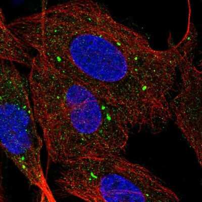
Scientific images may be resized, cropped, or adjusted for brightness or contrast for presentation purposes. These updates are limited to presentation and do not affect the underlying scientific interpretation of the data. Where available, additional source data may be provided upon request.
Loading...
Key Product Details
Species Reactivity
Human
Applications
Immunocytochemistry/ Immunofluorescence
Label
Unconjugated
Antibody Source
Polyclonal Rabbit IgG
Format
BSA Free
Loading...
Product Specifications
Immunogen
This SR-BI Antibody was developed against a Recombinant Protein corresponding to amino acids: PRSLSFNMWKEIPIPFYLSVYFFDVMNPSEILKGEKPQVRERGPYVYREFRHKSNITFNNNDTVSFLEYRTFQFQPSKSHGSESDYIVMPNILVLGA
Clonality
Polyclonal
Host
Rabbit
Isotype
IgG
Scientific Data Images for SR-BI Antibody - BSA Free
Immunocytochemistry/ Immunofluorescence: SR-BI Antibody [NBP2-54750]
Immunocytochemistry/Immunofluorescence: SR-BI Antibody [NBP2-54750] - Immunofluorescent staining of human cell line U-2 OS shows localization to vesicles.Applications for SR-BI Antibody - BSA Free
Application
Recommended Usage
Immunocytochemistry/ Immunofluorescence
0.25-2 ug/ml
Application Notes
ICC/IF, Fixation Permeabilization: Use PFA/Triton X-100.
Formulation, Preparation, and Storage
Purification
Affinity purified
Formulation
PBS (pH 7.2) and 40% Glycerol
Format
BSA Free
Preservative
0.02% Sodium Azide
Concentration
Concentrations vary lot to lot. See vial label for concentration. If unlisted please contact technical services.
Shipping
The product is shipped with polar packs. Upon receipt, store it immediately at the temperature recommended below.
Stability & Storage
Store at 4C short term. Aliquot and store at -20C long term. Avoid freeze-thaw cycles.
Background: SR-BI
At least 5 different human splice variants have been identified with theoretical molecular weights ranging from 60.9 kDa for the canonical sequence to 45 kDa for an N-terminally truncated variant (SR-BII) (3). The extracellular domain of human SR-BI (CLA-1) shares 80%, 80%, 89%, 86% and 84% aa sequence identity with mouse, rat, porcine, rabbit, and bovine SR-BI, respectively. SR-BI is highly glycosylated and removal of N-linked glycosylation reduces lipid transport.
References
1. Linton MF, Tao H, Linton EF, Yancey PG. (2017) SR-BI: A Multifunctional Receptor in Cholesterol Homeostasis and Atherosclerosis. Trends Endocrinol Metab. 28(6):461-472. PMID: 28259375
2. Hoekstra M, Sorci-Thomas M. (2017) Rediscovering scavenger receptor type BI: surprising new roles for the HDL receptor. Curr Opin Lipidol. 28(3):255-260. PMID: 28301373
3. Shen WJ, Asthana S, Kraemer FB, Azhar S. (2018) Scavenger receptor B type 1: expression, molecular regulation, and cholesterol transport function.
Long Name
Scavenger Receptor Class B, Member I
Alternate Names
CD36L1, CLA1, HDLQTL6, SCARB1, SR-B1, SRBI
Gene Symbol
SCARB1
Additional SR-BI Products
Product Documents for SR-BI Antibody - BSA Free
Certificate of Analysis
To download a Certificate of Analysis, please enter a lot or batch number in the search box below.
Product Specific Notices for SR-BI Antibody - BSA Free
This product is for research use only and is not approved for use in humans or in clinical diagnosis. Primary Antibodies are guaranteed for 1 year from date of receipt.
Related Research Areas
Customer Reviews for SR-BI Antibody - BSA Free
There are currently no reviews for this product. Be the first to review SR-BI Antibody - BSA Free and earn rewards!
Have you used SR-BI Antibody - BSA Free?
Submit a review and receive an Amazon gift card!
$25/€18/£15/$25CAN/¥2500 for a review with an image
$10/€7/£6/$10CAN/¥1110 for a review without an image
Submit a review
Protocols
Find general support by application which include: protocols, troubleshooting, illustrated assays, videos and webinars.
- Appropriate Fixation of IHC/ICC Samples
- Cellular Response to Hypoxia Protocols
- ClariTSA™ Fluorophore Kits
- Detection & Visualization of Antibody Binding
- ICC Cell Smear Protocol for Suspension Cells
- ICC Immunocytochemistry Protocol Videos
- ICC for Adherent Cells
- Immunocytochemistry (ICC) Protocol
- Immunocytochemistry Troubleshooting
- Immunofluorescence of Organoids Embedded in Cultrex Basement Membrane Extract
- Immunohistochemistry (IHC) and Immunocytochemistry (ICC) Protocols
- Preparing Samples for IHC/ICC Experiments
- Preventing Non-Specific Staining (Non-Specific Binding)
- Primary Antibody Selection & Optimization
- Protocol for VisUCyte™ HRP Polymer Detection Reagent
- Protocol for the Fluorescent ICC Staining of Cell Smears - Graphic
- Protocol for the Fluorescent ICC Staining of Cultured Cells on Coverslips - Graphic
- Protocol for the Preparation and Fluorescent ICC Staining of Cells on Coverslips
- Protocol for the Preparation and Fluorescent ICC Staining of Non-adherent Cells
- Protocol for the Preparation and Fluorescent ICC Staining of Stem Cells on Coverslips
- Protocol for the Preparation of a Cell Smear for Non-adherent Cell ICC - Graphic
- TUNEL and Active Caspase-3 Detection by IHC/ICC Protocol
- The Importance of IHC/ICC Controls
- View all Protocols, Troubleshooting, Illustrated assays and Webinars
Loading...