Jess Automated Western System
Jess Provides Powerful Multiplex Western Results
The Jess™ System, powered by Simple Western™ technology, is designed to deliver fast, high‑sensitivity, multiplex Western analysis with integrated chemiluminescent and two-colors (NIR and IR) fluorescent detection. Jess automates protein size separation and immunodetection for up to 25 capillaries per run, providing fully analyzed results in as little as 3 hours and eliminating the variability and labor‑intensive steps of traditional Western blots.
By combining automation, high sensitivity, and robust multiplexing, Jess transforms Western blot analysis and brings this essential workflow into the 21st century.
How Does Jess Work?
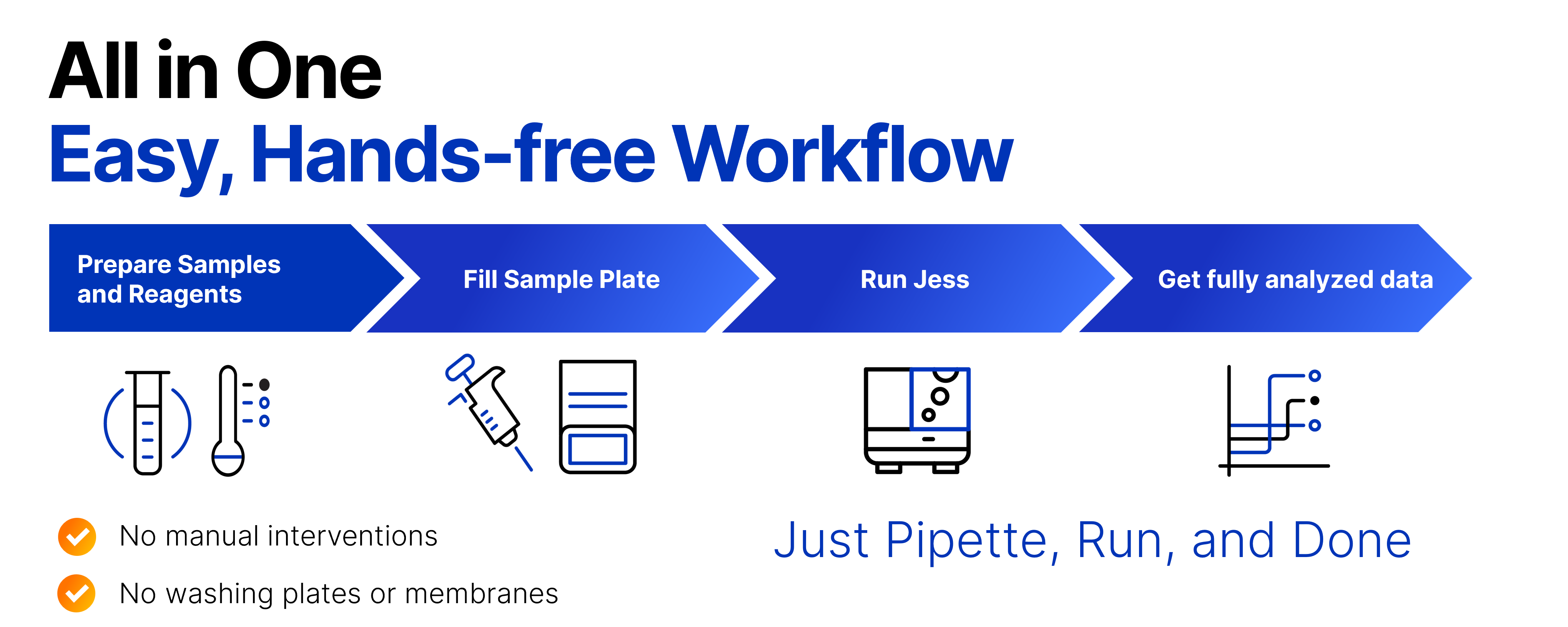
Jess automates the protein separation and immunodetection steps of traditional Western blotting, removing many tedious and error‑prone manual processes. After samples and reagents are loaded into the microplate, the system manages protein separation by size along with all antibody additions, incubations, washes, and detection steps. Fully quantitative results are generated in approximately 3 hours, supported by integrated chemiluminescent and fluorescent detection for picogram‑level sensitivity, built‑in total protein normalization, and highly reproducible data with molecular weight characterization. Jess delivers both protein quantitation plus specificity and size resolution, giving you comprehensive data in a single run.
Unboxing Jess with CatSci
Discover how Bioscience Team Leader Ryan Mordue from CatSci Ltd. unboxed and set up Jess. This video highlights just how incredibly easy it is to integrate Jess into an active research environment.
Hearing from the Experts: From Bench to Bedside
Discover how Jess transformed Dr. Soni’s research by providing the high sensitivity necessary to detect and quantify EV-associated proteins.
How Can Jess Help You?
Fast Time to Consistent, Reliable Results
With Jess, it's pipette, run & done! Samples, antibodies, and reagents are loaded into the plate, and the system automates every assay step—from protein separation and immunoprobing to detection and data analysis—inside a fully enclosed capillary environment. With only 30 minutes of experimental setup and approximately 3 hours of hands‑free run time, Jess delivers intra-assay CVs <15%, giving you the consistency you need to be confident in your data.
Figure, Right: Multiplexed analysis of total AKT and pAKT with total protein normalization using Jess. (A) Lysates from Jurkat cells either untreated (-) or treated (+) with calyculin A were analyzed at 0.25 mg/mL and 0.15 mg/mL final concentrations, respectively. Each lysate was probed for total AKT with IR (green bands) and pAKT with NIR (red bands). (B) The Total Protein Assay was performed in the same lanes shown in panel A.

Detect Low and High Abundance Proteins with Confidence
With just 3 µL of sample, Jess allows you to get down to picogram-level sensitivity with chemiluminescent detection or dual color (NIR / IR) fluorescent detection. It enables you to detect high and low abundance proteins in the same sample, even when they have identical molecular weights.

More Data from Every Sample with RePlex
RePlex™ assay enables two sequential immunoassays (IA) or an IA + total protein assay (TPA) within the same capillary, maximizing data output from each sample load and providing deeper biological insight without additional sample consumption.
With the RePlex feature, Jess offers flexible multiplexing options that allow you to do multi-target detection and protein normalization in the same capillary.
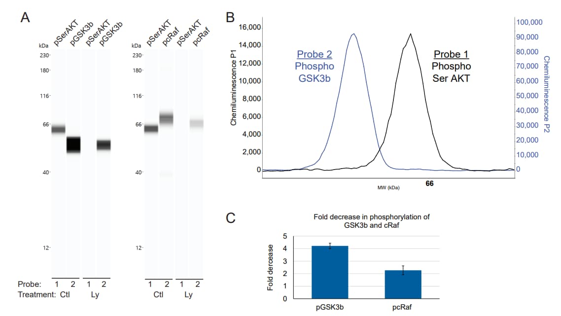
Figure, Right: RePlex detection of concomitant phosphorylation for AKT and downstream targets in the same capillary. (A) Lane view of AKT, GSK3b and cRaf phosphorylation in control (Ctl) and LY294002-treated (LY) Jurkat lysates. (B) RePlex enables use of the same secondary antibody in Probe 1 and Probe 2, with no carryover signal, as demonstrated by detection of phosphorylated AKT and GSK3b using rabbit-derived primary antibodies in sequential probes of the same sample. (C) Quantitation shows a decrease in GSK3b and cRaf phosphorylation in LY294002-treated Jurkat lysates. Error bars represent standard deviations of the means.
Gain Fully Quantitative Analysis
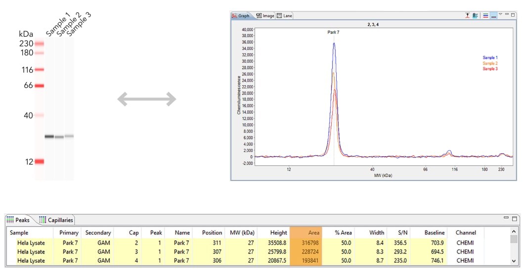
With Jess, protein quantification is a breeze. At the conclusion of your run, use the lane view option to compare band intensity or dive deep for fully quantitative analysis of protein size and concentration. With a few clicks, you’ll be analyzing immunoassay-like standard curves and precisely quantifying your protein.
Normalization with Total Protein Using RePlex
Total protein normalization provides a more accurate and reliable way to quantify protein expression by correcting differences in total protein load and avoiding the variability seen with housekeeping proteins. With RePlex, Jess offers total protein normalization using both chemiluminescent and NIR detection, enabling flexible and consistent analysis across a wide range of targets. Same‑time detection within the capillary delivers highly consistent, dependable results without any blot staining or wash steps—modernizing normalization and strengthening confidence in every data set.
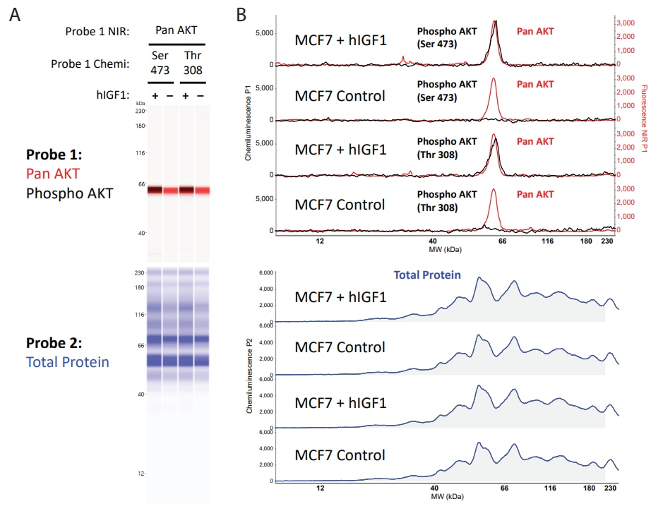
Figure, Right: Automated immunoassay and total protein detection in a single capillary of AKT phosphorylation in MCF7 lysates treated and untreated with hIGF1. (A) Phosphorylated AKT (phospho AKT) isoforms and pan AKT were detected in Probe 1 using chemiluminescence and NIR fluorescence, respectively, while total protein signal was detected in Probe 2. (B) Example graph views of phospho AKT and pan AKT (top), and total protein signal (bottom) for samples in panel A
Flexible Imaging for Traditional Western Blot Membranes
Jess includes an integrated imaging system that supports imaging of traditional western blotting membranes, offering added flexibility for labs that need both fully automated capillary‑based Western analysis and conventional blot imaging in a single instrument.

Jess’s imaging system allows for imaging of traditional Western blotting membranes
Jess Certified Service Provider Program
Find an Expert to Process Your Samples with Jess
The CSP program combines dedicated expertise with our instrument portfolio including Jess to create a robust solution for effective, reliable sample analysis. See the service providers that have successfully completed Bio-Techne’s CSP training program and are certified to run your samples on Jess.
Instrument Specifications
| 23 kg (50 lbs) Description | Total Protein with Chemiluminescence Detection | Chemiluminescence Detection | Fluorescence Detection |
|---|---|---|---|
| Sample required + | 0.3-1.2 µg | 0.3-1.2 µg | 2-4 µg |
| Volume required | 3 µL/well | ||
| Size range | Molecular weight (MW) ladder ranges from 2-440 kDa | ||
| Sizing CV | <10% | ||
| Intra-assay CV | <15% | ||
| Inter-assay CV | N/A | <20%* | <20% |
| Resolution (± percent difference in MW) | ± 10% for MW >20 kDa ± 15-20% for MW <20 kDA | ||
| Quantitation CV | <20% (total protein, chemiluminescence and fluorescence) | ||
| Dynamic range | 2-3 logs | 3-4 logs | |
| Sensitivity | ng | Low pg | High pg |
| Capillary | 5 cm, 100 µm, 400 nL | ||
| Runtime | <3 hours RePlex: 5 hours | ||
| Capillaries per run | 13 or 25 | ||
| Weight | 23 kg (50 lbs) | ||
| Dimensions (closed) | 0.33 m x 0.36 m x 0.52 m (1.07' x 1.15' x 1.71') | ||
| Power | US/CAN 120 V AC, 60 hz, 4.2 amps Europe 240 AC, 50 Hz, 2.1 amps Japan 100 AC, 50/60 Hz, 5.0 amps | ||
| Operating temperature | 18-24 °C | ||
| Operating humidity | 20-60% relative, non-condensing | ||
+ Sample Required is 3 µL/well
*Inter-assay CV is calculated with normalization to the system control protein
** Percent peak area
How to Set-Up Jess Training Kits
Watch this step-by-step video to learn how to run and set up Jess training kit.
Webinars
Going Beyond the Limits of Traditional Westerns: Detecting Low-Abundance Proteins in Your Model System
Beyond the Blot: Achieving Reproducible, Publication-Ready Protein Expression Data
Interrogating Cell Signaling Pathways in iPSC-Derived Microglia to Understand Alzheimer's Pathology
From ugly blots to publication‑ready data
Discover how Simple Western™ Jess™ is advancing neurodegeneration research.

"Using Jess has been extremely rewarding, especially due to how easy it was to transfer our western blotting assays to a fully automated format on Simple Western and how well CST antibodies perform on the platform."
Chris LaBreck, Ph.D., Senior Product Scientist, Cell Signaling Technology (CST)

"It saves so much time! It takes away so much of the pipetting and manipulation error. You just load your samples and wait for results, and in the meantime, you can dedicate yourself to other activities."
Nicolas G. Bazan, M.D., Ph.D., Director, LSUHSC Neuroscience Center of Excellence
"Our clients expect high-quality data with rapid turnarounds. Jess allows me to accurately assess drug compound potency, and with its high throughput ability I can screen multiple compounds quickly and efficiently."
Rachel Doidge Ph.D., Senior Research Scientist, Aurelia Bioscience
 Powered by Bioz