Anti-Mouse Ig (H+L) Comp-Bead 3 Population (7.5 um) Kit
Novus Biologicals | Catalog # NBP3-00499
Loading...
Key Product Details
Applications
Flow Cytometry
Kit Type
Kit
Product Summary for Anti-Mouse Ig (H+L) Comp-Bead 3 Population (7.5 um) Kit
The Anti-Mouse Ig (H+L) Compensation Beads are designed to capture fluorophore-conjugated antibodies, serving as accurate and consistent compensation controls in multicolor flow cytometry.
Particle size: 7.0 -7.9 micron
Particle material: polystyrene beads coated with goat anti-mouse IgG
- Bind to antibodies of mouse, rat, and hamster origin and are immunoglobulin light chain independent
- Compatible with UV- to near IR-excited fluorophores
- Exhibit low autofluorescence regardless of excitation wavelength (e.x. violet laser or red laser) or detection bandpass
- Contains 3 populations (negative, low, high) for comprehensive quality control of fluorophores
Particle size: 7.0 -7.9 micron
Particle material: polystyrene beads coated with goat anti-mouse IgG
Loading...
Scientific Data Images for Anti-Mouse Ig (H+L) Comp-Bead 3 Population (7.5 um) Kit
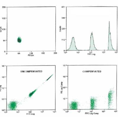
Anti-Mouse Ig (H+L) Comp-Bead 3 Population (7.5 um) Kit [NBP3-00499] - Histogram of flow cytometry data showing fron and side scatter and the three populations in the FITC channel. Dot plots show uncompensated and compensation populations in the FITC Log X PE Log.
Kit Contents for Anti-Mouse Ig (H+L) Comp-Bead 3 Population (7.5 um) Kit
- Comp Beads (high) - 5 mL
- Comp Beads (low) - 5 mL
- Comp Beads (negative) - 5 mL
Formulation, Preparation, and Storage
Formulation
PBS, 0.2% BSA
Preservative
0.02% Sodium Azide
Concentration
Concentration is not relevant for this product. Please see the protocols for proper use of this product.
Shipping
The product is shipped with polar packs. Upon receipt, store it immediately at the temperature recommended below.
Storage
Store at 4C. Do not freeze.
Background: Anti-Mouse Ig (H+L) Comp-Bead 3 Population (7.5 um)
Compensation beads are a highly desirable alternative to traditional cell-based single-color compensation. Positive beads can come pre-loaded with anti-IgG for conjugated antibody capture of mouse, rat, or hamster origin. Uncoated (negative) beads ensure standardization of the autofluorescence in each channel. Beads are preferable to cell-based compensation for several reasons:
1) No precious sample is wasted, and more sample can be used for experimental acquisition.
2) Since 100% of positive beads are able to capture the antibody, the exact experimental fluorochrome can be used regardless of a marker's cellular expression. Dimly expressed markers may not be bright enough for compensation using cells.
3) Reduced autofluorescence of beads allows for more precise calculation of spectral overlap.
Alternate Names
Comp Beads, Compensation Beads, Compensation Capture Beads, Flow Cytometry Compensation
Additional Anti-Mouse Ig (H+L) Comp-Bead 3 Population (7.5 um) Products
Product Documents for Anti-Mouse Ig (H+L) Comp-Bead 3 Population (7.5 um) Kit
Certificate of Analysis
To download a Certificate of Analysis, please enter a lot or batch number in the search box below.
Product Specific Notices for Anti-Mouse Ig (H+L) Comp-Bead 3 Population (7.5 um) Kit
This product is for research use only and is not approved for use in humans or in clinical diagnosis. Kits are guaranteed for 6 months from date of receipt.
Customer Reviews for Anti-Mouse Ig (H+L) Comp-Bead 3 Population (7.5 um) Kit
There are currently no reviews for this product. Be the first to review Anti-Mouse Ig (H+L) Comp-Bead 3 Population (7.5 um) Kit and earn rewards!
Have you used Anti-Mouse Ig (H+L) Comp-Bead 3 Population (7.5 um) Kit?
Submit a review and receive an Amazon gift card!
$25/€18/£15/$25CAN/¥2500 Yen for a review with an image
$10/€7/£6/$10CAN/¥1110 Yen for a review without an image
Submit a review
Protocols
View specific protocols for Anti-Mouse Ig (H+L) Comp-Bead 3 Population (7.5 um) Kit (NBP3-00499):
Shake vigorously or vortex before use. Sonication will help to increase the number of singles. Use for routine quality control of mouse antibody conjugates used in flow cytometry. Add 1 drop from each bottle to a centrifuge tube, followed by the addition of the conjugate being evaluated. After exposure to appropriate mouse antibody conjugates, three different fluorescent peaks shall be resolved by flow cytometry.
Find general support by application which include: protocols, troubleshooting, illustrated assays, videos and webinars.
- 7-Amino Actinomycin D (7-AAD) Cell Viability Flow Cytometry Protocol
- Extracellular Membrane Flow Cytometry Protocol
- Flow Cytometry Protocol for Cell Surface Markers
- Flow Cytometry Protocol for Staining Membrane Associated Proteins
- Flow Cytometry Staining Protocols
- Flow Cytometry Troubleshooting Guide
- Intracellular Flow Cytometry Protocol Using Alcohol (Methanol)
- Intracellular Flow Cytometry Protocol Using Detergents
- Intracellular Nuclear Staining Flow Cytometry Protocol Using Detergents
- Intracellular Staining Flow Cytometry Protocol Using Alcohol Permeabilization
- Intracellular Staining Flow Cytometry Protocol Using Detergents to Permeabilize Cells
- Propidium Iodide Cell Viability Flow Cytometry Protocol
- Protocol for Liperfluo
- Protocol for the Characterization of Human Th22 Cells
- Protocol for the Characterization of Human Th9 Cells
- Protocol: Annexin V and PI Staining by Flow Cytometry
- Protocol: Annexin V and PI Staining for Apoptosis by Flow Cytometry
- Troubleshooting Guide: Fluorokine Flow Cytometry Kits
- View all Protocols, Troubleshooting, Illustrated assays and Webinars
Loading...